WBCs and RBCs
1/32
There's no tags or description
Looks like no tags are added yet.
Name | Mastery | Learn | Test | Matching | Spaced |
|---|
No study sessions yet.
33 Terms
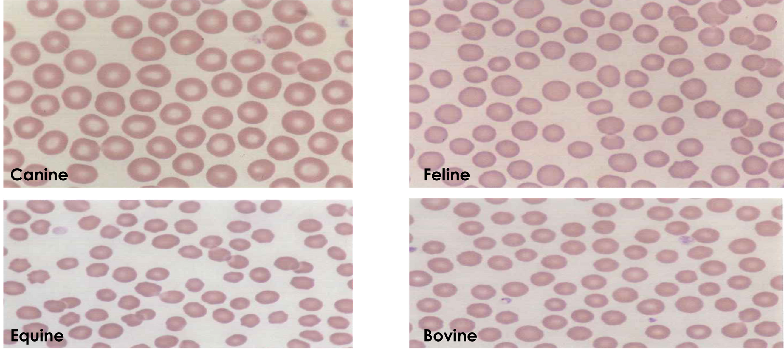
Normal RBCs
Avian RBC
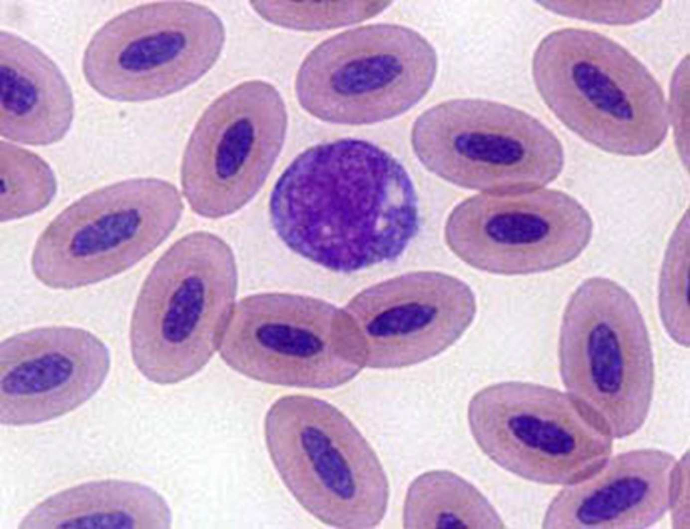
Camelid RBC
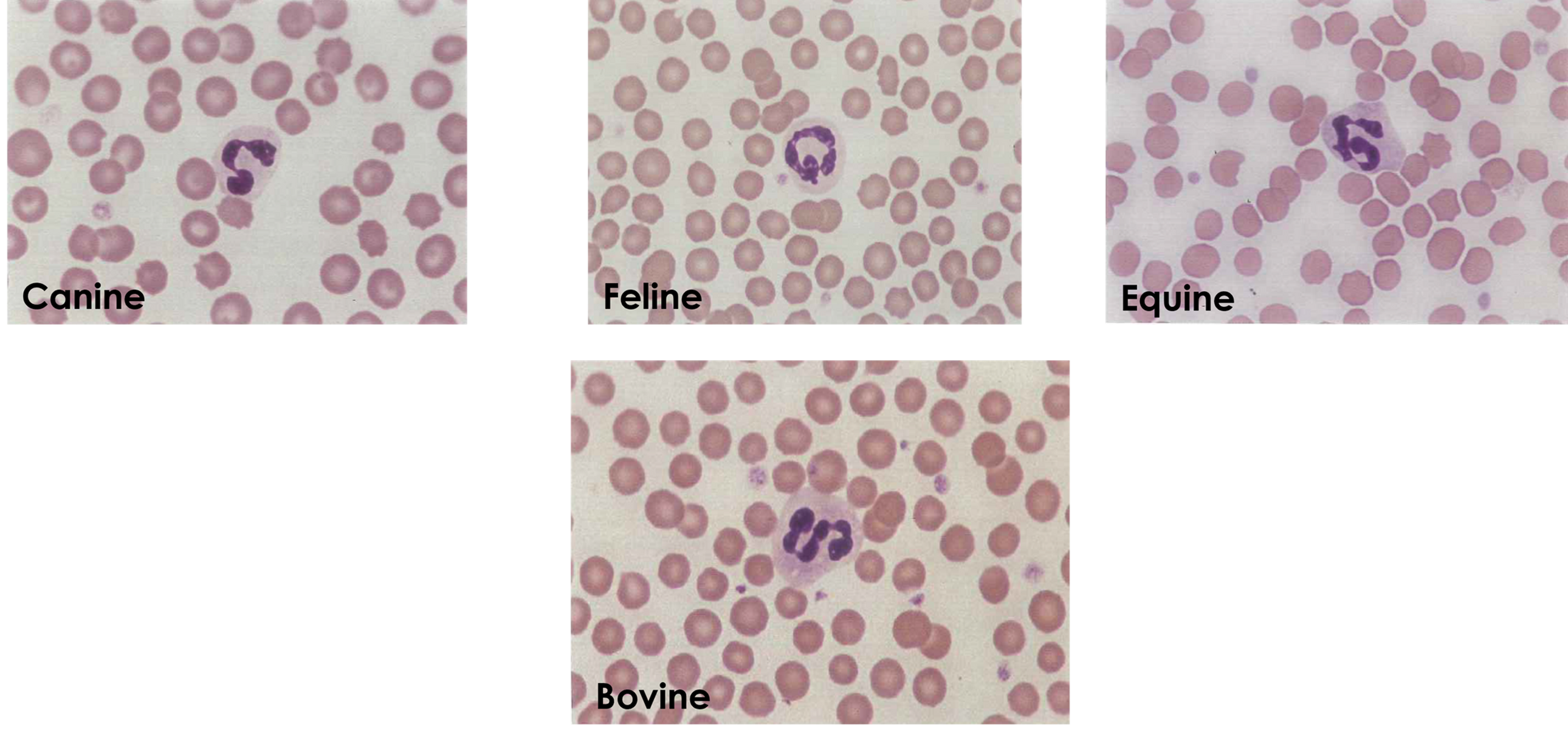
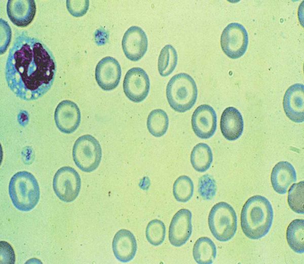
Neutrophils
-Most common WBC in all domestic species except ruminants
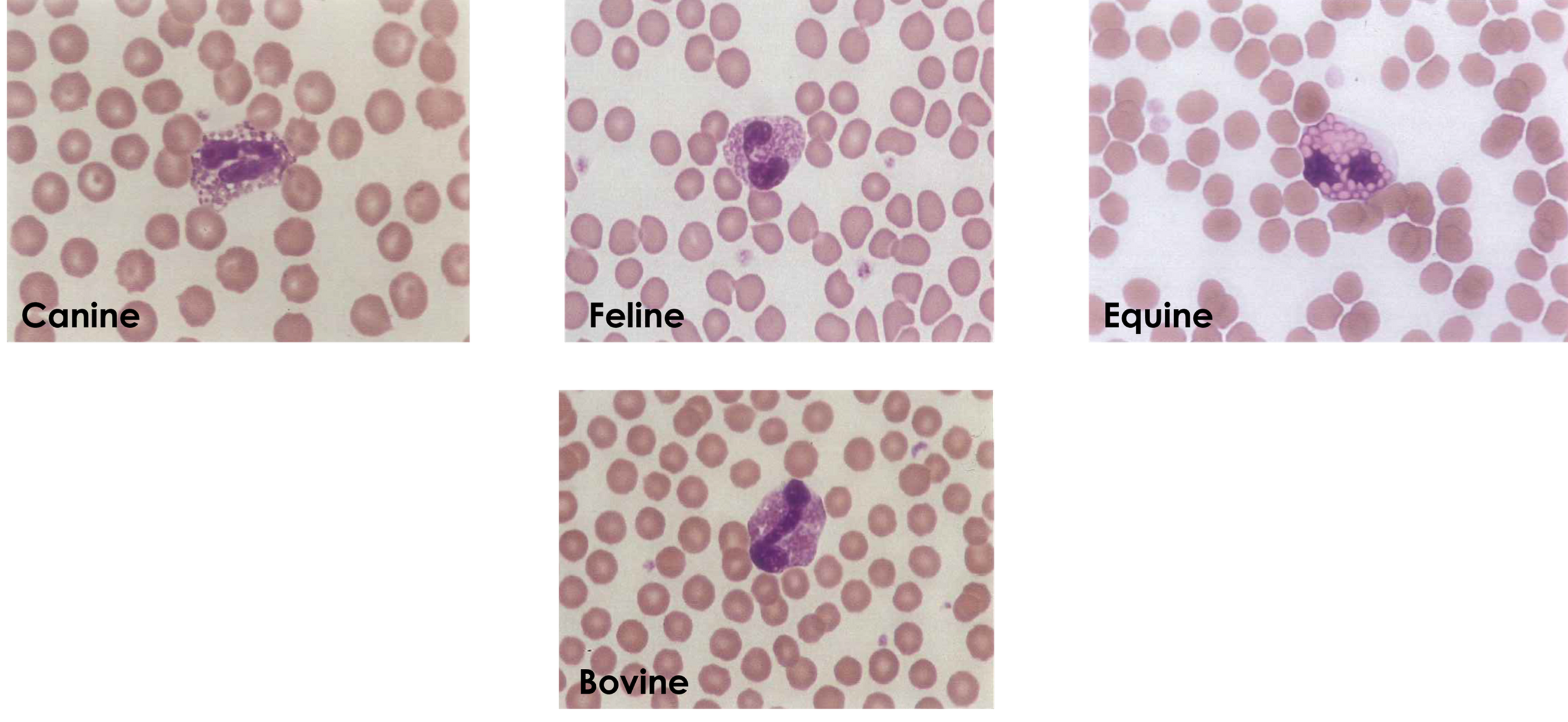
Eosinophils
-About the same size as a neutrophil
-Absent or present in very low numbers (normal)
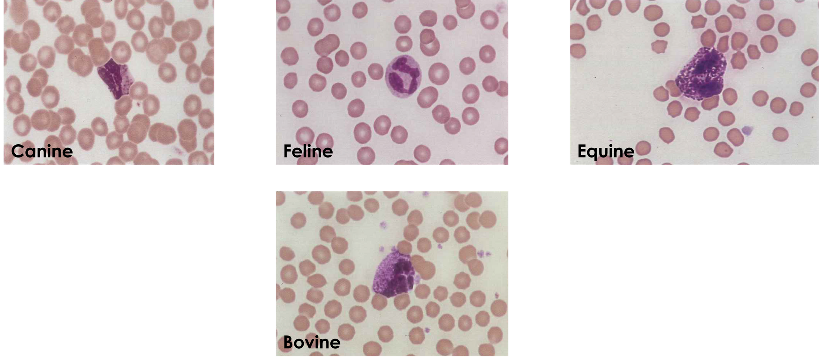
Basophils
-Rarely seen in normal blood smears
-Common in equine
-Slightly larger or same size as neutrophil
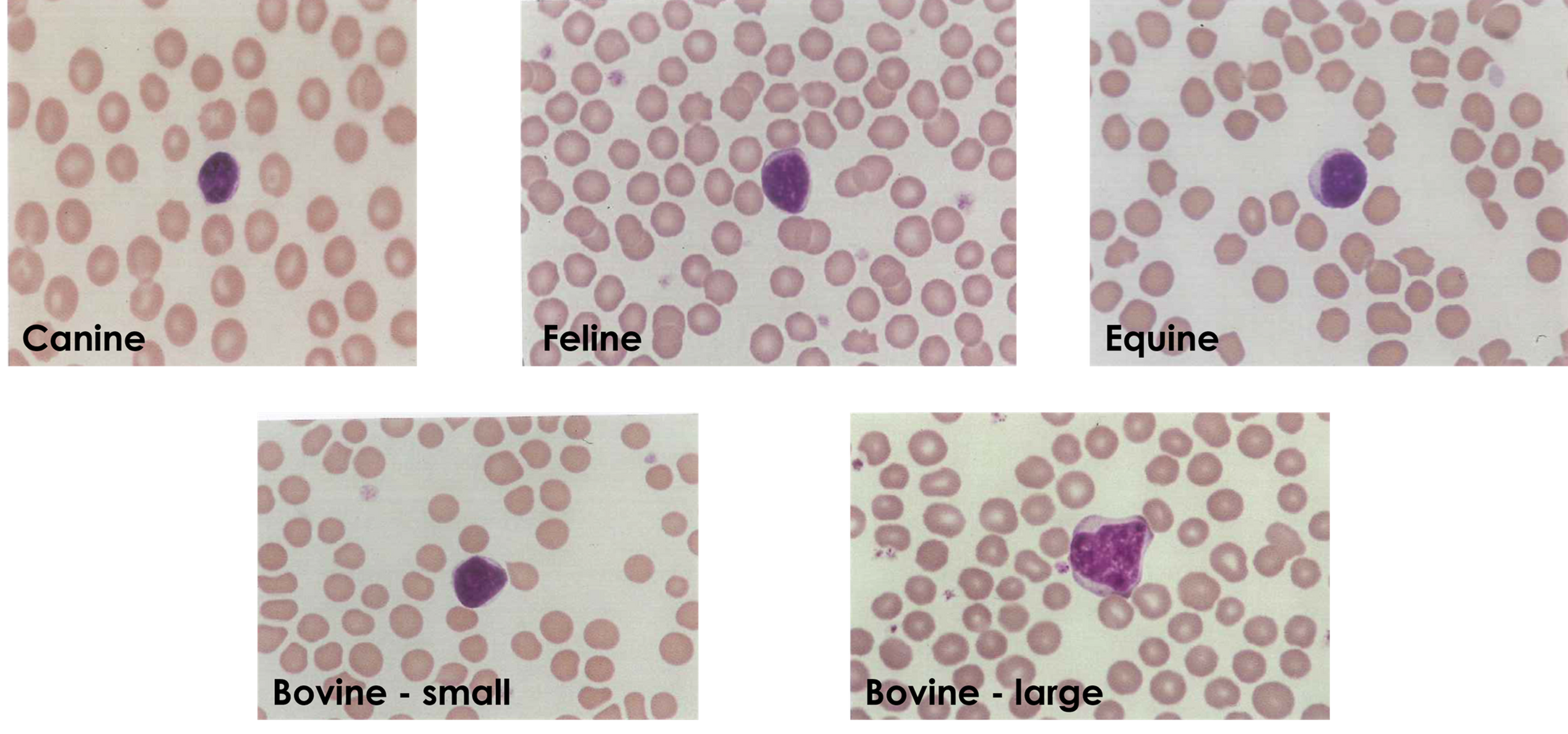
Lymphocytes
-Most common WBC in ruminants and lab animals
-Smallest of WBCs
-Minimal cytoplasm
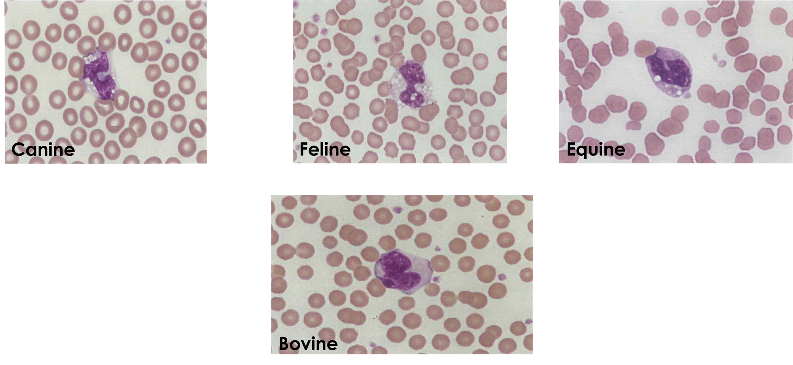
Monocytes
-Largest of all WBCs
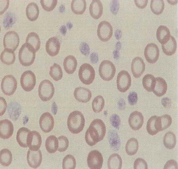
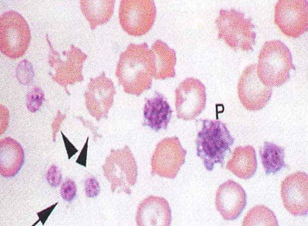
Platelets (Thrombocytes)
-Fragments of cytoplasm of megakaryocytes in bone marrow
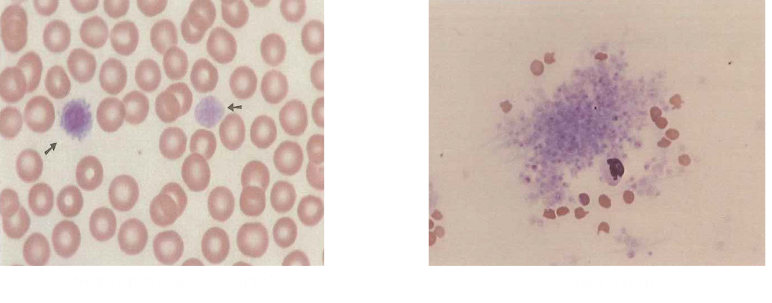
Macro platelet -common in cats
Platelet clump -common in dogs
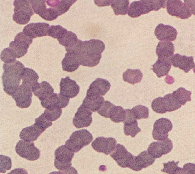
Rouleaux Formation (abnormal)
-Most common in horses
-May be an artifact of delay, refrigeration, or inflammation
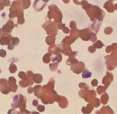
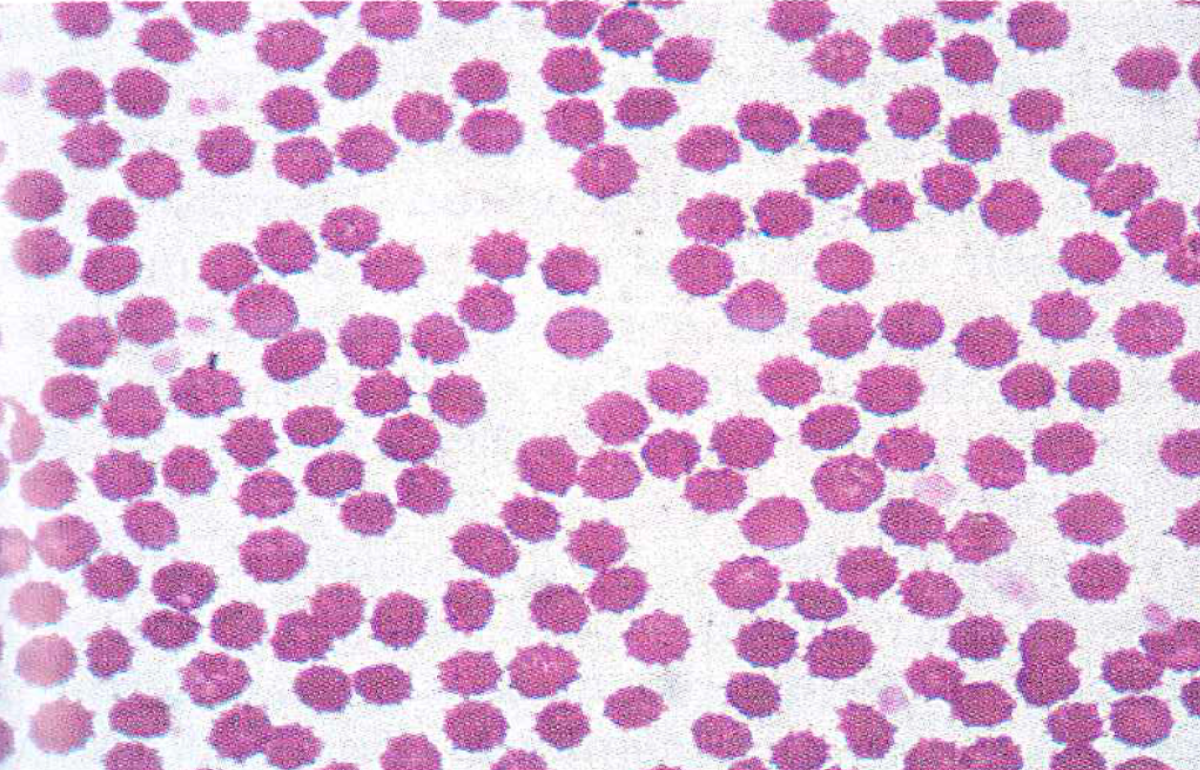
Agglutination (abnormal)
-Antibody coats the RBC surface causing clumping
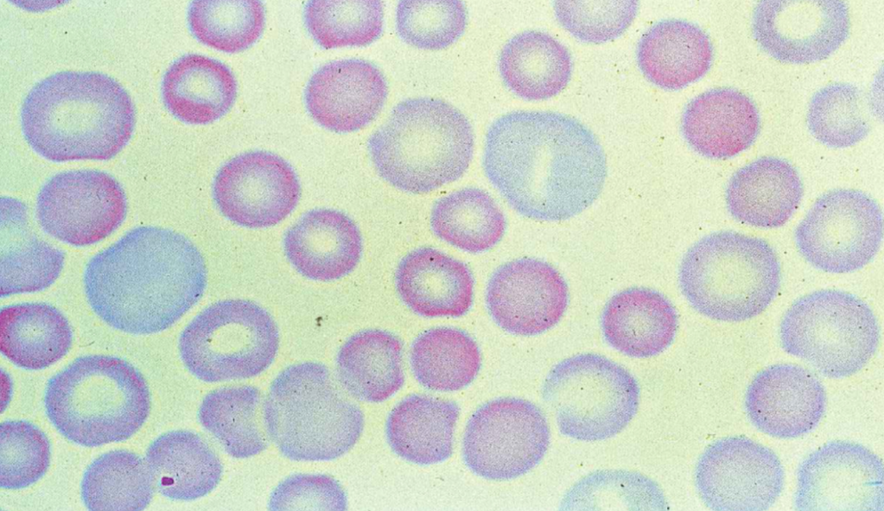
Anisocytosis (abnormal)
-Variation in RBC size
Hypochromic (abnormal)
-Increased central pallor and less heme

Torocyte (abnormal)
-Normal color but middle of cell is absent
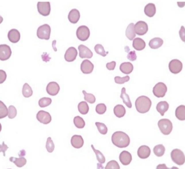
Poikilocytosis (abnormal)
-Variation in cell shape
-Common in healthy goats and neonate ruminants
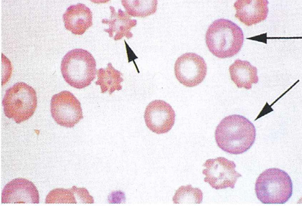
Echinocytes (abnormal)
-Crenated RBCs
-Surrounding solution is hypertonic
-Most common shape variation of RBCs
Acanthocytes (abnormal)
-Projections over entire surface
-Associated with liver and kidney disease and rattlesnake bites in dogs
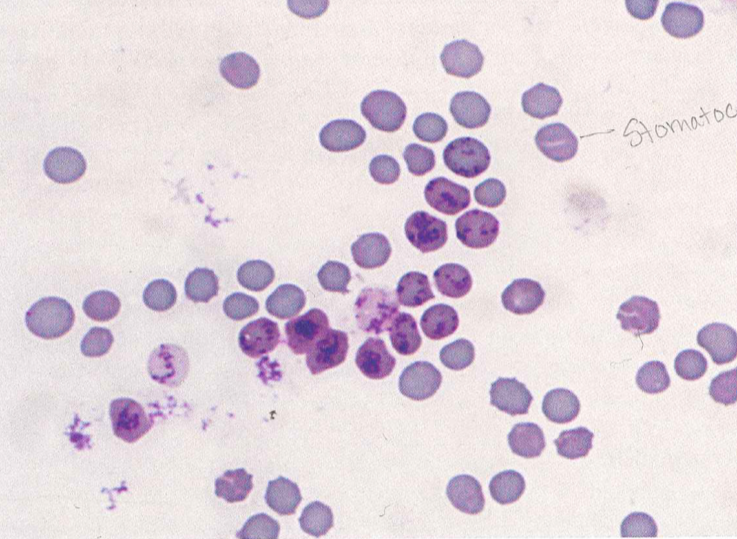
Stomatocytes (abnormal)
-Elongated, often curved, central pale area resembling a mouth
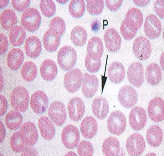
Knizocytes (abnormal)
-Bar cells
-Darker staining central area which extends across cell with pale on either side
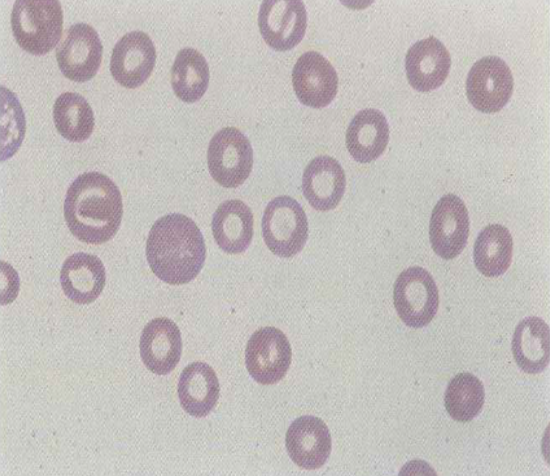
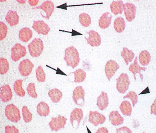
Codocytes (abnormal)
-Target cells
-Thicker, darker staining center surrounded by lighter staining area and dark periphery
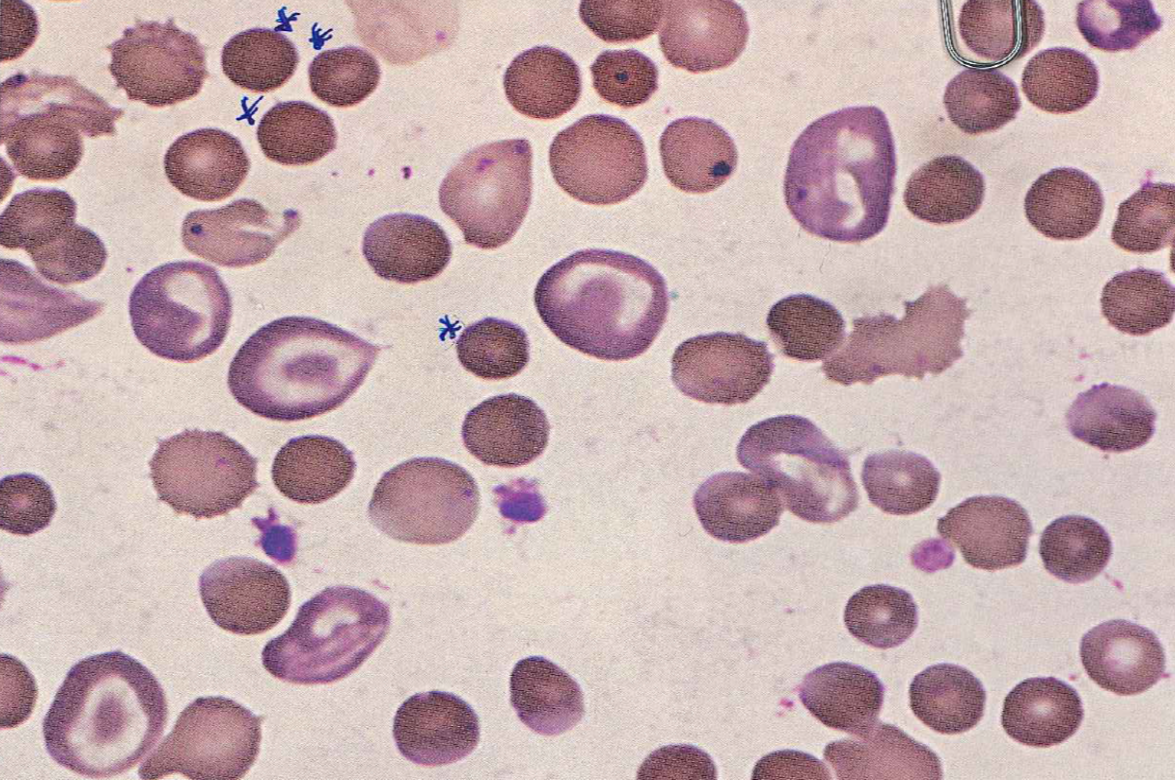
Spherocytes (abnormal)
-Thickened/rounded, smaller, darker staining (hyperchromic)
-Seen with autoimmune diseases and splenic disorders
Schistocyte (abnormal)
-irregular RBC fragment due to mechanical damage to cell membrane
Keratocyte (abnormal)
-Helmet cell
-RBC with portion removed
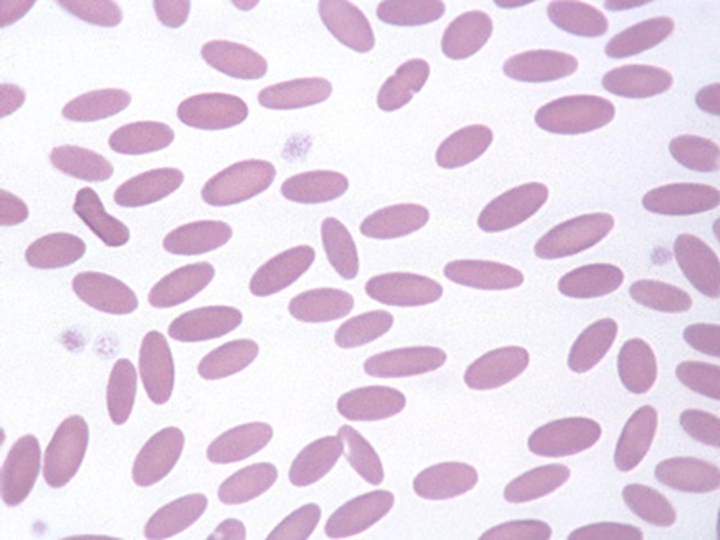
Ovalocyte/Elliptocyte
-Oval shaped RBC
-Normal for camelids
-Nucleated forms in birds, fish, amphibians, reptiles

Dacryocyte (abnormal)
-Teardrop shaped RBC

Fusocyte/Accuminocyte (abnormal)
-Spindle shaped that tapers to a point at both ends
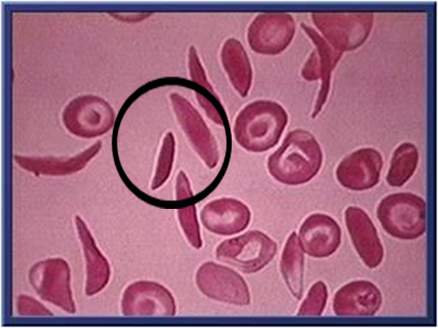
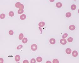
Drepanocyte (abnormal)
-Sickle cell
-Elongated, thread-like cell
-Seen in deer (cervids)
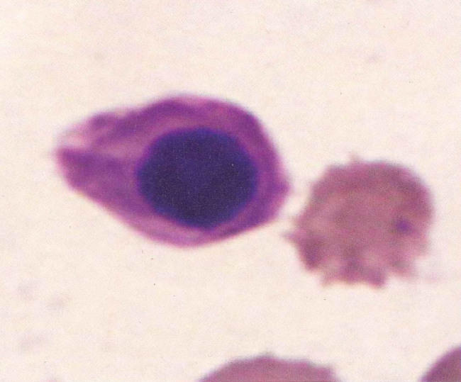
Nucleated RBC (abnormal)
-Anemia (increased RBCs from bone marrow due to increased destruction or loss of RBC in circulation)
Reticulocytes
-Immature RBCs
-Polychromic and macrocytic
-Dark stain: NMB; Bluish stain: Diff Quik
-Cats: Aggregate and Punctate
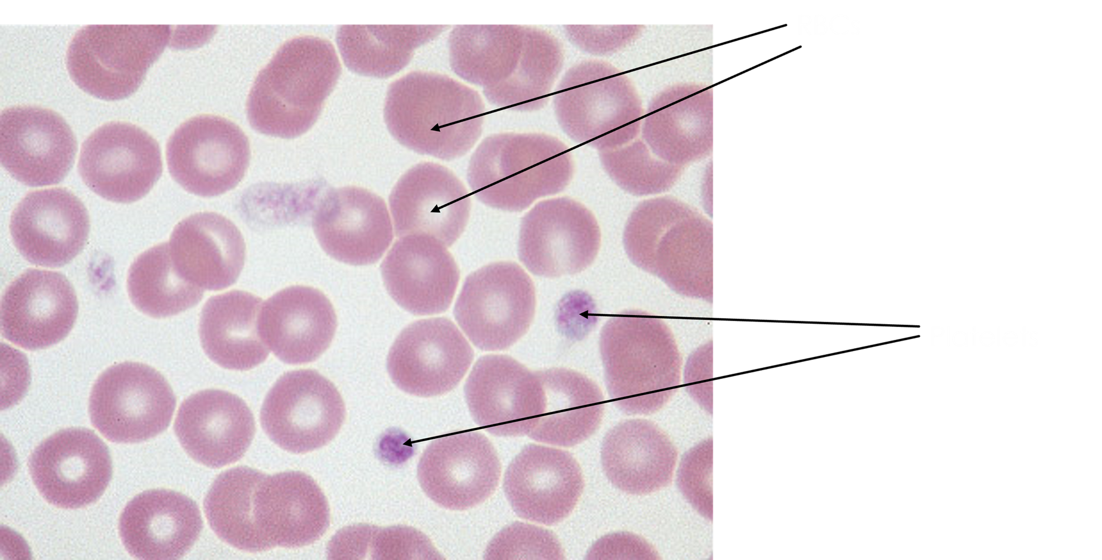
Howell-Jolly Body
-Stains well with Diff Quik
-Small retained portion of nucleus
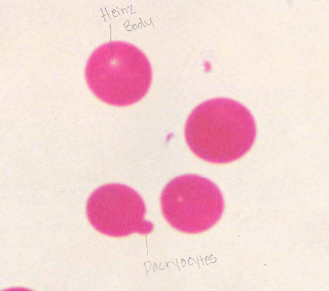
Heinz Body
-Stains well with NMB
-Small, round, refractile denatured hemoglobin that is bright and colorless with slightly darkened edges
-Single or multiple within the cell or surface
Basophilic stippling
-Stains well with Diff Quik
-Sometimes seen secondary to regenerative bone marrow response to anemia
-Heavy metal toxicity (lead poisoning)