ANHB2214 Cardiovascular system
1/13
There's no tags or description
Looks like no tags are added yet.
Name | Mastery | Learn | Test | Matching | Spaced | Call with Kai |
|---|
No analytics yet
Send a link to your students to track their progress
14 Terms

Identify:
atrium myocardium
ventricle myocardium +lumen
valve
connective tissue
artery
vein
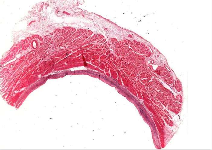
epicardium
endocardium
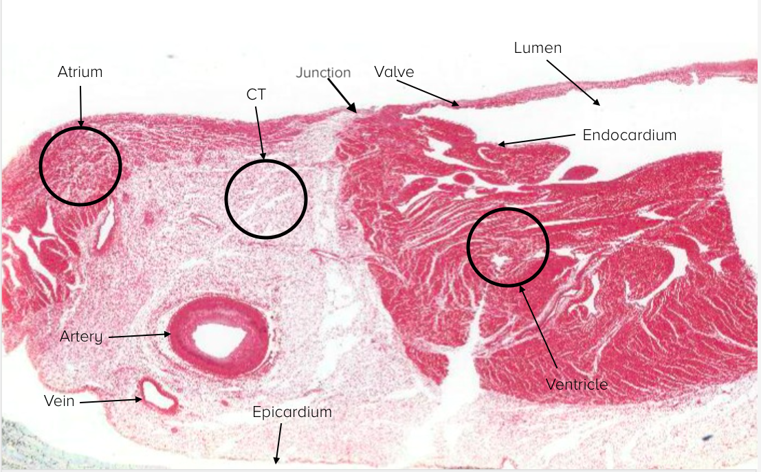
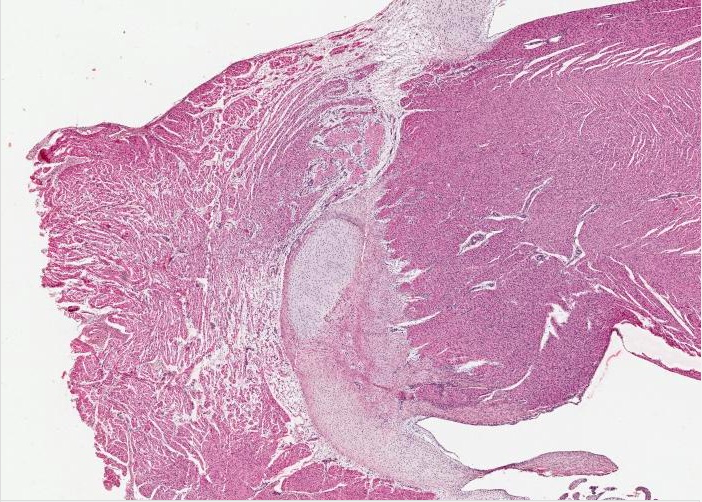
If this image was rotated 90 degrees clockwise, then it would be in its ordinary orientation. The atrium would be at the top and the ventricle below, the outside of the heart to the left and the lumen of the chambers to the right. The heart valve is the thin line of tissue extending downwards. Note that it attaches to the wall of the heart at the junction between the atrium and the ventricle. Note also that the muscle mass of the ventricle does not actually contact that of the atrium, a point of some physiological importance.
A substantial ring of connective tissue lies between the upper and lower chambers of the heart. The prominent round structure is a section of a coronary artery. A small vein can also be seen.
The main layer of both chambers of the heart is the myocardium. The atrium has a much thinner myocardial layer than does the ventricle. The endocardium lines the lumen of the chambers. The epicardium (visceral layer of serous pericardium) is a single layer of mesothelial cells on the outer surface of the heart and has a large number of fat cells within underlying adipose tissue, making it stain palely. This epicardium continues over the vessels entering and leaving the heart then reflects back to form the parietal layer of serous pericardium. The potential space (pericardial cavity) created between the visceral and parietal layers of the serous pericardium contains pericardial fluid.
ATRIOVENTRICULAR JUNCTION:
The heart valve attaches to the wall of the heart at the junction between the cardiac muscle layers (myocardium) of the atrium and the ventricle. Note also that the myocardium of the ventricle does not actually contact the myocardium of the atrium, as a septum of connective tissue creates an intermediary tissue layer, a point of some physiological importance.
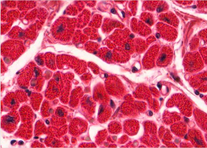
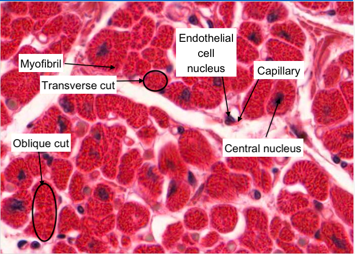
Cardiac muscle cells (cardiomyocytes) comprising the myocardium of the atrium are smaller than those in the ventricle. Why do you think that is so? In this image you can see most individual cardiac muscle fibres cut transversely (some with a central nucleus visible) and a few other fibres have been sectioned obliquely. The dark red "dots" within the muscle cytoplasm represents the contractile myofibrils. Note the occasional small blood capillary with a clear lumen lined by endothelium and often you can see a single endothelial cell nucleus.
What is this?
How do you know? (main features)
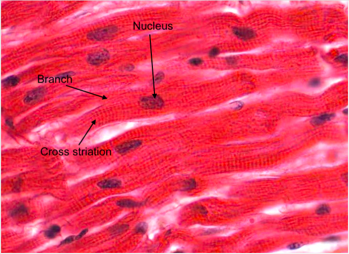
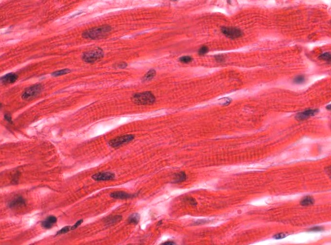
In this image you can see some individual cardiac muscle fibres cut longitudinally and in some places you can see where they branch and exhibit obvious cross striations. Occasionally, you will see a fibre that has been cut through the nucleus. Cardiac muscle fibres have normally only a single nucleus located in the centre of the fibre.
What is this? How do you know?
The epicardium is a fairly thick layer of loose connective tissue (mostly adipose tissue) with a single layer of mesothelial cells on the free surface (not visible in this image). Aggregations of fat cells are a dead giveaway for distinguishing the epicardium from the endocardium.
What is this? identify main features
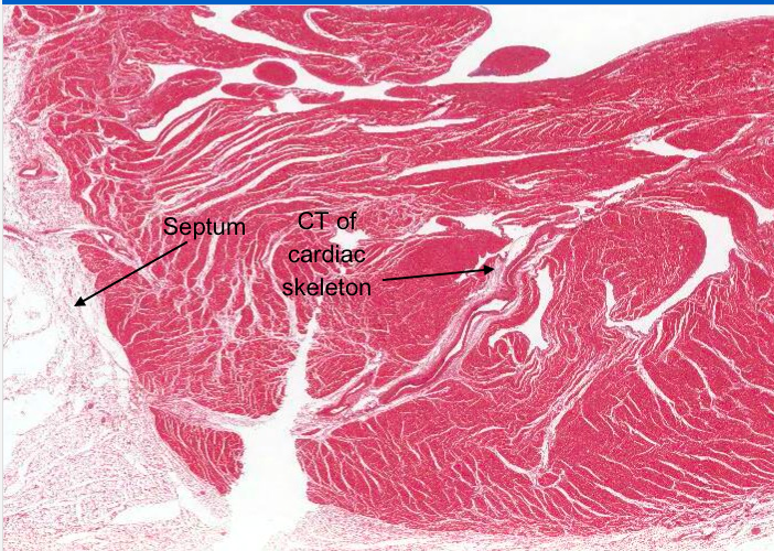
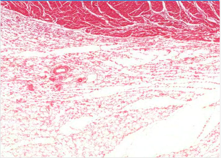
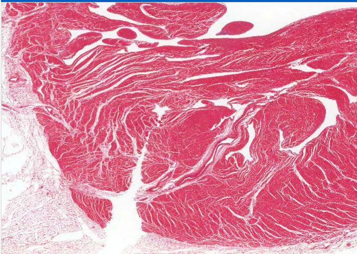
The main layer of the heart wall is the myocardium, which corresponds to the tunica media of an artery. It is a thick band of darkly stained cardiac muscle. Whilst the clear spaces throughout the cardiac muscle are mosty artifactual resulting from tissue shrinkage it does isolate lighter stained connective tissue components of the cardiac skeleton which also house a large blood vessel (collapsed).
The light stained region on the left side of this image would be part of the septum separating the cardiac muscle mass of the atrium from that of the ventricle.
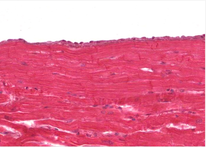
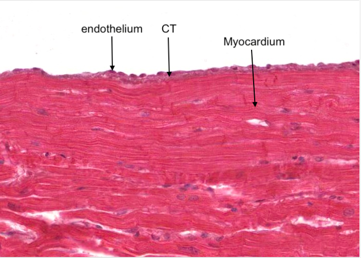
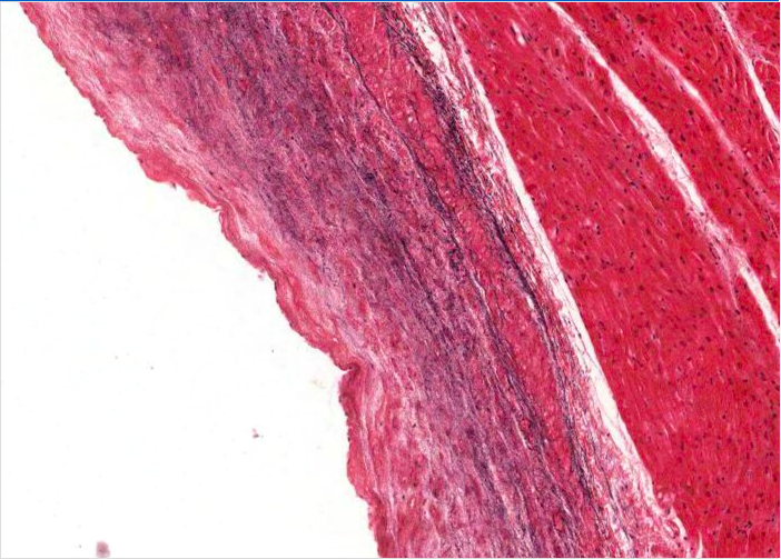
The endocardium is a thin layer, consisting of an endothelium and some underlying connective tissue. Often there may be two or three layers of connective tissue separated by a thin layer of smooth muscle. It overlies the myocardium and lines the chambers of the heart and surfaces of heart valves.
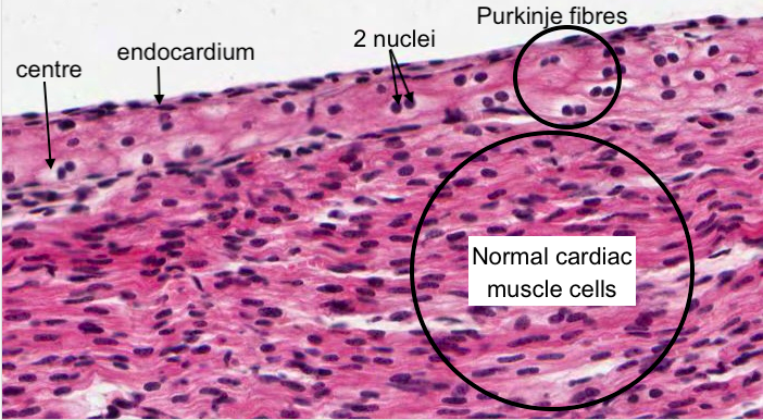
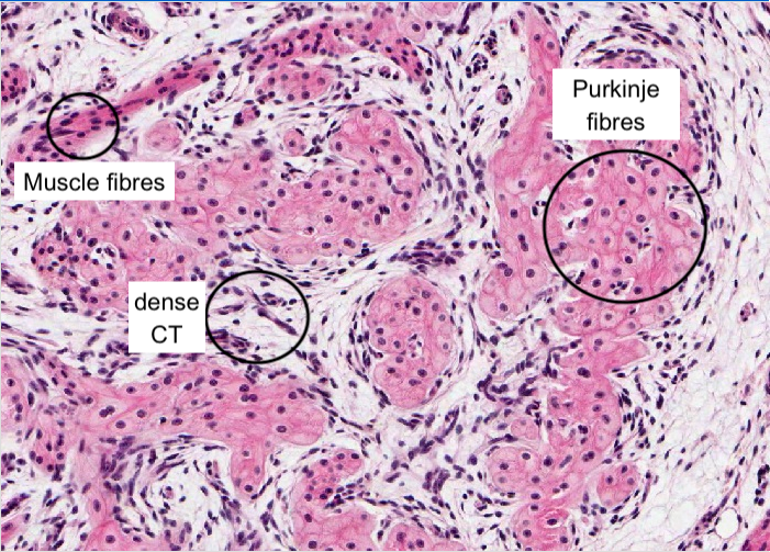
A modified type of cardiac muscle cell, the Purkinje fibre, forms the conduction system of the heart. Bands of these fibres run from the atrium down along the interventricular septum to the apex of the heart to make the ventricles contract from the bottom up. They can best be seen just under the endocardium.
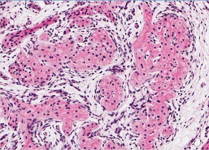
They may be distinguished from normal cardiac muscle cells by the following criteria (depending on whether you can view them sectioned transversely or longitudinally):
Purkinje fibers are wider and lighter-staining in H&E.
Except for the nucleus, the central part of a Purkinje fibre appears empty when stained with H&E. This is due to an abundance of glycogen, which does not stain well in H&E.
Myofibrils are confined to the periphery of a Purkinje fibre.
Purkinje fibres are often arranged in intimate groups of four or five. Such a group is surrounded by connective tissue.
A substantial portion of Purkinje fibres have two nuclei.
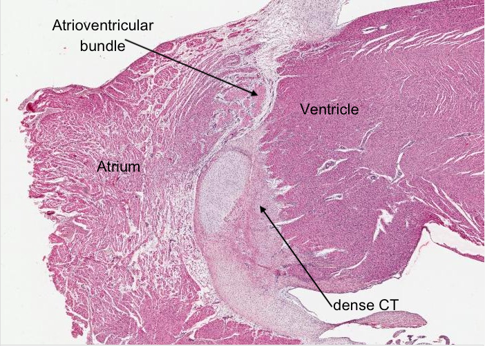
The membranous part of the interventricular septum contains dense connective tissue and part of the conducting system of the heart - the atrioventricular bundle (of Purkinje fibres). There is no cardiac muscle present except maybe for some muscle fibres from the atrium and ventricle forming their attachments to this connective tissue skeleton.
The membranous part of the interventricular septum contains dense connective tissue and part of the conducting system of the heart - the atrioventricular bundle (of Purkinje fibres). There is no cardiac muscle present except maybe for some muscle fibres from the atrium and ventricle forming their attachments to this connective tissue skeleton.
How is the atrium different to the ventricle?
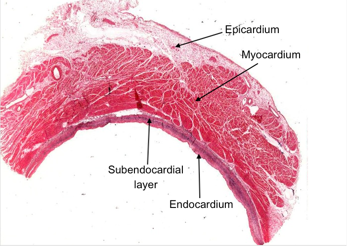
The atrium has the same three layers as the ventricle i.e. epicardium, myocardium and endocardium. However, the myocardium is thinner and the endocardium thicker In the atrium with a well recognised subendocardial layer.
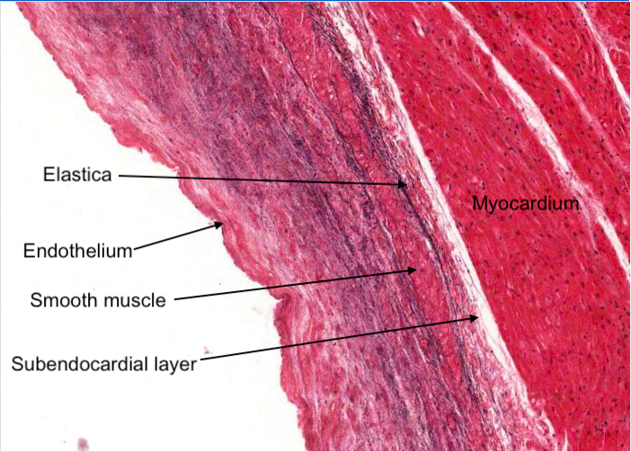
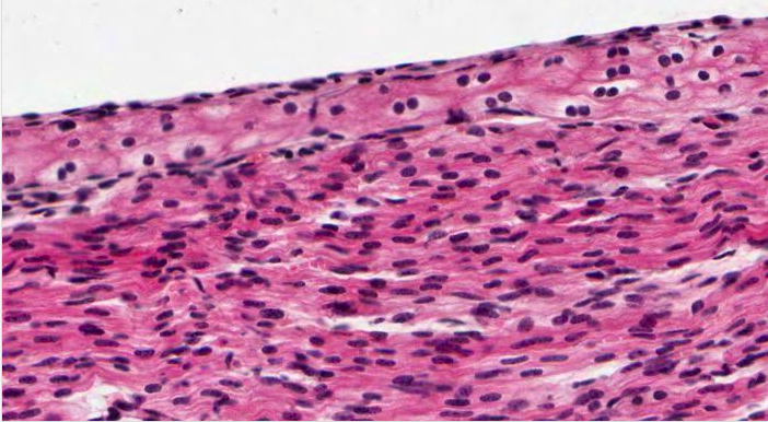
Examine the endocardium of the atrium. It is very well developed. It has an inner layer of endothelium and a deeper layer of connective tissue, elastica and smooth muscle. A further deeper layer is the "subendocardial layer" which becomes continuous with the connective tissue supporting the myocardium.
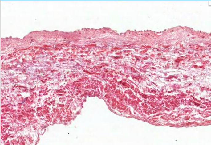
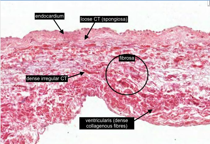
Heart valves are attached to the fibrous rings forming the openings of each valve and each contain a central core of connective tissue. An endocardium lines both surfaces. The central "fibrosa" is dense irregular connective tissue (collagenous) and is connected to the dense irregular connective tissue that surrounds the fibrous rings around each valve opening and the connective tissue elements separating all four chambers of the heart.
Loose connective tissue (the "spongiosa") is on the atrial or blood vessel side of each valve. This layer acts as a "shock absorber" when the valve closes.
The ventricularis is the dense layer of collagenous fibres and elastic fibres layer adjacent to the ventricle or atrial surface of each valve and covered with endothelium. In the atrioventricular valves it is continuous with the chorda tendineae and ultimately to the papillary muscles.
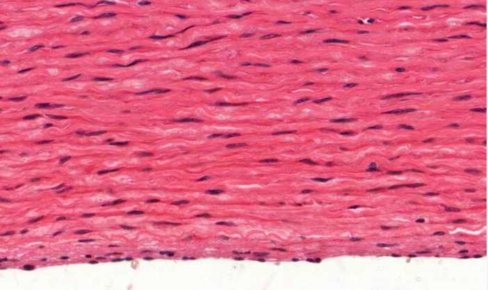
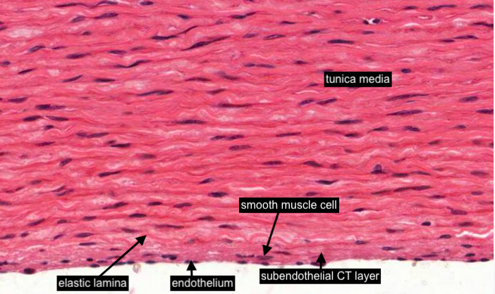
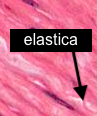
In an elastic artery the thin tunica intima may have the occasional smooth muscle cell in the subendothelial connective tissue layer. Look carefully to notice the rather wiggly internal elastic lamina which is similar to the elastic laminae present in the tunica media but it is the closest elastic lamina to the lumen. The nucleus of each endothelial cell is flattened. The bulk of this image shows the tunica media.

You would have already noticed that the tunica media is the thickest layer in the elastic artery (aorta). Although hard to see in H&E sections (imaged here), compared to aorta stained with the elastin stain you will observe in a later item, elastica appears as a blank pale wavy band, because it is made up of a hydrophobic protein and hence relatively impervious to aqueous dye molecules.
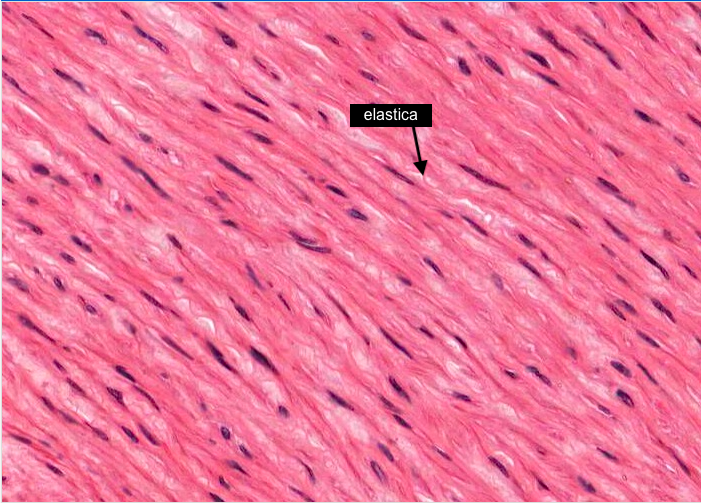
There are many layers of "wiggly" or "contracted" elastic membranes (elastic lamellae) interspersed amongst layers of smooth muscle cells each with an elongated nucleus. The cytoplasm of the smooth muscle cells extends past the ends of their nuclei and stains darkly,
Remember there are no fibroblasts in the tunica media. Collagen fibres are also present but hard to discern from the smooth muscle albeit they are lighter-stained, wavy wisps of tissue in this image. This distinction will become obvious in specially stained sections of the tunica media in items that follow.