ANHB2212 - The Articular System
1/96
There's no tags or description
Looks like no tags are added yet.
Name | Mastery | Learn | Test | Matching | Spaced | Call with Kai |
|---|
No analytics yet
Send a link to your students to track their progress
97 Terms
Joint
Any point where 2 bones make contact, irrespective of the degree of mobility
Arthrology
tudy of joint structure and function
What is the proper name for the elbow joint?
ulnohumeral/radiohumeral joint
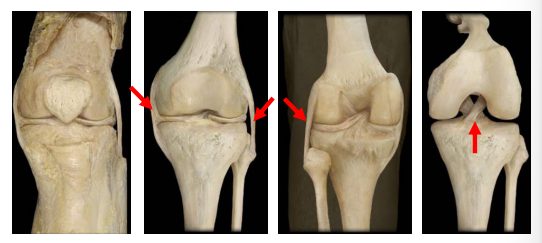
What is the proper name for the knee joint?
tibiofemoral joint
What is the proper name for the hip joint?
coxal joint
What is the proper name for the ankle joint?
talocrural
How are joints classified histologically?
According to the dominant type of connective tissue:
• Fibrous
• Cartilaginous
• Bony
• Synovial
How are joints classified functionally?
• Synarthrosis: immovable
• Diarthrosis: freely movable
• Amphiarthrosis: somewhat moveable
Fibrous Joints
Adjacent bones are joined by collagen fibres (dense regular connective tissue) that:
• emerge from one bone
• cross the space between them and
• penetrate into the other bone
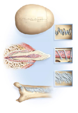
What are the 3 types of fibrous joints?
sutures
gomphoses
syndesmoses
What type of joint connects the cranial bones?
sutures
What type of joint connects teeth to socket?
gomphoses
What type of joint is in the interosseous membranes?
syndesmoses
Describe the mobility of the 3 types of fibrous joints.
sutures and gomphoses = synarthroses due to short fibres
syndesmoses = amphiarthroses due to long fibres
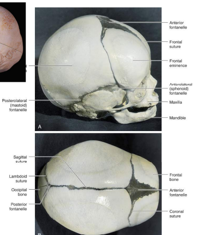
What 3 things does the fetal cranium consist of?
bones
sutures
fontanelles
Fontanelles
Broader areas of connective tissue that eventually become sutures, allowing for brain growth and deformation during birth:
anterior
anterolateral
posterior
What are the functions of syndesmoses joints?
maintain integrity between long bones
resist forces that attempt to separate the two bones
areas of muscle attachmen
What is the least mobile syndesmoses joint?
tibiofibular syndesmosis
What is the most mobile syndesmoses joint?
radioulnar syndesmosis
Function of the tibiofibular syndesmosis
provides strength and stability to the leg and ankle during weight-bearing
Function of the radioulnar syndesmosis
permits rotation of the radius bone during forearm movements (pronation & supination)
What are the 2 types of cartilaginous joints?
synchondrosis and symphysis
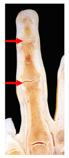
Synchondroses
The primary cartilaginous joint where bones are joined by hyaline cartilage, and allows no movement (synarthrosis). It can be temporary or permanent,
What type of joint is the epiphyseal plate?
temporary synchondrosis and synostosis
What is the function of the epiphyseal plate?
To facilitate bone lengthening during development at the epiphyseal plate. The epiphyseal plate connects the diaphysis (shaft of the bone) with the epiphysis (end of the bone) in children
What is the function of permanent synchondrosis?
To connect bones without movement (synarthrosis)
What type of joint is the first sternocostal joint and the anterior ends of the other 11 ribs?
permanent synchondrosis
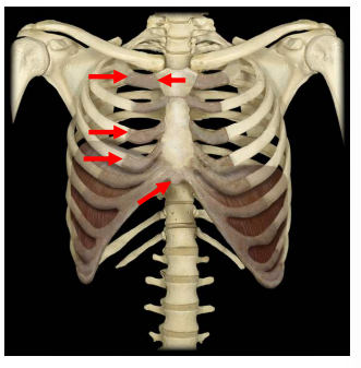
Symphysis
The secondary cartilaginous joint that allows limited movement (amphiarthrosis) by connecting bones by fibrocartilage to resist pulling and bending forces. It can be narrow or wide.
What type of joint are the intervertebral discs?
wide symphysis
What type of joint is the pubic symphysis?
narrow symphysis
What type of joint is the manubriosternal joint?
symphysis or synchondrosis and sometimes synostosis
Why is slight mobility needed in the pubic symphysis?
childbirth
Why is slight mobility needed in the manubriosternal joint?
breathing
Why is slight mobility needed in the intervertebral discs?
movement between adjacent vertebrae
cushioning during high-impact activity
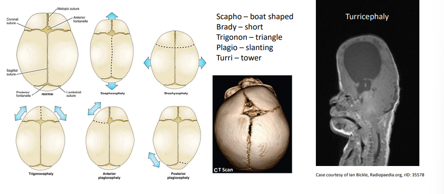
Bony Joints / Synostosis
An immobile joint where the gap between bones has ossified. It can occur at both cartilaginous & fibrous joints.
What issues can occur due to premature suture closure?
Deformations of the skull including Turricephaly.
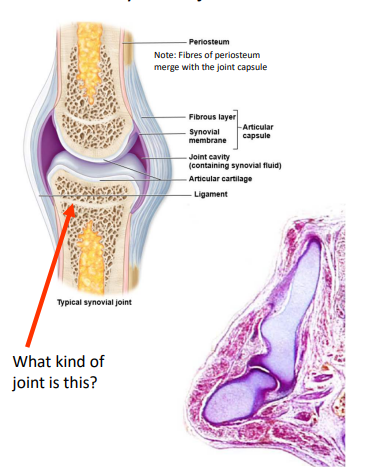
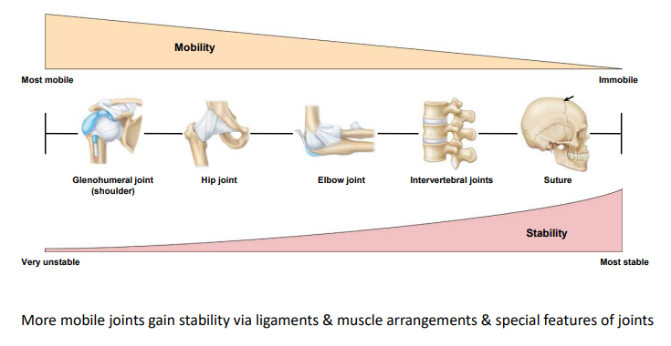
What are features of synovial joints?
most complex joints
most movable joints (diarthroses)
Articular capsule (fibrous joint capsule – dense irregular CT)
Articular cartilage (hyaline cartilage) – no nerve supply
Synovial fluid (produced by the synovial membrane)
What is the function of synovial joints?
to prevent friction between the articulating bones
What is the range of motion of synovial joints impacted by?
• Structure of the articular surface of the bones
• Strength and tautness of joint capsules & ligaments
• Actions of muscles and tendons
Degrees of Freedom
how many anatomical planes a joint can move through
What are the 3 types of synovial joints and their degrees of freedom?
1 DOF = Uniaxial/Monoaxial Joint
2 DOF = Biaxial Joints
3 DOF = Multiaxial Joints
What are the 2 types of multiaxial joints?
ball and socket
planar/gliding
Ball and Socket Joint
An articulation between the rounded head of one bone (ball) and the concavity of another (socket). Permits flexion/extension, abduction/adduction, and rotation
What are the 2 examples of a ball and socket joint?
hip and shoulder joints
Is the socket of the glenoid cavity of the shoulder shallow or deep? why?
Shallow to allow for extensive range of motion at the shoulder
Is the socket of the acetabulum of the hip shallow or deep? why?
Deep to constrain femoral movement.
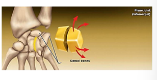
Planar/Gliding Joint
An articulation between bones that are both flat and of similar size. Multiaxial because it permits many movements BUT surrounding ligaments and bones usually restrict this joint to small, tight motions
What type of joint is the intercarpal and intertarsal joints?
Planar joint
What type of joint is the zygapophyseal joint of the vertebrae
planar joint
What are the 2 types of biaxial joints?
Condyloid/Ellipsoid
Saddle
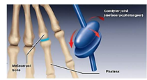
Condyloid/Ellipsoid Joint
An articulation between the shallow depression of one bone and the rounded structure of another bone or bones
What type of joint is the metacarpophalangeal joint?
condyloid
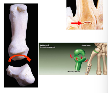
Saddle Joint
An articulation between two bones that are saddle-shaped, i.e. concave (inward curve) in one direction and convex (outward curve) in another
What type of joint is the first carpometacarpal joint of the thumb? why?
Saddle joint permits the thumb to flex and extend (within the plane of the palm) & abduct and adduct (perpendicular to the palm). This dexterity gives humans the characteristic trait of “opposable” thumbs.
What type of joint is the sternoclavicular joint?
saddle joint
What are the 2 types of uniaxial joints?
hinge
pivot
Hinge Joint
An articulation between the convex end of one bone and the concave end of another (not hemispherical)
What type of joint is the interpharyngeal joint?
hinge joint
What type of joint is the elbow joint?
hinge joint
What type of joint is the knee joint?
hinge joint
What type of joint is the ankle joint?
hinge joint
Pivot Joint
An articulation within a ligamentous ring between the rounded end of one bone and another bone. Permits rotation within this ring.
What type of joint is the atlantoaxial joint between C1 (atlas) and C2 (axis) of the vertebrae?
pivot joint
What type of joint is the proximal radioulnar joint?
Pivot Joint
Where does the radial head sit in the proximal radioulnar joint and why?
Head of the radius sits in the annular radial ligament to hold the bone in place during pronation and supination
What is the joint structure trade-off?
Labrum
Rings of fibrocartilage are only found in ball and socket joints (on the socket). It functions to deepen the joint socket.
Intra-articular discs
Thin plates of fibrocartilage connected to the fibrous joint capsule
What are the functions of the intra-articular discs?
• separate synovial cavities to allow for separate movements to occur in each space – each bone articulates with the disc (e.g. elevation / depression; protraction / retraction)
• increase joint stability
• aid in directing the flow of synovial fluid to areas of the articular cartilage that experience the most friction
• permit more even distribution of forces between the articulating surfaces of bones
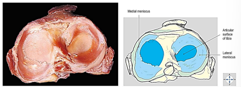
Menisci
Crescent-shaped incomplete intra-articular discs (fibrocartilage) found in the knee joint.
What is the function of menisci?
joint stability (increases joint concavity)
separate synovial cavity
distribution of synovial fluid
Movements: Between femur and menisci = flexion & extension
Where does rotation of the leg occur in the knee joint?
Between the tibia and menisci during flexion at the knee
Capsular Ligaments
Ligaments derived from the articular capsule that give joints strength and stability. They can be intracapsular or extracapsular.
What type of capsular ligament is the cruciate ligament of the knee?
intracapsular
What type of capsular ligament is the lateral collateral ligament of the knee?
extracapsular
Describe general joint development
Joint development starts with the condensation of mesenchyme within the developing bone – the Interzone (~week 6). This condensed mesenchyme differentiates into dense fibrous tissue. Interzonal mesenchyme then remains the same (fibrous joints) or differentiates (cartilaginous and synovial joints). The dense fibrous tissue also gives rise to intra-articular diss, menisci and enclosed joint ligaments.
Describe the development of fibrous joints in relation to interzonal mesenchyme.
interzonal mesenchyme remains as dense fibrous tissue
Describe the development of cartilaginous joints in relation to interzonal mesenchyme
interzonal mesenchyme differentiates into hyaline cartilage (synchondroses) or hyaline and fibrocartilage (symphyses)
Describe the development of synovial joints in relation to interzonal mesenchyme
• the cells in the interzone begin to flatten and cavitation occurs at the joint location.
• the interzonal mesenchyme differentiates:
- At the ends of adjacent bones into articular cartilage
- Centrally, the mesenchyme undergoes cavitation = synovial cavity
- Peripherally, to form the joint capsule and other ligaments
- Where the mesenchyme lines the joint capsule, it forms the synovial membrane (secretes synovial fluid)
What are 4 special features of joints?
labra
intra-articular discs
menisci
capsular ligaments
What are the two ligaments of the radioulnar joint?
Radial collateral ligament
Annular ligament
What is the function of the annular ligament?
Holds the radial side of the elbow together and holds the rotating head of the radius in place in the radial notch of the ulna.
What is the function of the radial collateral ligament?
Holds the humerus and the radial head securely together.
Opposition
A combination of abduction, flexion, and medial rotation, all occurring at the carpometacarpal joint of the thumb.
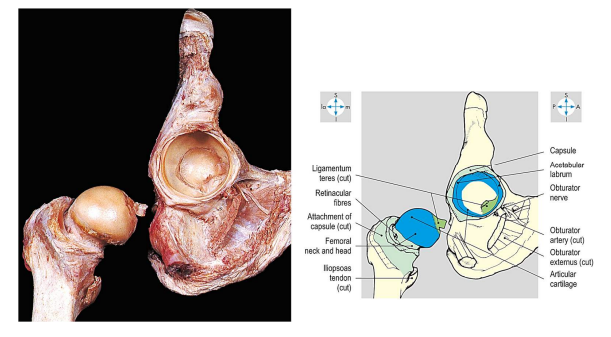
Describe the 2 ligaments of the hip joint capsule.
The capsule is thin on the underside. Everywhere else it’s thick and very strong. This part of the capsule behind is called the ischio-femoral ligament. This anterior part of the capsule, which is the thickest, is known as the ilio-femoral ligament. The fibers of these capsular ligaments become tight when the joint is extended.
Describe the 2 menisci articular surfaces of the tibia.
The lateral meniscus is much more mobile than the medial one, partly because its two ends are attached close together, partly because of a big difference in the mobility of the joint capsule around the edge, the lateral one is almost a circle, the medial one is more C-shaped. In cross section, each meniscus is thick at the outer edge and thin at the inner edge. The two ends of each meniscus are attached to the inter-articular area of the tibia, the medial ones far apart, the lateral ones close together.
What are the two pairs of ligaments of the knee joint?
tibia (medial) and fibular (lateral) collateral ligaments
posterior and anterior cruciate ligaments
Cruciate ligaments
They prevent forward (posterior cruciate ligament) and backward (anterior cruciate ligament) movement of the femur and tibia. By preventing backward and forward movement, the cruciate ligaments ensure that the condyles of the femur stay in one place, as they roll on the condyles of the tibia. Without them, the femur would roll off the back of the tibia in flexion, and would roll off the front of it in extension.
Collateral Ligaments
The fibular collateral ligament stands out from the side of the knee joint. Unlike its tibial counterpart, it doesn’t blend with the joint capsule and is not attached to a meniscus. When the knee joint is extended, both the collateral ligaments are tight. When it’s flexed, they become less tight. The function of the collateral ligaments is to keep the femoral and tibial condyles together,and thus to prevent the knee joint from bending from side to side.
Describe the joint capsule of the temporomandibular joint.
Most of the capsule is thin and loose, to allow the various movements that we’ll see. On the lateral aspect the capsule is thickened by this lateral ligament.
Bicondylar Joint
two distinct surfaces on one bone articulate with corresponding distinct surfaces on another bone
What are the 2 bicondylar joints?
tibiofemoral joint (knee joint)
temporomandibular joint (jaw)
What tissue subtype covers the surface of bones?
dense irregular connective tissue (periosteum)
How does the metacarpophalangeal of the fingers and thumb differ?
fingers = condyler = biaxial
thumb = oblong condyle = uniaxial
What movement occurs between the mandibular condyle and the intra articular disc?
depression/elevation
What movement occurs between the intra-articular disc and the temporal bone?
protraction/retraction
What type of capsular ligament is the medial collateral ligament of the knee?
capsular