Looks like no one added any tags here yet for you.
Sherrington’s Law of reciprocal inhibition
Agonist / Antagonist relationship
When one muscle contracts, its antagonist relaxes to allow smooth movement
This produces “Pseudoparesis” (fake weakness) in situations of both acute and chronic muscle imbalance syndromes
Inhibited = Weak/Psuedoparetic
Upper Trapezius
Sternocleidomastoid
Levator Scapulae
Pectoralis Major (upper fibers)
Pectoralis Minor
Cervical Erector Spinae
Scalenes
Facilitated = Tight/Hypertonic
Mid/Lower Trapezius
Latissimus Dorsi
Rhomboids
Deep Cervical Flexors
Pectoralis Minor
O: Anterior ends of the third to fifth ribs
I: Coracoid Process of Scapula
A: Protracts and downwardly rotates the pectoral girdle at the AC and SC joints.
Iliopsoas
O: Transverse processes of L1-L5 vertebrae, vertebral bodies of T12-L5 vertebrae, and adjacent intervertebral discs.
I: Lesser trochanter of the femur
A: Flex thigh at hip; flexes trunk
Anterior and Middle Scalene
Origin
Anterior: anterior tubercles of transverse processes of C3-C6
Middle: Transverse processes of C2-C7
Insertion
Anterior: Scalene tubercle of Rib 1
Middle: Superior surface of Rib 1
Sternocleidomastoid
Origin
Sternal Head: anterior surface of the manubrium of sternum
Clavicular Head: medial one third of the clavicle
Insertion
Mastoid Process of temporal bone and superior nuchal
line of occipital bone.
What attaches to the coracoid process?
a – pectoralis minor
b – coracobrachialis and biceps b
c – coracoacromial ligament
d – coracoclavicular ligaments
Rhomboid Major and Minor
Origin
Major: spinous processes of T2 to T5
Minor: spinous processes of C7 to T1
Insertion
Major: medial border of scapula
(between spine and inferior angle)
Minor: medial border of scapula
(at spine)
Levator Scapula
Origin: Transverse processes of C1- C4
Insertion: Medial border of scapula, between superior angle and superior portion of spine of scapula
Deltoid
Origin: Lateral one-third of clavicle, acromion and spine of scapula
Insertion: Deltoid tuberosity
Pectoralis Major
Origin: medial half of clavicle, sternum and cartilage of first through sixth ribs
Insertion: crest of the greater tubercle of humerus
(Anterior Axillary Fold)
Latissimus Dorsi
Origin: Inferior angle of scapula, spinous processes of T6-12, last 3 or 4 ribs, thoracolumbar aponeurosis and posterior iliac crest
Insertion: intertubercular groove of the humerus
(posterior axillary fold)
Teres Major
Origin: Inferior angle and the lower one-third of lateral border of the scapula
Insertion: crest of the lesser tubercle of the humerus
(posterior axillary fold)
Biceps Brachii
Origin
Short Head: coracoid process of scapula
Long Head: supraglenoid tubercle of scapula
Insertion
Tuberosity of the radius and aponeurosis of the biceps brachii
Triceps Brachii
Origin
Long Head: infraglenoid tubercle of scapula
Lateral Head: posterior surface of proximal half of humerus
Medial Head: posterior surface of distal half of the humerus
Insertion
Olecranon process of the ulna
Brachioradialis
Origin: Proximal two-thirds of the lateral supracondylar ridge of humerus
Insertion: styloid process of radius
FOOSH injury
•Scaphoid fracture (and possible avascular necrosis)
•Somatic dysfunction of radial head
•Fracture of distal radius (usually Colle’s Fracture)
Medial Collateral Ligament
Attaches from medial epicondyle of femur to medial margin and surface of tibia.
Less flexible that lateral collateral ligament and more prone to injury.
Has attachment to medial meniscus and joint capsule.
Lateral Collateral Ligament
Attaches from lateral epicondyle of femur to head of fibula.
Rectus Femoris
Origin: Anterior Inferior Iliac Spine (AIIS)
Insertion: Tibial Tuberosity (via the patella and patellar ligament)
Vastus Lateralis
Origin: Medial lip of linea aspera
Insertion: Tibial Tuberosity (via the patella and patellar ligament)
Vastus Medialis
Origin: Medial lip of linea aspera
Insertion: Tibial Tuberosity (via the patella and patellar ligament)
Vastus Intermedius
Origin: Anterior and lateral shaft of the femur
Insertion: Tibial tuberosity (via the patella and patellar ligament)
Adductor Magnus
Origin: Inferior ramus of the pubis, ramus of the ischium, and ischial tuberosity
Insertion: Medial lip of linea aspera and adductor tubercle
Biceps Femoris
Origin Short Head: Lateral lip of linea aspera
Long Head: Ischial tuberosity
Insertion: Head of Fibula
Tensor Fascia Lata
Insertion: Iliotibial tract (which then inserts distally to the tibial tubercle, AKA Gerdy’s tubercle)
Origin: Iliac crest, posterior to the ASIS
Semitendinosus
Origin: Ischial tuberosity
Insertion: Proximal, medial shat of the tibia at the pes anserinus tendon
Semimembranosus
Origin: Ischial tuberosity
Insertion: Posterior aspect of medial condyle of tibia
Gluteus Maximus
Origin: Coccyx, edge of sacrum, posterior iliac crest, sacrotuberous and sacroiliac ligaments
Insertion: Iliotibial tract (upper fibers) and gluteal tuberosity (lower fibers)
Gluteus Medius
Origin: gluteal surface of ilium, between posterior and anterior gluteal lines, just below the iliac crest
Insertion: Lateral aspect of greater trochanter
Gluteus Minimus
Origin: Gluteal surface of the ilium between the anterior and inferior gluteal lines
Insertion: Anterior aspect of greater trochanter
Piriformis
Origin: Anterior surface of sacrum
Insertion: Superior aspect of greater trochanter
Iliopsoas Group
Origin
Iliacus: Iliac fossa
Psoas major: bodies and transverse processes of L1-L5
Insertion Lessor trochanter
Sartorius
Origin: Anterior Superior Iliac Spine (ASIS)
Insertion: Pes Anserinus Tendon
What makes up the Pes Anserinus Tendon?
Sartorius, Gracilis, and Semitendinosus
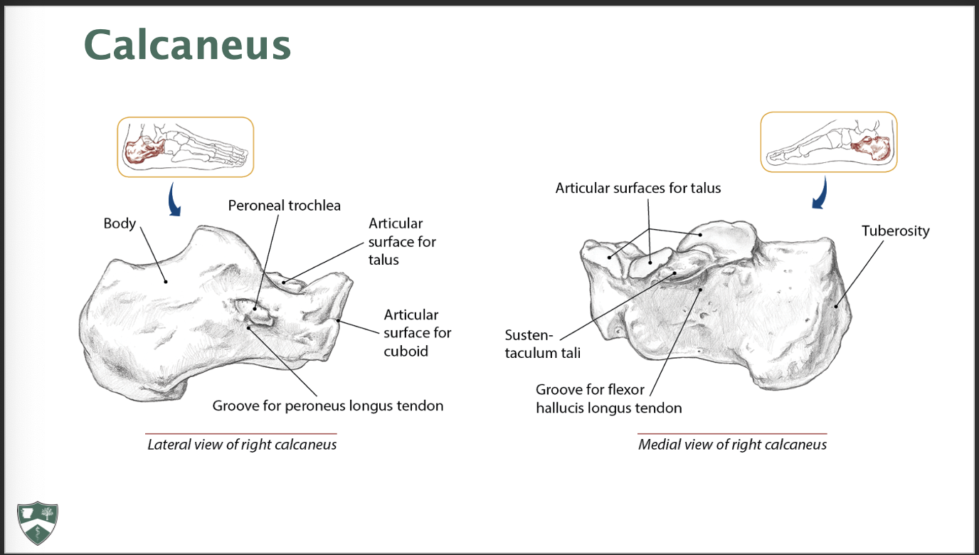
Parts of the Calcaneus
Gastrocnemius
Origin: Condyles of femur (posterior surfaces)
Insertion: Calcaneous via calcaneal tendon (AKA Achilles Tendon)
Peroneus Longus
Origin: Head of fibular and proximal two thirds of lateral fibula
Insertion: Base of first metatarsal and medial cuneiform
Peroneus Brevis
Origin: Distal two thirds of lateral fibula
Insertion: Tuberosity of fifth metatarsal
Tibialis Anterior
Origin: Lateral condyle of tibia; proximal, lateral surface of tibia and interosseous membrane
Insertion: Medial cuneiform and base of the first metatarsal
Extensor Digitorum Longus
Origin: Lateral condyle of tibia; proximal, anterior shaft of fibula and interosseous membrane
Insertion: Middle and distal phalanges of second through fifth toes
Extensor Hallucis Longus
Origin: Middle, anterior surface of fibula and interosseous membrane
Insertion: Distal phalanx of first toe
What is important about the lumbosacral fascia?
It plays a vital role in the mechanical stability of the lower back, by transferring load between the trunk and the lower limbs
What do you always start with before treating lymphatics?
Thoracic Inlet/Outler release