Chapter 16 - The Autonomic Nervous System
1/13
There's no tags or description
Looks like no tags are added yet.
Name | Mastery | Learn | Test | Matching | Spaced | Call with Kai |
|---|
No analytics yet
Send a link to your students to track their progress
14 Terms
Autonomic Nervous System
–Visceral efferent (motor) output for control of smooth (involuntary) muscle, cardiac muscle, and glands
Smooth muscle - in walls of digestive organs, bladder, blood vessels (arteries), and lung bronchi
Cardiac muscle - heart
Glands - sweat glands, salivary glands, digestive glands of stomach and intestines
–Has 2 divisions:
Sympathetic nervous system
Parasympathetic nervous system
Most organs receive both sympathetic and parasympathetic inputs
–These inputs have opposite actions (excitatory versus inhibitory) on organ activity
Excitatory actions: increases heart rate, increases digestive organ activity, increases gland secretion, causes contraction of smooth muscle (bladder contraction; vasoconstriction, with decreased blood flow)
Inhibitory actions: decreases heart rate, decreases digestive organ activity, decreases gland secretion, causes relaxation of smooth muscle (vasodilation, with increased blood flow)
–Neither system is purely excitatory or inhibitory
Organ response determined by functional role of that organ during increased sympathetic activity (“fight or flight”) versus parasympathetic activity (“rest and digest”)
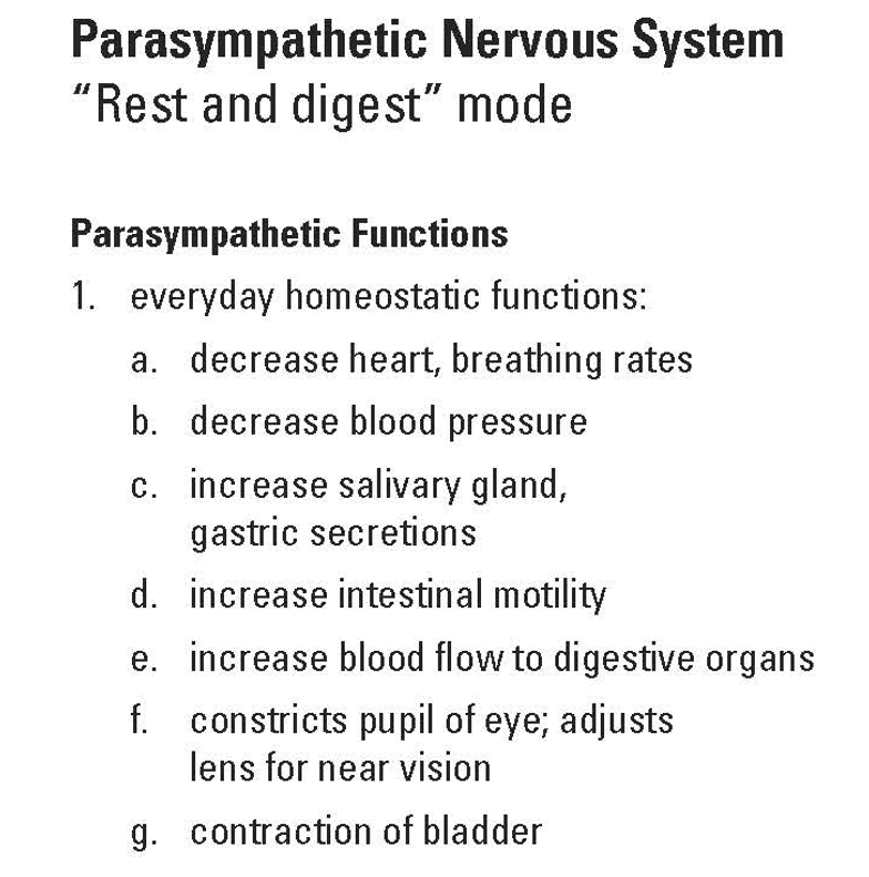
Parasympathetic system
(“rest & digest” mode)
–Parasympathetic system is active when body is well fed and at rest
Inhibitory actions:
Slows heart/breathing rates
Vasodilation for increased blood flow to digestive organs
Excitatory actions:
Increased digestive organ activity
Increased digestive gland secretions
Contraction of bladder (urination)
–Has organ-specific responses
Can selectively activate input to individual organs
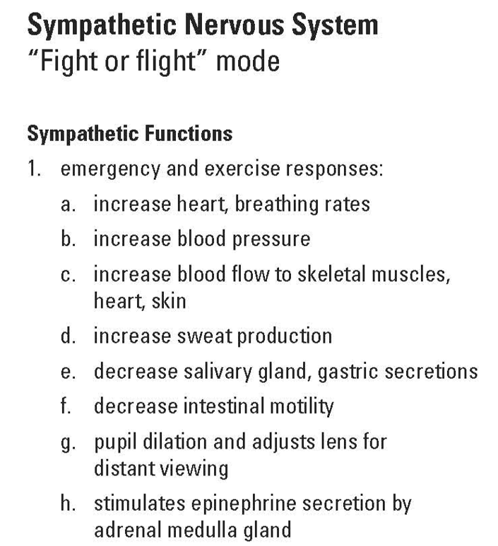
Sympathetic system
(“fight or flight” mode)
–Sympathetic system is active during exercise or when body is in danger
Inhibitory actions:
Decreases activities of digestive organs and glands
Vasodilation - increases blood flow to heart, skeletal muscles, and skin
Excitatory actions:
Increases heart/breathing rates
Increases blood pressure
Vasoconstriction - decreases blood flow to digestive organs
Increases sweat gland secretions
Stimulates epinephrine secretion by adrenal gland
–Sympathetic system produces wide-spread (full) body response
If body is in danger, all body systems must be fully and rapidly mobilized (if needed to meet the danger) or inactivated (if not needed)
Full body sympathetic response also reinforced by secretion of epinephrine (adrenaline) from adrenal (medulla) gland
→ Epinephrine secreted into blood and carried to all parts of body
→ Epinephrine produces same body responses as does sympathetic nervous system activation
Sympathetic Functions
Autonomic System Anatomy
Both sympathetic and parasympathetic systems consist of a two neuron chain
–Preganglionic neuron - first neuron in chain
Neuron cell body located within CNS (brain stem or spinal cord)
Axon exits CNS - synapses onto postganglionic neuron
–Postganglionic neuron - second neuron in chain
Neuron cell body located in a ganglion (collection of neurons found outside of CNS)
Receives synaptic input from preganglionic neuron
Axon of postganglionic neuron travels to target organ/tissue
Since both sympathetic and parasympathetic systems consist of a two neuron chain, there are four types of autonomic neurons:
–Preganglionic sympathetic neurons
–Postganglionic sympathetic neurons
–Preganglionic parasympathetic neurons
–Postganglionic parasympathetic neurons
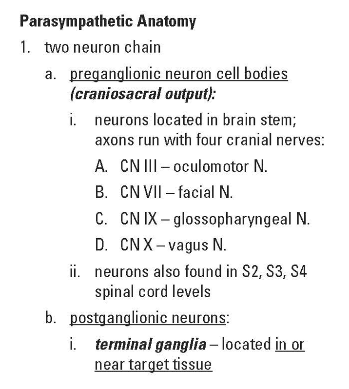
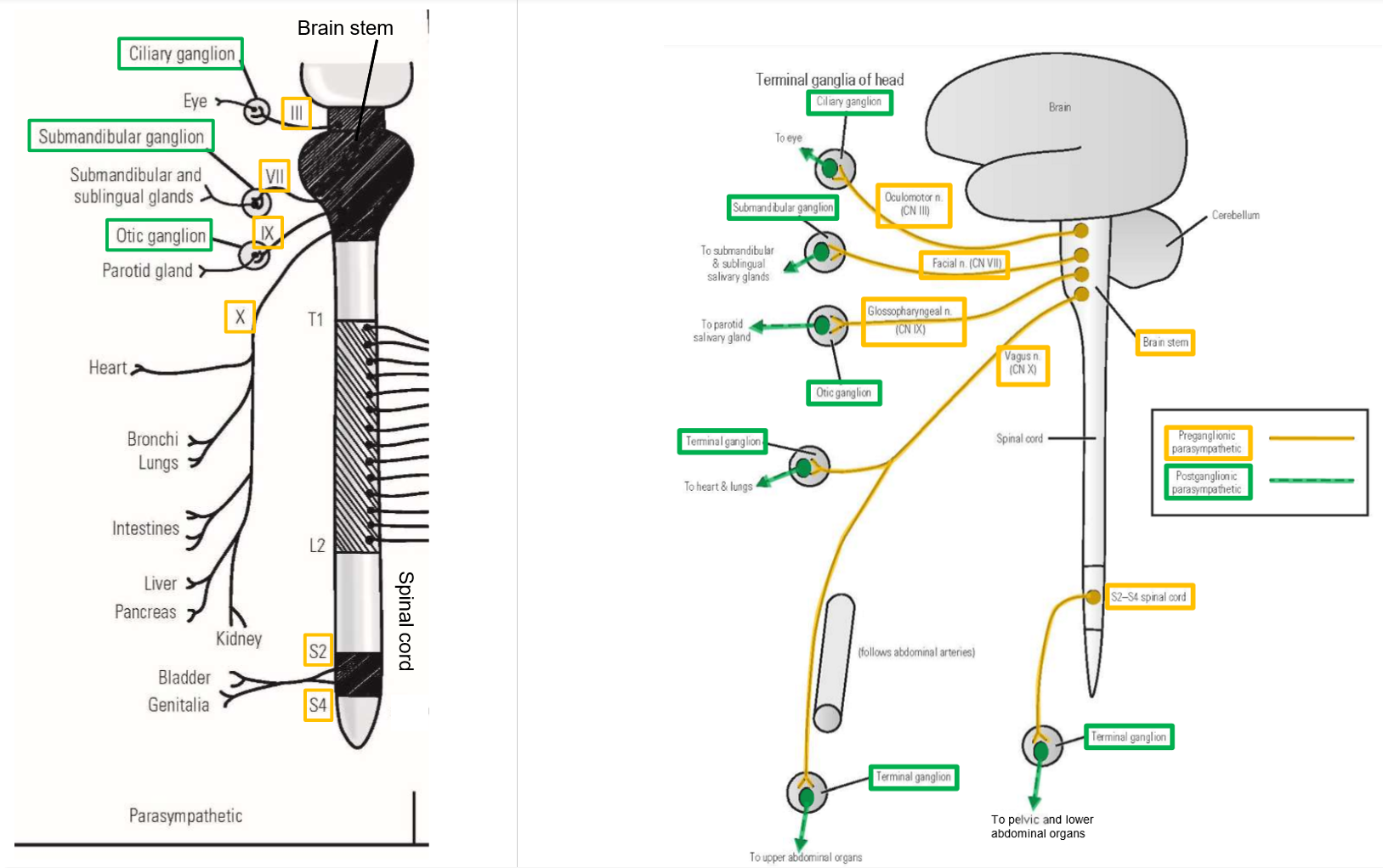
Parasympathetic Nervous System (Anatomy)
–Begins with craniosacral output
Preganglionic parasympathetic neuron cell bodies are located in either brain stem or S2-S4 sacral spinal cord
–Postganglionic parasympathetic neuron cell bodies located in terminal ganglia
Terminal ganglia are located in or near their target organ
→ Terminal ganglia of head are located near their target organ - these ganglia are individually named
→ Terminal ganglia of body are located embedded in wall of target organ - these ganglia are not individually named (just called terminal ganglia)
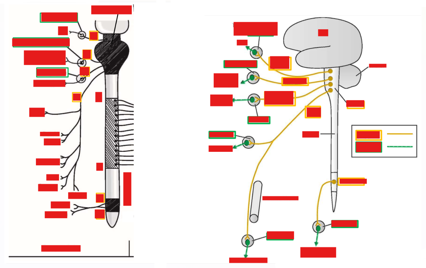
Individual names of Terminal ganglia of head in Parasympathetic Nervous System
Parasympathetic nervous system - begins with craniosacral output
–Preganglionic parasympathetic neuron cell bodies located in either brain stem or S2-S4 sacral spinal cord
–Brain stem output:
Axons from preganglionic parasympathetic neurons are carried by only four cranial nerves (III, VII, IX, or X)
–S2-S4 spinal cord output:
Axons from preganglionic parasympathetic neurons travel with S2, S3, or S4 spinal nerves
–Postganglionic parasympathetic neuron cell bodies located in terminal ganglia
Terminal ganglia are located in or near their target organ
–Terminal ganglia of head are located near their target organ and are individually named:
Ciliary ganglion - located immediately posterior to eye
→ Receives preganglionic input via oculomotor nerve (CN III)
→ Postganglionic parasympathetic neurons of ciliary ganglion supply the eye
–Terminal ganglia of head are individually named:
•Submandibular ganglion - located in posterior oral cavity
Receives preganglionic input via facial nerve (CN VII)
Postganglionic parasympathetic neurons of submandibular ganglion supply the submandibular and sublingual salivary glands
•Otic ganglion - located posterior to mandible
Receives preganglionic input via glossopharyngeal nerve (CN IX)
Postganglionic parasympathetic neurons of otic ganglion supply the parotid salivary gland
–Terminal ganglia of body are small and embedded within wall of target organ (these are not individually named)
Terminal ganglia for heart, lungs, and upper abdominal organs receive preganglionic parasympathetic input via the vagus nerve (CN X)
Terminal ganglia for lower abdominal and pelvic organs receive preganglionic parasympathetic input via the S2-S4 spinal nerves
–Craniosacral output
Preganglionic parasympathetic neurons located in brain stem (axons run with only four cranial nerves - III, VII, IX, X) or located in S2-S4 spinal cord (axons travel with S2, S3, or S4 spinal nerves)
–Postganglionic parasympathetic neurons located in terminal ganglia
Only terminal ganglia of head are individually named
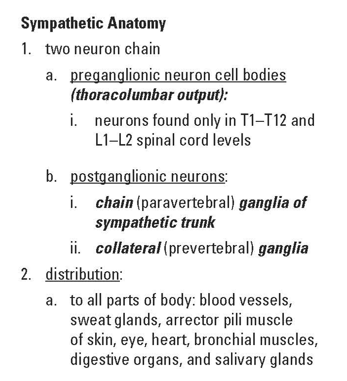
Sympathetic Nervous System Anatomy
–Begins with thoracolumbar output
Preganglionic sympathetic neuron cell bodies are located in T1-T12 thoracic and L1-L2 lumbar spinal cord
→ Preganglionic axons exit spinal cord with T1-L2 spinal nerves
–Postganglionic sympathetic neuron cell bodies located in either chain ganglia or collateral ganglia
Chain ganglia supply head, body trunk, and limbs
Collateral ganglia supply abdominal and pelvic organs
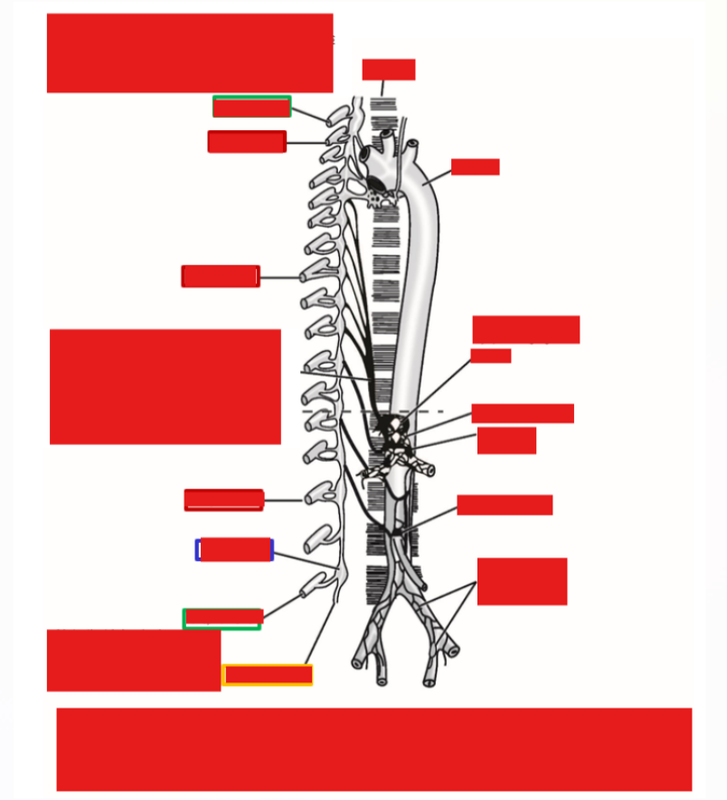
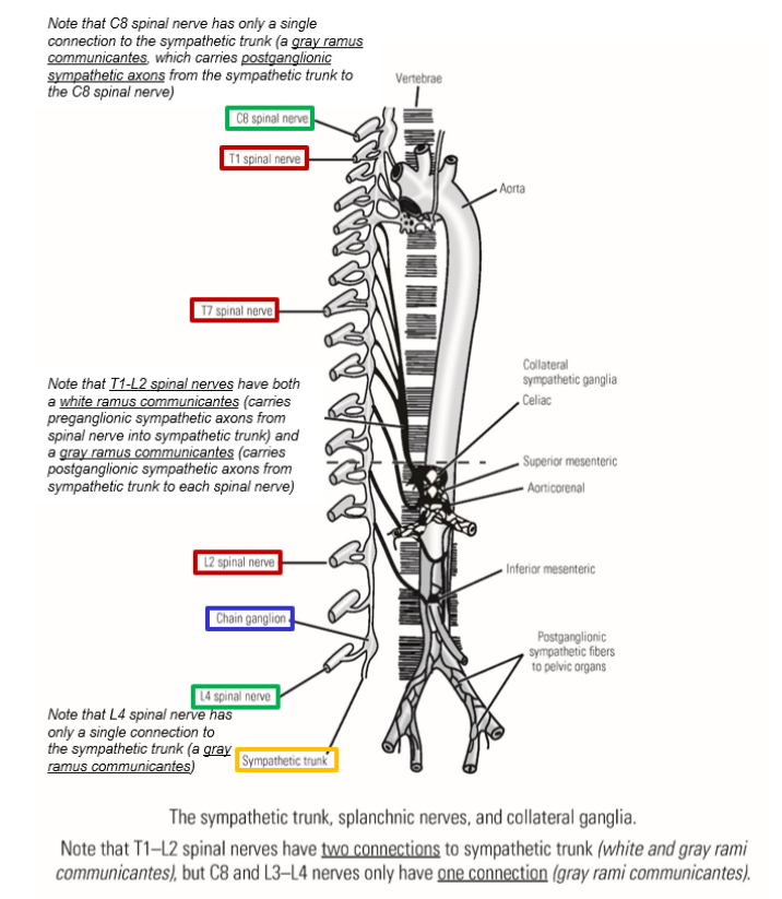
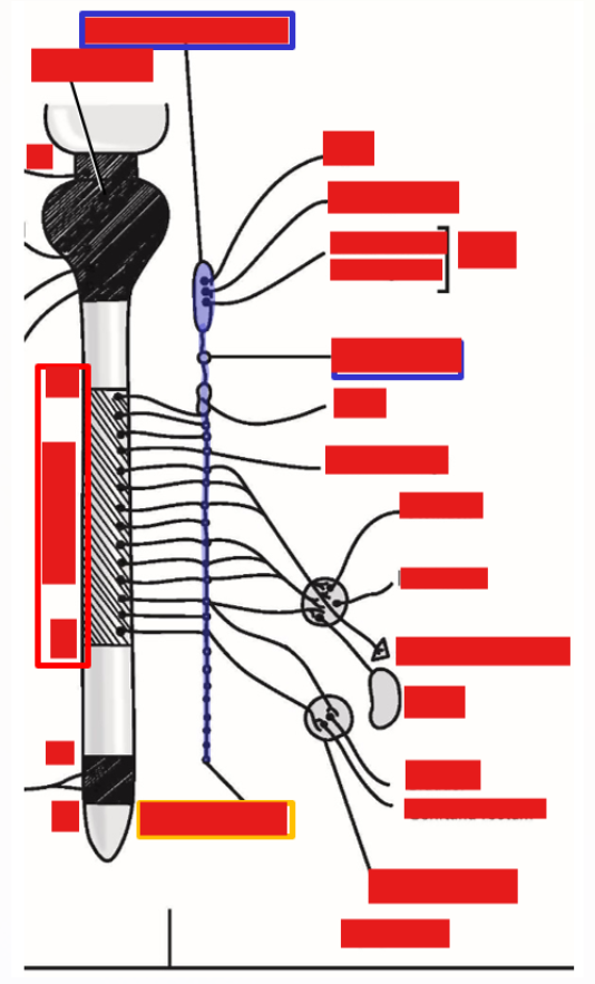
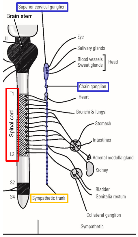
Sympathetic Nervous System Pathway
–Sympathetic trunk
Consists of a series of interconnected chain ganglia
→ Interconnected by nerve fibers (axons) running up or down within sympathetic trunk
Sympathetic trunk located along lateral sides of vertebral column (both right & left sides)
–Extends from just below base of skull to lower sacrum
Superior cervical ganglion
→ Largest of all chain ganglia
→ Located at superior end of sympathetic trunk
–Sympathetic innervation provided to body in 3 ways:
Innervation to blood vessels and sweat glands of body trunk and limbs
→ Stimulates sweating and increased blood flow to skin for heat loss
Innervation to blood vessels, sweat glands, and salivary glands of head
→ Stimulates sweating and increased blood flow to the skin for heat loss
→ Decreases salivary gland secretion
Innervation to abdominal and pelvic organs (see Chapter 36)
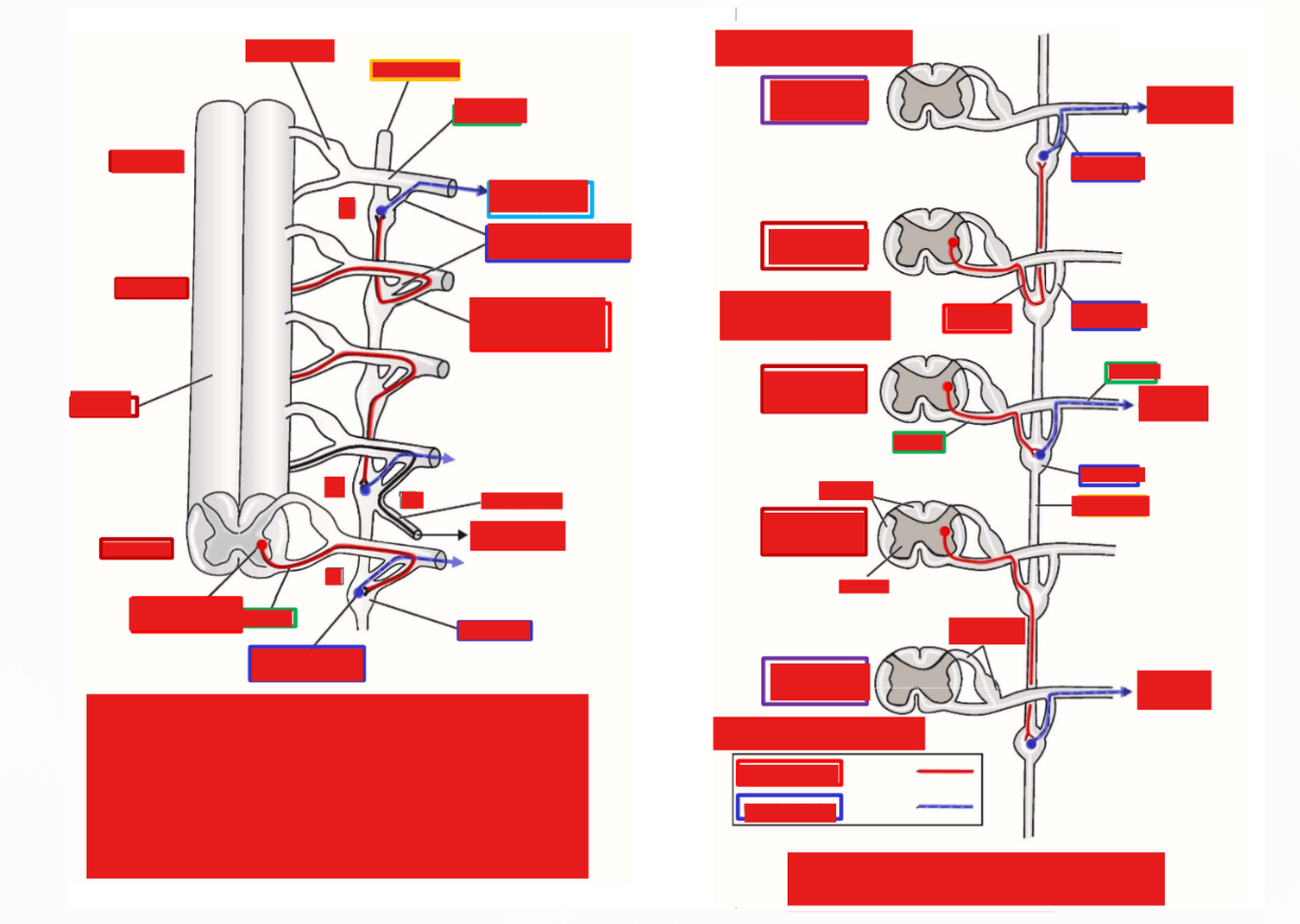
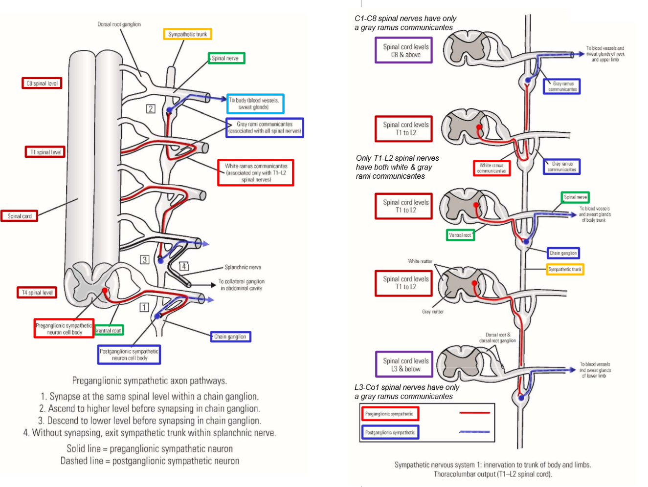
Sympathetic Innervation to Blood Vessels-Sweat Glands of Body Trunk and Limbs
Preganglionic sympathetic neurons are located in T1-L2 spinal cord
Neuron cell bodies located in gray matter of spinal cord
Axons exit spinal cord via ventral root
→ Ventral root carries only efferent axons
Axons run with T1-L2 spinal nerves
Axons of preganglionic sympathetic neurons leave spinal nerve and pass into sympathetic trunk
Axons run through a white ramus communicantes
→ These carry only preganglionic sympathetic axons from spinal nerve into sympathetic trunk
T1-L2 spinal nerves are only nerves that give off white rami communicantes (these are the only spinal nerves that carry preganglionic sympathetic axons)
Within sympathetic trunk
Axon of preganglionic sympathetic neuron will travel to a chain ganglion where it will synapse onto a postganglionic sympathetic neuron
Axons may terminate in chain ganglion at same level as entry, or may travel up or down within sympathetic trunk to reach chain ganglion at higher or lower level from entry
→ Ascending/descending axons form interconnections between chain ganglia of sympathetic trunk
Axons of postganglionic sympathetic neurons then leave sympathetic trunk and join into a spinal nerve
Axon runs through a gray ramus communicantes
→ These carry only postganglionic sympathetic axons from the sympathetic trunk to a spinal nerve
All spinal nerves (C1-Co1) receive a gray ramus communicantes
All spinal nerves carry postganglionic sympathetic axons
Axons of postganglionic sympathetic neurons are carried by all spinal nerves
If spinal nerve joins into a plexus, postganglionic sympathetic axons pass through plexus and continue with all peripheral nerves of limbs
Postganglionic sympathetic axons go to blood vessels and sweat glands of skin
Stimulate sweating and vasodilation (increased blood flow to skin) for heat loss (heat from muscle activity)
Sympathetic trunk functions to expand short range of sympathetic output (from T1-L2 spinal cord) into much longer structure (sympathetic trunk) that allows for connections with all spinal nerves (C1-Co1)
Sympathetic trunk runs entire length of vertebral column - allows for connections with all spinal nerves