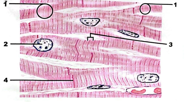
Histology
1/48
Earn XP
Description and Tags
Name | Mastery | Learn | Test | Matching | Spaced | Call with Kai |
|---|
No analytics yet
Send a link to your students to track their progress
49 Terms
what is this … ?
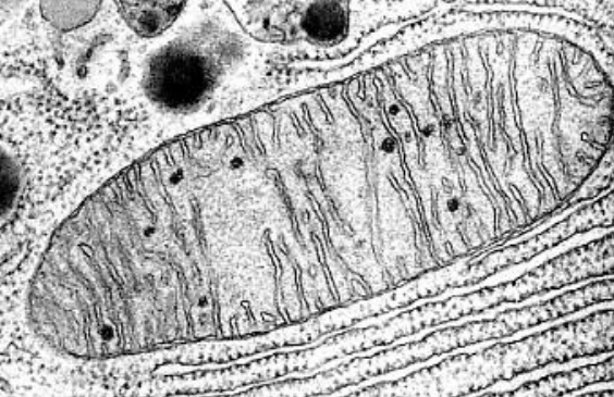
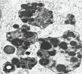
Mitochondria
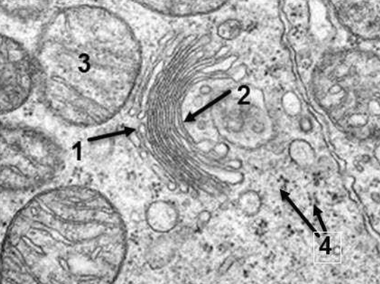
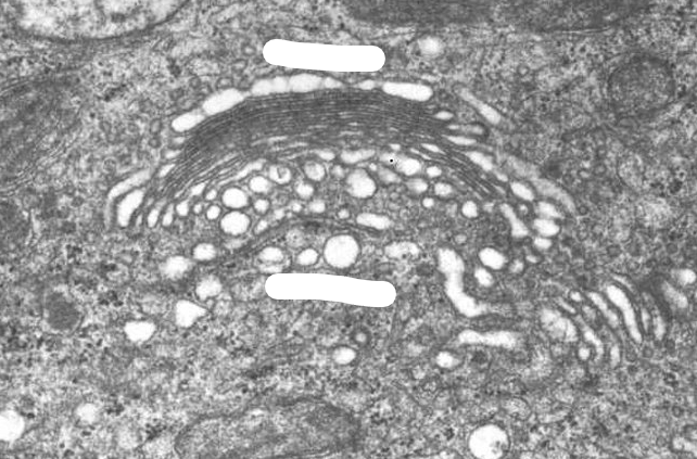
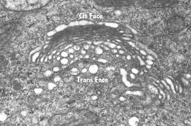
1- cis face of Golgi.
2-Trans face of Golgi.
3. Mitochondria.
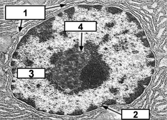
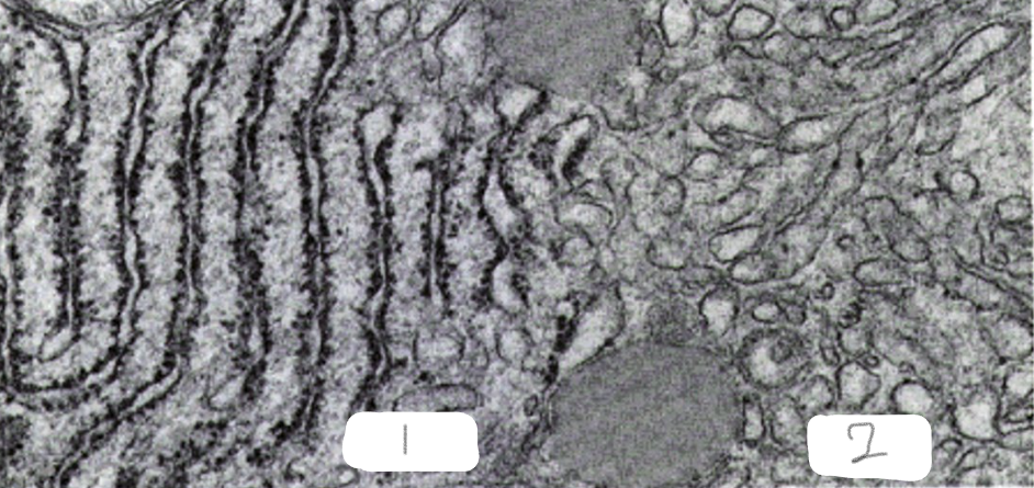
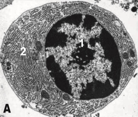
1-RER
2-Nucler Envelope
3-chromatin
4-Nucleolus
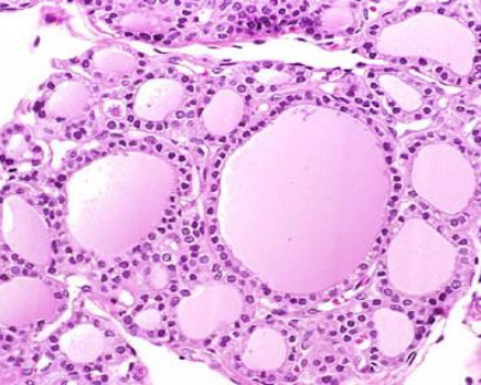
Simple cuboidal epithelium
Thyroid follicles
Simple squamous epithelium
Endothelium
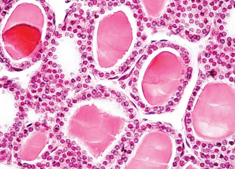
Simple cuboidal epithelium
(Thyroid follicles)
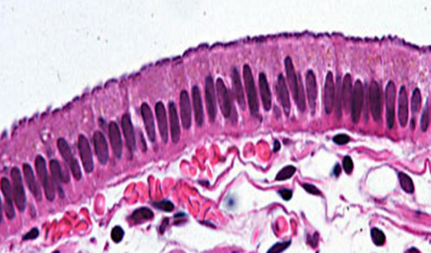
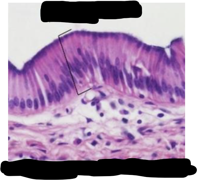
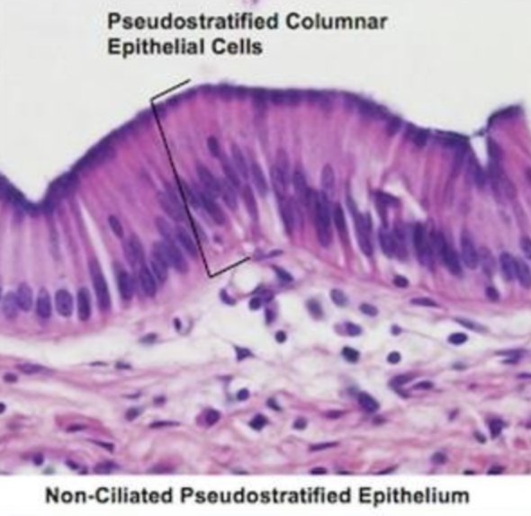
Simple columnar
epithelium non ciliated
(Gall bladder)
RER
RER
SER
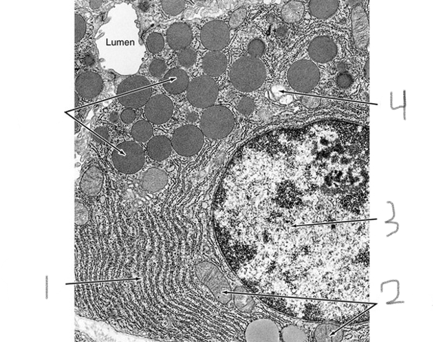
1-RER
2-Mitochondria
3-Nucleus
4-Lysosomes
Golgi apparatus
Lysosomes
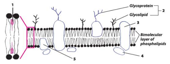
cell membrane
1: cholesterol molecule.
2: Glycocalyx.
3: transmembrane integral protein.
4: peripheral protein .
5: integral membrane protein
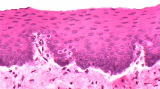
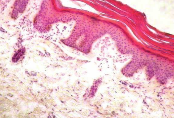
Non keratinized stratified squamous epithelium
(Mouth cavity)
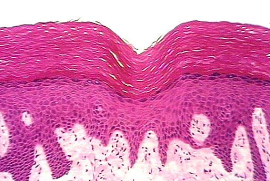
keratinized stratified squamous epithelium
(skin)
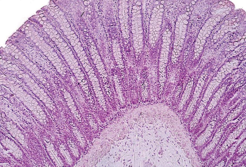
Large intestine
Simple tubular glands with goblet cells
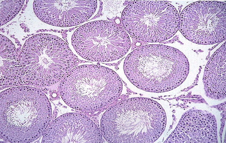
somniferous tubules
Germinal epithelium
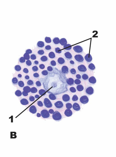
Mast cell
1- Nucleus
2- Basophilic granules
3-lymphocytes
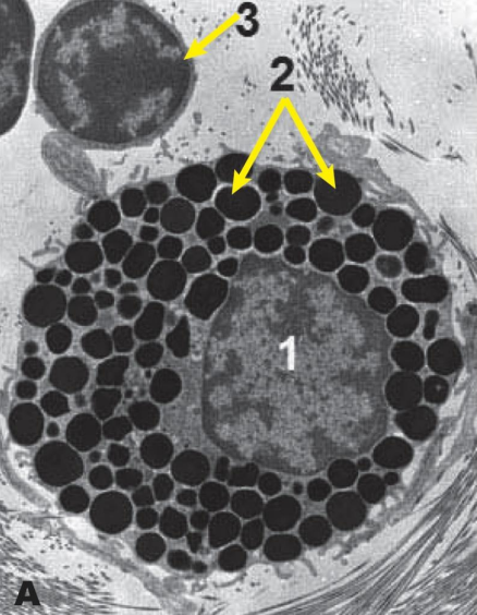
Mast cell
1- Nucleus
2- Basophilic granules
Plasma cell
1-Nucleus with cart-wheel chromatin pattern
2-RER
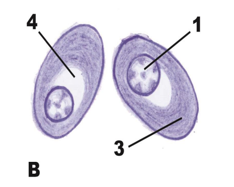
Plasma cell
1- Nucleus with cart-wheel chromatin pattern
3- Basophilic cytoplasm
4-Negative Golgi image
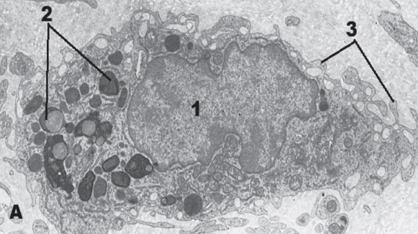
Macrophage
1.Eccentric kidney shaped nucleus
2.Numerous lysosomes
3.Multiple pseudopodia
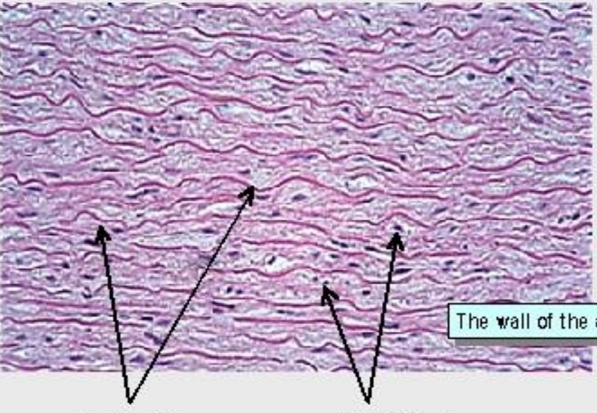
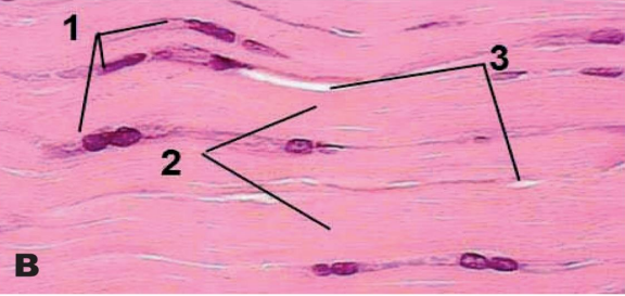
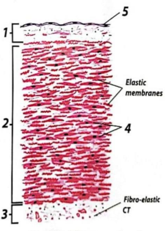
Elastic CT (H&E)
the 2 arrows to the left are elastic fibers
the others are fibroblast
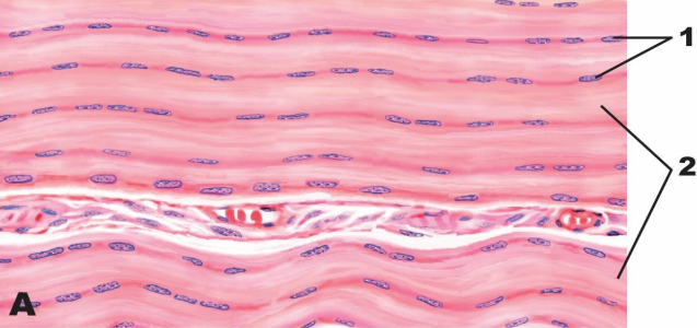
(White fibrous CT)
1-Rows of fibroblasts (tendon cells)
2-Wavy bundles of collagen fibers
(White fibrous CT)
1-Rows of fibroblasts (tendon cells)
2-Wavy bundles of collagen fibers
3-Little intercellular substance
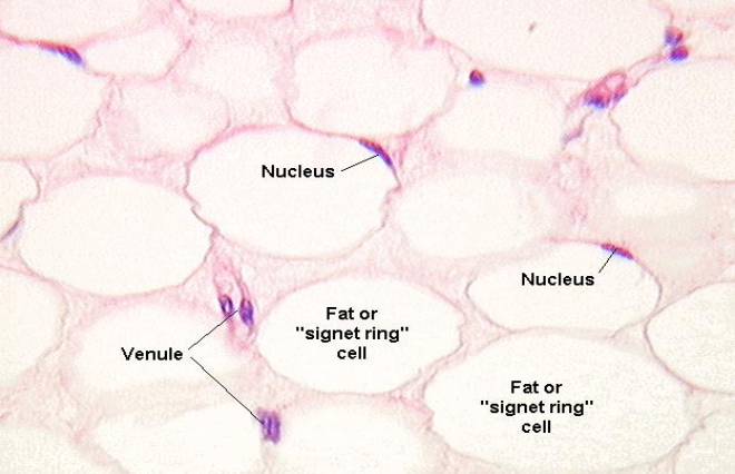
Adipose CT
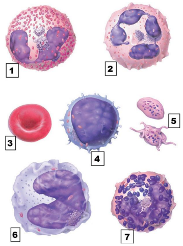
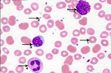
1- Eosinophil. 2- Neutrophil. 3- Erythrocyte. 4- Lymphocyte. 5- Blood platelets. 6- Monocyte. 7- Basophil.
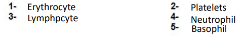
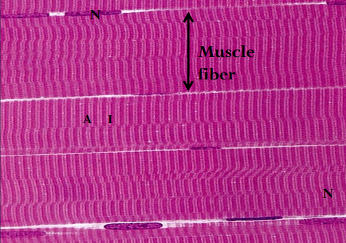
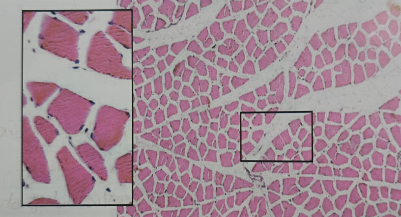
skeletal muscle
skeletal muscle
Skeletal muscle
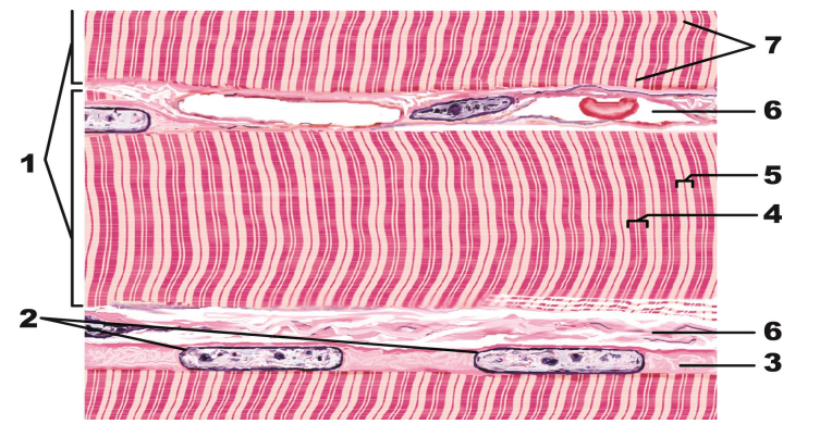
1. Skeletal muscle fibers. 2. Peripheral subsarcolemmal nuclei. 3. Sarcoplasm. 4. A band. 5. I band. 6. Endomysium. 7. Z lines.
smooth muscle
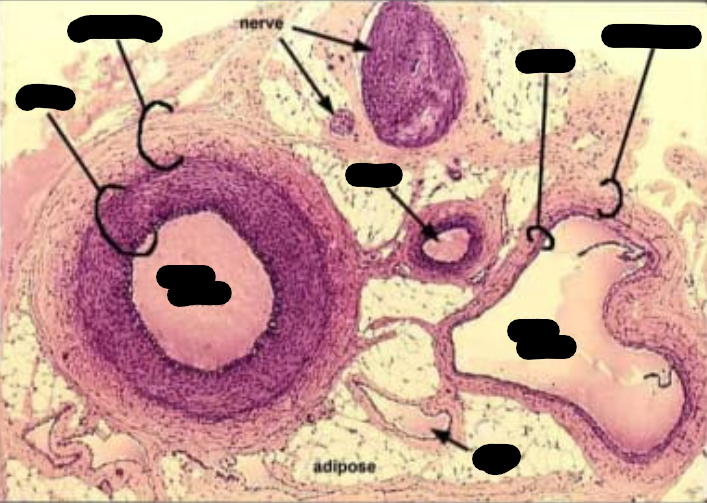
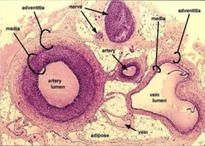
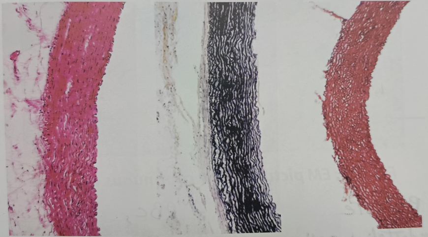
blood vessel
1- Tunica intima 2- Tunica media 3- Tunica adventitia 4- Smooth muscle fibers 5- Endothelium
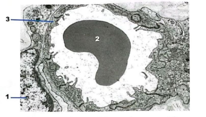
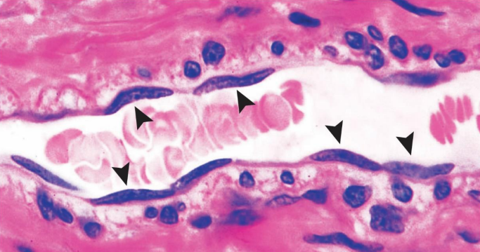
Continuous blood capillaries
1- Pericyte
2- RBCs
3- endothelial cell (capillary wall cell)
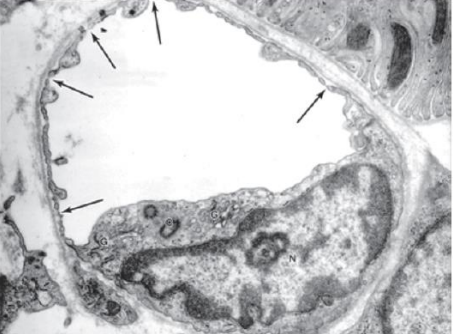
Fenestrated capillary
the arrows point towards (Fenestrations)
Thick skin
Mention 2-3 diagnostic features of Thick skin
Dermo-epidermal interdigitation are regular and deep
large number of sweet glands
absence of hair follicle and sebaceous glands
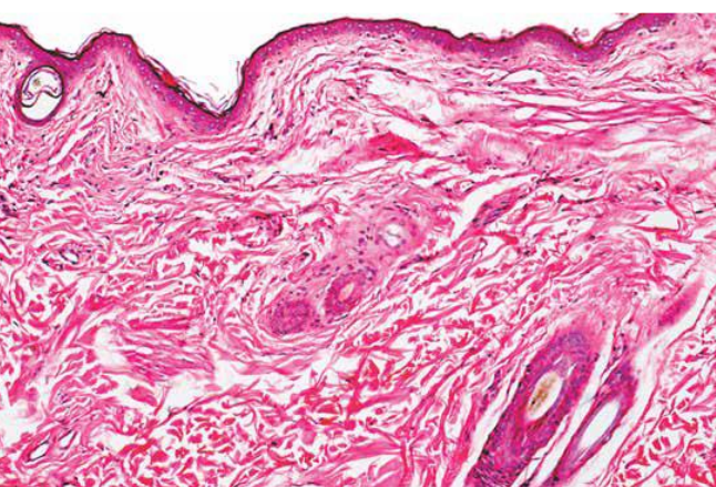
thin skin
Mention 2-3 diagnostic features of thin skin
little amount of sweet glands
presence of hair follicles and sebaceous glands
Dermo-epidermal interdigitation are irregular and shallow
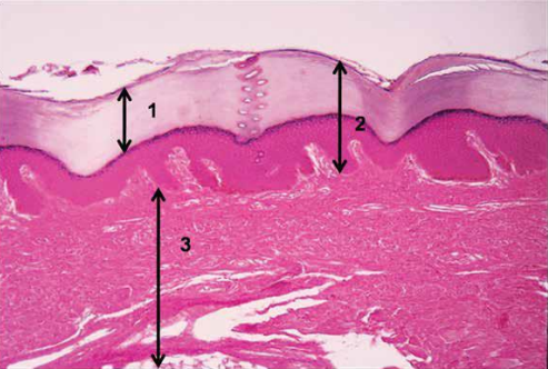
Thick skin
1- Stratum corneum.
2- Epidermis
3- Dermis.
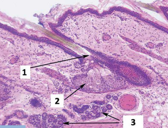
LM picture of thin skin
1- Hair follicle. 2- Sebaceous gland. 3- Sweat gland.


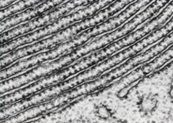
LM of perkinji fibers
H&E V.V.G Orcein stains
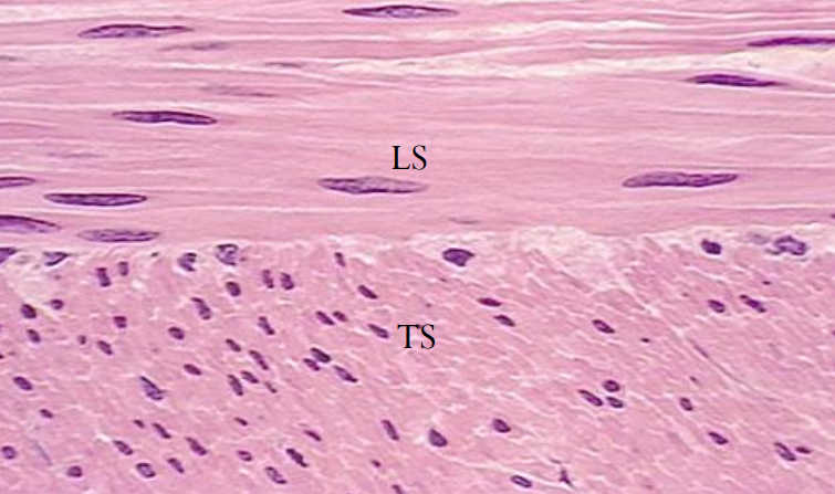
TS LM of skeletal muscle
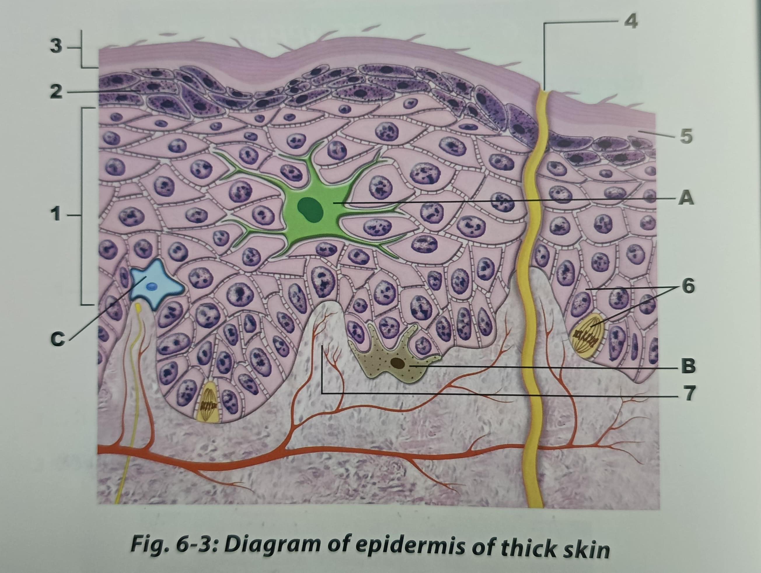
thick skin
1-S.spinosem
2-S.granulium
3-S.cornem
4.swear pores
5.s.lucidum
6-S.basel
7-Nerve
A-Langerhans cell
B-melanocyte
C-Markell cell