chapter 20 AnP 2
1/97
There's no tags or description
Looks like no tags are added yet.
Name | Mastery | Learn | Test | Matching | Spaced | Call with Kai |
|---|
No analytics yet
Send a link to your students to track their progress
98 Terms
for each of the following organs, provide two clues that would help identify it under a microscope
tongue
salivary gland
pancreas
liver
tongue → papilae with taste buds
salivary glands → roundish lobules with connective tissue septa
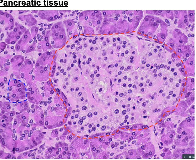
pancreas → island of pink within sea of purple
liver → hexagonal plates with central veins
why would you expect to find blood vessels associated with islets of langerhans
to carry hormones (insulin and glucagon) to their target tissues
after eating a candy bar and drinking soda, what pancreatic hormone would be released into the blood
insulin
the space between the lingual papillae can narrow as we age. Why would that affect our sensation of taste
there will be less interaction with food molecules as we age
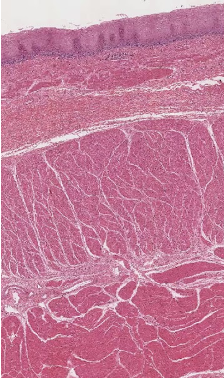
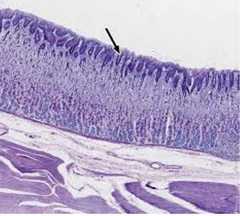
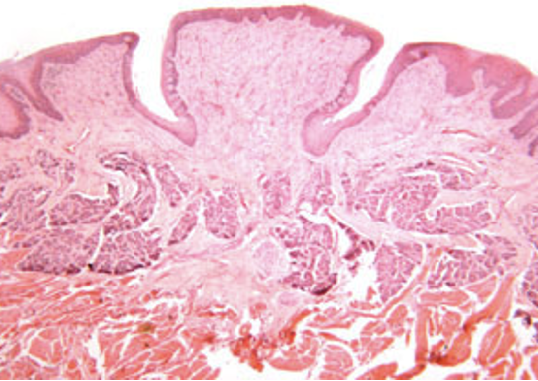
this is a picture of the
esophagus
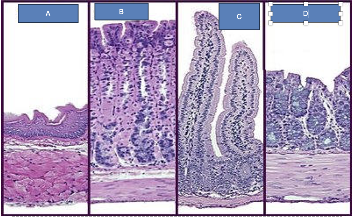
label
A. esophagus
B. stomach
C. small intestine
D. large intestine
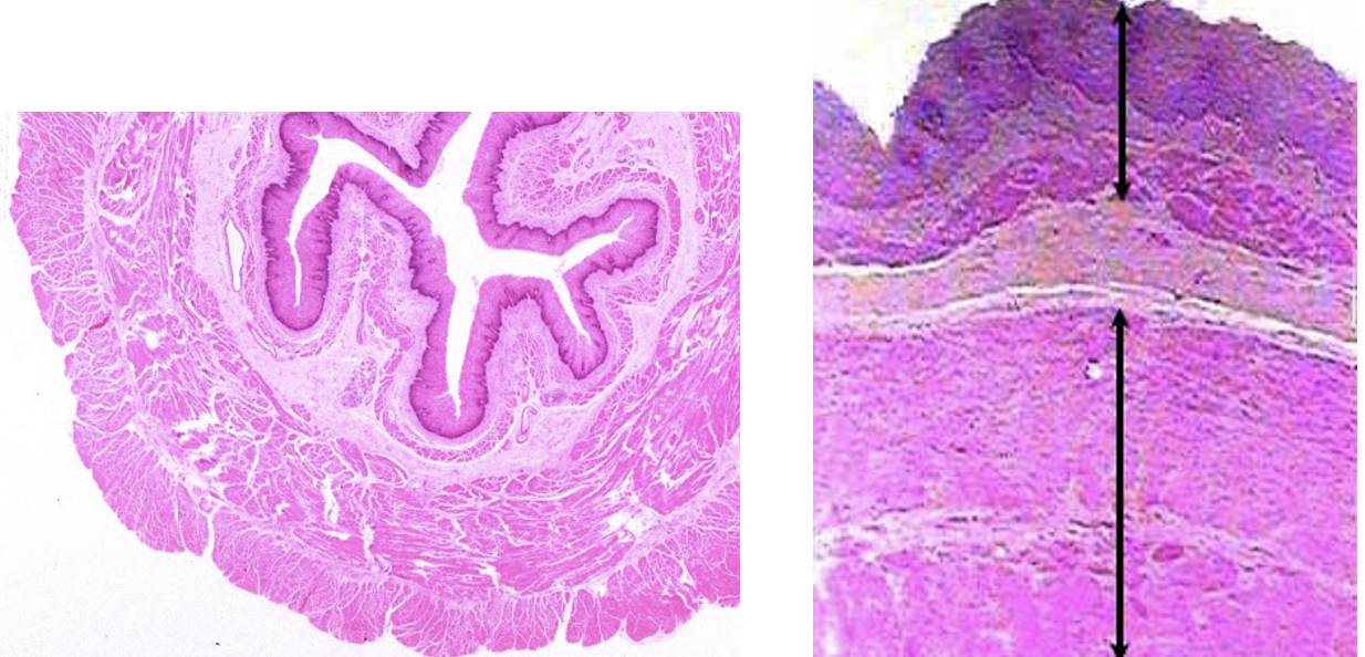
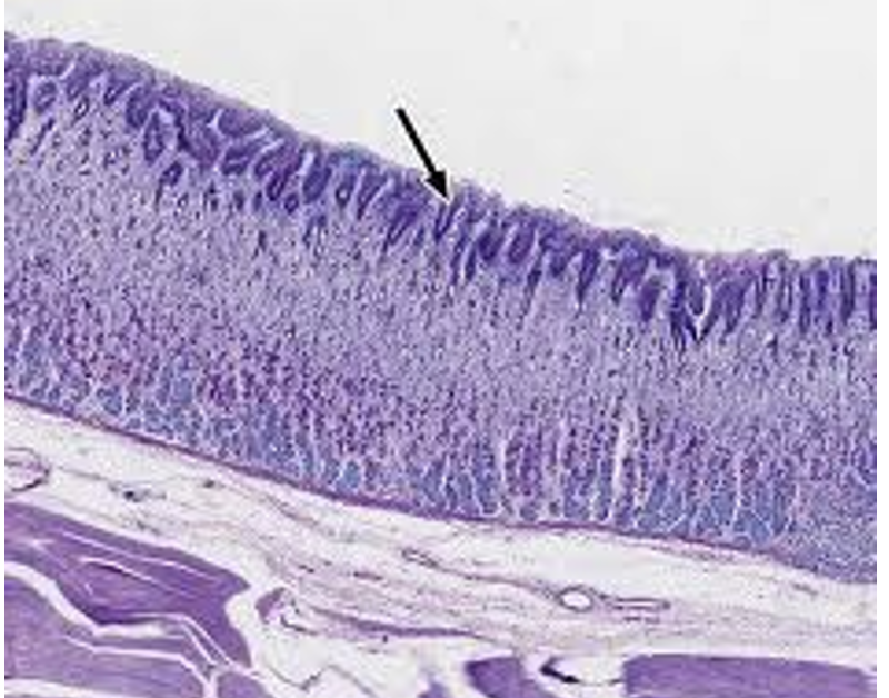
what is this
the esophagus
what is this?
the stomach
what is this
stomach
what is this?
the stomach
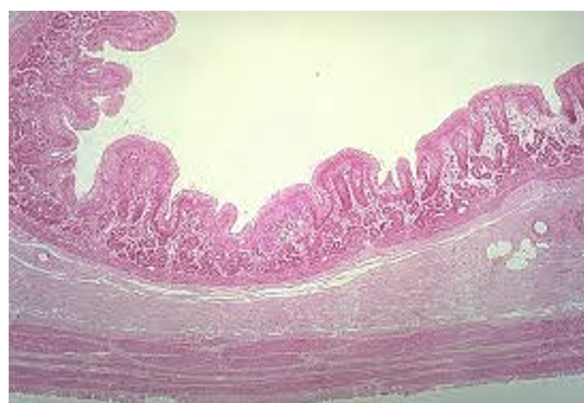
what is this
the stomach
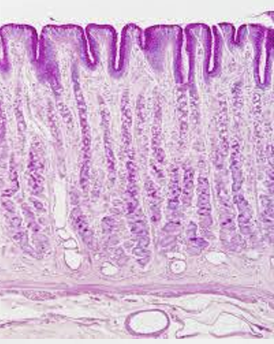
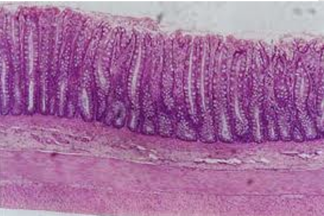
what is this
the small intestine
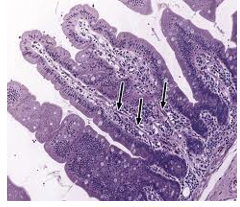
what is this
the small intestine
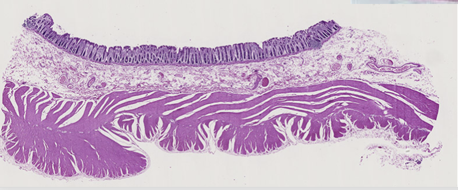
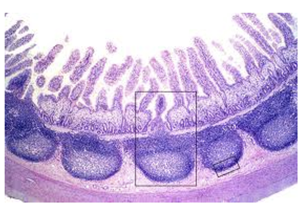
what is this
the colon of the large intestine
what is this
the colon of the large intestine
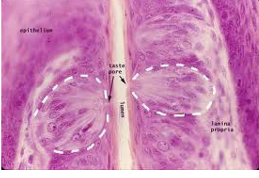
what is this
taste bud on the tongue
what is this
the tongue
what is this
the tongue
label
this is the pancreas
acinus
Islets of Langerhans
duct
what is this
the liver
what do accessory organs do?
Play a role in digestion even though food does not actually pass through them

what is this a picture of?
a tongue

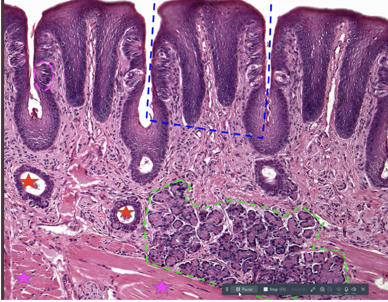
the tongue has notable projections on its superior surface is the blue box? What is it and what. isits function
this is the papillae. They provide a source of friction for the tongue, which is helpful when eating soft foods
the papillae contain
taste buds

what is the pink circle on the side? What is its function
these are taste buds. They are involved in a sense of gustation (being able to taste something)
the epithelium of the papillae is ______. What does this do
non-keratinized stratified squamous. This provides protection against chemical, thermal and abrasive injuries that foodstuffs can occasionally cause
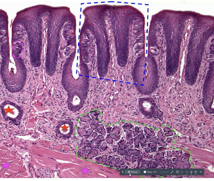
what is the pink star? what does it do
these are the skeletal muscle fibers inferior to the papilar. These are responsible for changing the shape and position of the tongue. They also project in many different directions. This gives the tongue great flexibility and the capacity to move in multiple directions
interspeed among the muscle cells are numerous
lingual salivary glands

what is the green and its function
this is the lingual salivary glands. they constantly secrete saliva in order to keep the oral epithelium moist
multilayered circular structures found on the lateral surfaces of the papillae.
taste buds
each taste buds contain furty slim receptors known as
gustatory cells that are involved in chemical detections and initiating the sensation of taste.
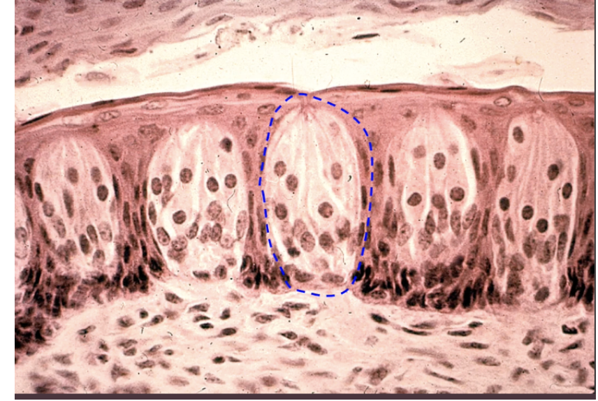
what is this?
a taste bud

what is this
parotid gland
what are septa on the parotid gland
inward extensions of connective tissue capsule that divide the gland into lobules. Supports and compartmentalizes it
The cells of each serous acinus are
pyramidal in shape and often have granules of material to be secreted
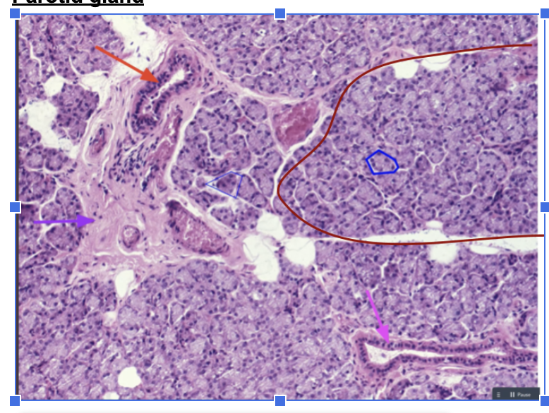
what is the blue?
secretory cells called the serous acini
The acini are surrounded by myoepithelial cells. This layer of epithelium does what
contracts to help move serous fluid out of the lumen of the serous acini and into the attached ducts
what is the black arrow pointing at
myoepithelial cells
fluid from serous acini flows into neighboring
intercalated ducts

what is purple and what is it lined by
intercalated ducts which are lined by simple squamous or cubodial epithelium
intercalated ducts join together to form larger
stratiated ducts
what is red? What are some characteristics
this is a stratified duct. These ducts are lined by simple colmnar epithelial cells which exhibit significant folding on their basal surface and thats why they have their name.
Striated ducts merge to form _____ + function
larger ducts which continue merging and eventually form the parotid excretory duct that carries saliva to the oral cavity
The epithelium of excretory ducts can increase from
simple columnar to pseudostratified columnar or even stratified columnar.
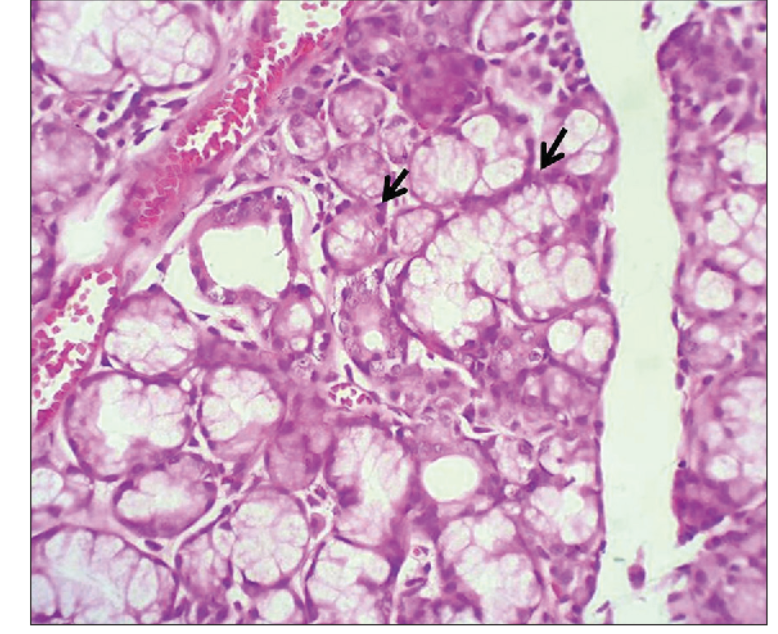
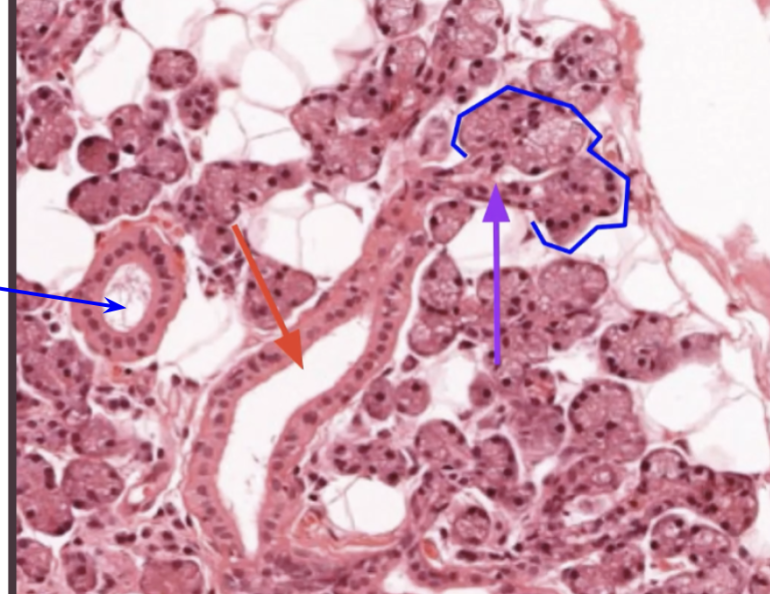
what is it?
this is the pancreas
in the pancreas, the partitions of connective tissue separate the pancreas into
lobules
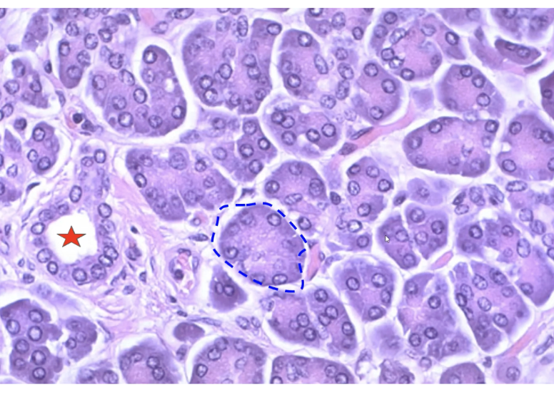
Each lobules will primarily consist of circular collections of cells stained a deep purple. These cells are _____ cells and each small collection of them is a ______
acinar cells; pancreatic acinus

what is blue
pancreatic acini

function of blue
this is the pancreactic acini. They produce a multitude of digestive enzymes and secrete the digestive enzymes into small ducts. including proteases, lipases, amylases, and nucleases
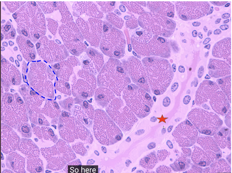
The acini secrete the digestive enzymes into small ducts that converge to form
large ducts that merge into the main pancreatic duct, which empties into the duodenum
Together the alkaline fluid from the duct cells and the enzymes from the acinar cells constitute
pancreatic juice
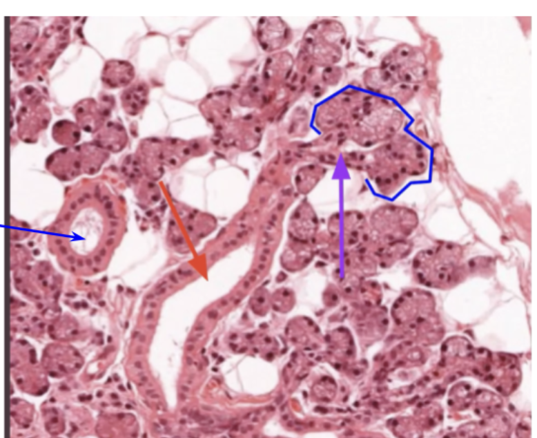
what is the red star? describe it
it is a pancreatic duct. They are lined by cuboidal epithelium which secretes an alkaline fluid.
exocrine function of pancreas
secretion of pancreatic juice
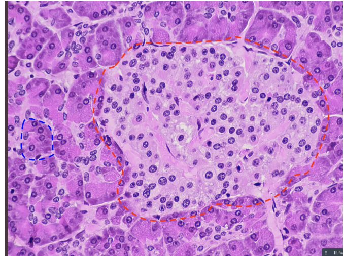
Scattered amidst the acini and making up 1% of the pancreas is the
islets of Langerhans which control blood glucose levels
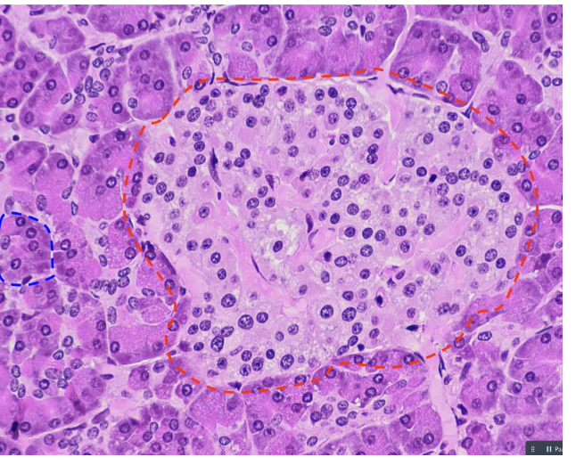
what is outlined in red
islets of langerhans. These are found clusters of cells that are light pink in color and have prominent nuclei. While they can’t be distinguished with the microscope, several discrete cell types can be found in these.
alpha cells in the islets of langerhans release
glucagon
beta cells in the islets of langerhans release
insulin
together, alpha and beta cells release hormones that
act in an antagonistic and negative feedback fashion to control plasma glucose levels
When plasma glucose levels rise → the beta cells release ____ and then
insulin. Insulin increases uptake of glucose by most body cells and lowers the levels of plasma glucose
When plasma glucose levels fall → alpha cells release glucagon and then
Glucagon increases glycogen breakdown and glucose release by the liver, raising the level of plasma glucose
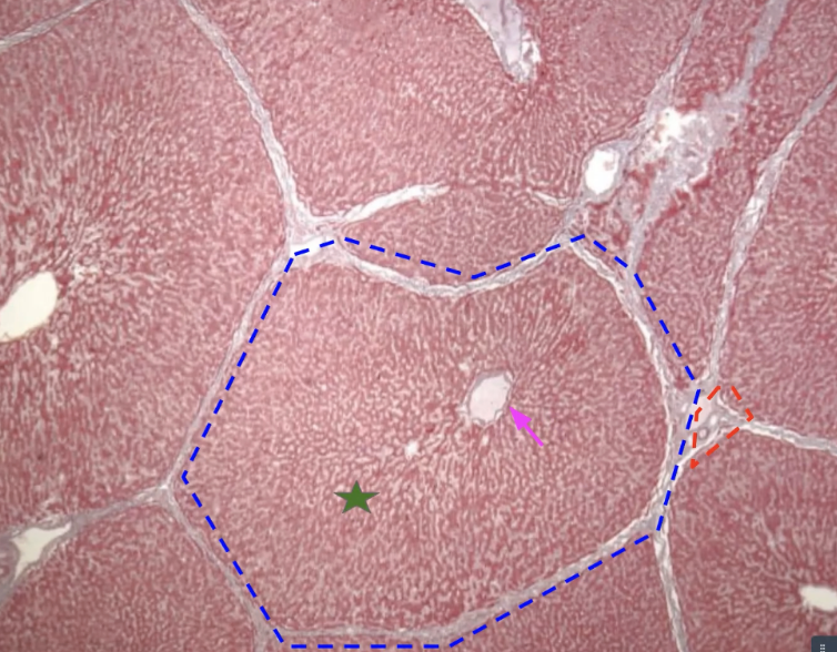
what is this? what is in blue
this is the liver. The blue is a liver lobule
liver lobules are composed primarily of
hepatocytes
functions of liver lobules
nutrient metabolism
vitamin and mineral storage
drug inactivation
Lobules also play a role in modifying the blood by
synthesizing plasma proteins and removing circulating pathogens, debris, aged RBCs, hormones, antibodies, and toxins.
The digestive functions of liver lobules
is the synthesis and secretion of bile
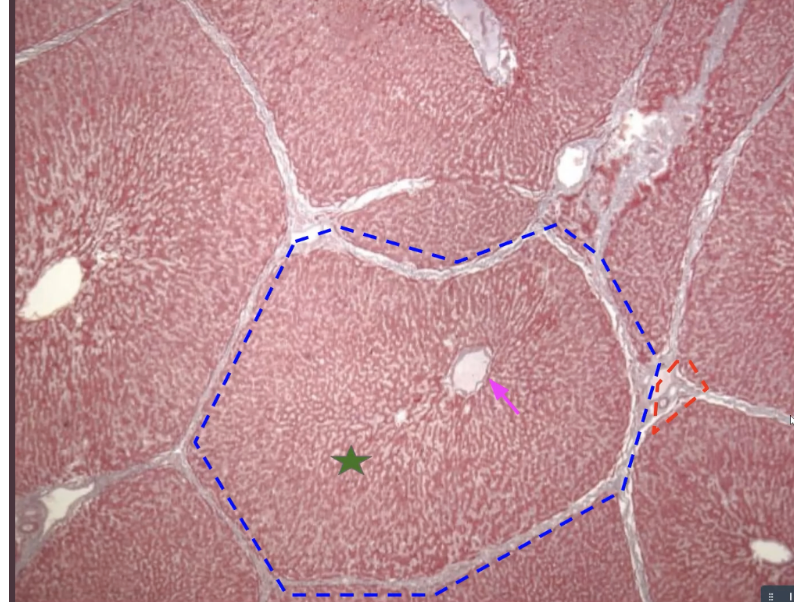
Adjacent lobules are separated from one another by connective tissue known as
interlobular septa
In the center of each lobule is a blood vessel known as the
central vein
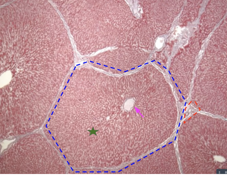
pink arrow with function
the central vein drain liver lobules taking blood onward towards hepatic vein
at each of the 6 corners of the lobule, there is a portal triad composed of
2 blood vessels and a bile duct
The bile duct in each triad collects bile
from the hepatocytes within the lobule and ultimately delivers it to the large left and right hepatic ducts that take bile away from the liver (duodenum or gallbladder)
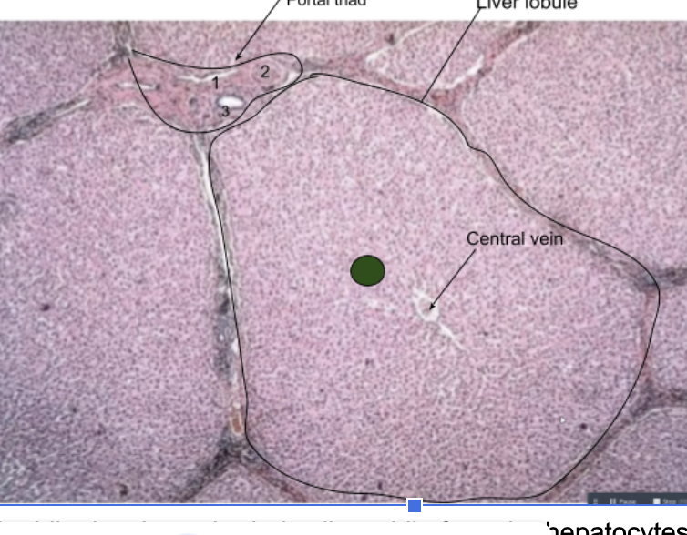
what is each part of the portal triad
branch of hepatic portal vein (portal venule)
arteriole: branch of hepatic artery
bile duct
the two blood vessels of the portal triad are
portal arteriole: a branch of the hepatic artery
portal venule (a branch of the hepatic portal vein)
the portal arteriole and portal venule bring blood
to the sinusoidal capillaries that run inbetween the hepatocytes and empty into the central vein.
The portal arteriole brings oxygen rich blood
to feed hepatocytes
The portal venule brings blood
from the hepatic portal system (draining the intestines, stomach, and spleen) which may be rich in nutrients, toxins, and pathogens. The hepatocytes can then eliminate any dangerous substances and gather any nutrients
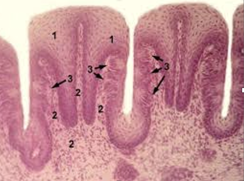
label everything
this is the tongue
blue box → stratified squamous epithelium papilae
pink circle → taste buds
green circle → seromucous salivary glands
red star → salivary glands
these make saliva
pink star → skeletal muscle
bands of ______ make up the bulk of the tongue
skeletal muscle
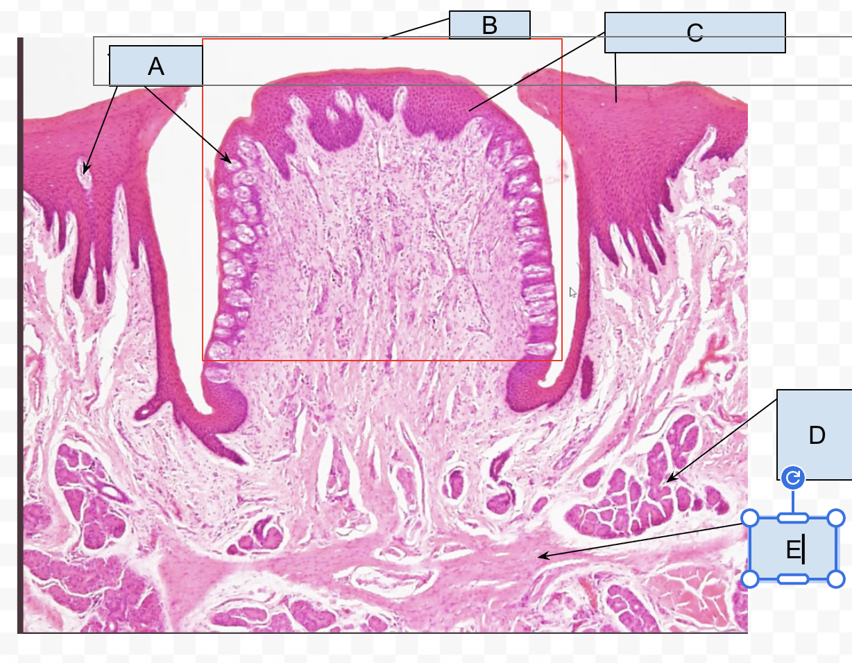
label the tongue
A. taste buds
B. papilae
C. stratified Squamous epitheium
D. lingual salivary glands
E. Skeletal muscle
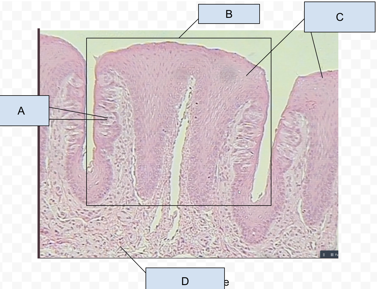
label
A. taste buds
B. papilae
C. stratified squamous epithelium
D. skeletal muscle tissue
in taste buds, there can sometimes be an opening called
the taste pour which lets chemicals in food interact with the cells and create the sensation of taste.
label the parotid gland
red arrow → stratified epithelium duct
blue → serocus acini
purple → connective tissue suptum
pink→ salivary duct of simple epithelium
a whole circle is a lobule
serous acini function
secrete saliva into intercalated ducts
stratified ducts merge to form larger ducts with stratified epithelium which eventually all merge to
exit the gland
Label
blue shape → serous acini
purple arrow → intercalated duct with simple epithelium
red arrow → stratified duct
blue arrow → stratified duct
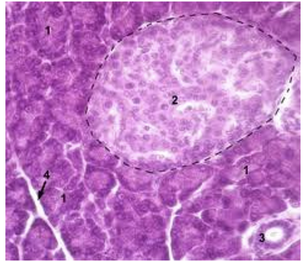
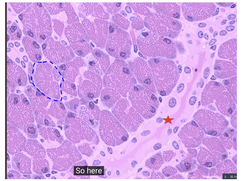
99% of the pancreas is composed of clusters of cells attached to small ducts. These clusters are known as
pancreatic acini
label
blue → pancreatic acini
red → islet of langerhans/pancreatic islets
label
Blue → pancreatic acini
Red → pancreatic duct
function of pancreatic duct
The duct carries the acinar enzymes and secretes an alkaline fluid as well. The high pH of pancreatic juice helps nutralize the acidity of gastric chyme
label
Red star → pancreatic duct
Blue star → pancreatic acini
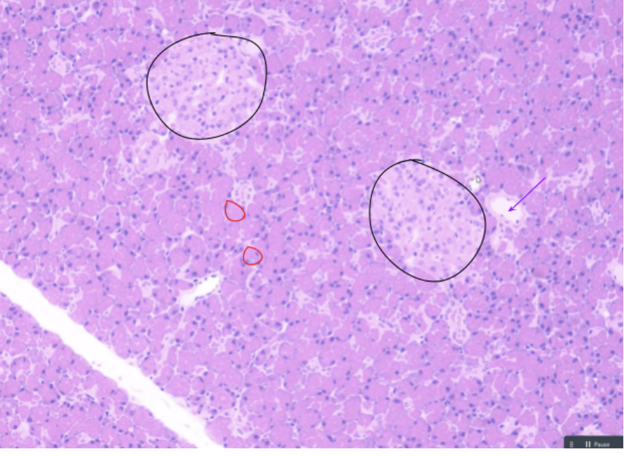
label
pancreas
Black circle → pancreatic islet
Red circles → acini
Purple arrow -> pancreatic duct
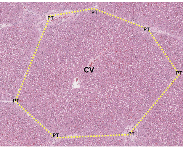
The liver is composed of thousands of roughly hexagonal structures called ______. Each liver lobule contains a lumen of a______surrounded by _______
liver lobules; central vein; hepatocytes and sinusoidal capillaries
Blood flows through the triad vessels
through the sinusoids to the central vein and is filtered along the way
the bile duct collects bile produced by
hepatocytes
label the liver
blue → liver lobule
green star → hepatocytes and sinusoidal capillaries
red → portal triad
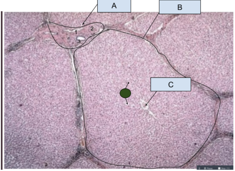
label liver
A. portal triad
B. Liver lobule
C. central vein
green → pink is hepatocyte while white is sinusoidal capillaries
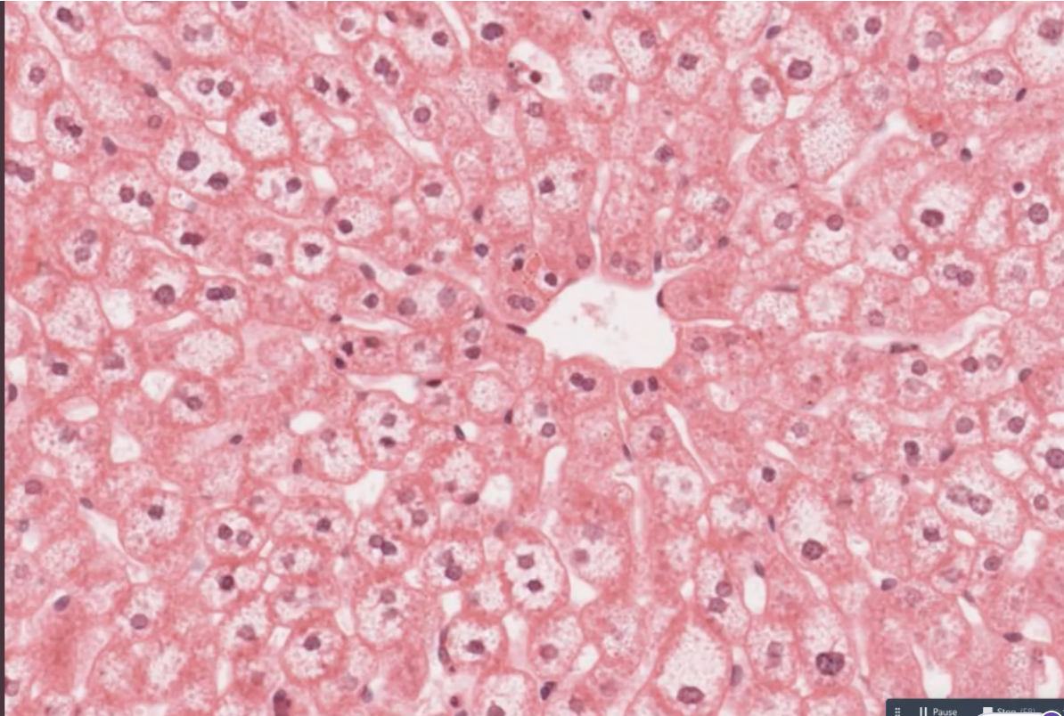
label the middle of the liver lobule
circle → central vein
pink → hepatocytes
white parts → sinusoidal capillaries
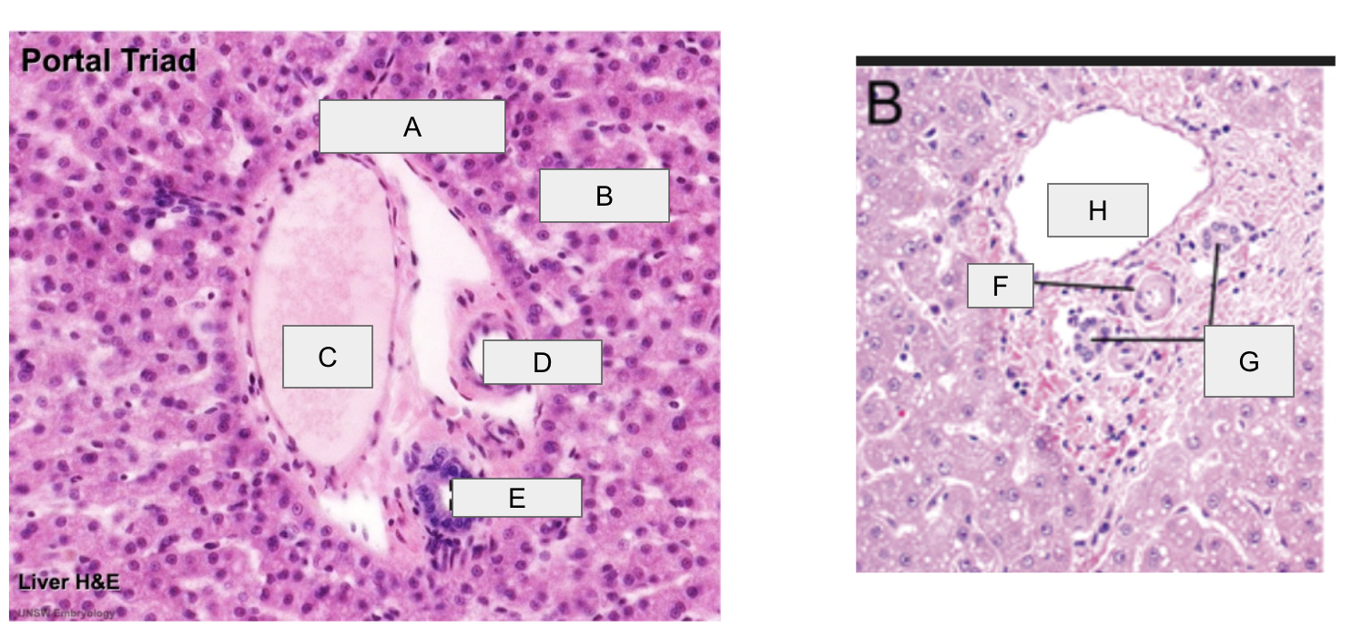
label portal triad
A. interlobular connective tissue
B. hepatocytes
C. portal vein
D. branch of hepatic artery
E. branch of bile duct
F. branch of hepatic artery
G. branch of bile duct
H. PV