Conjunctival Pigmentations
1/35
There's no tags or description
Looks like no tags are added yet.
Name | Mastery | Learn | Test | Matching | Spaced | Call with Kai |
|---|
No analytics yet
Send a link to your students to track their progress
36 Terms
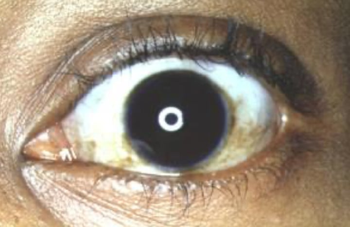
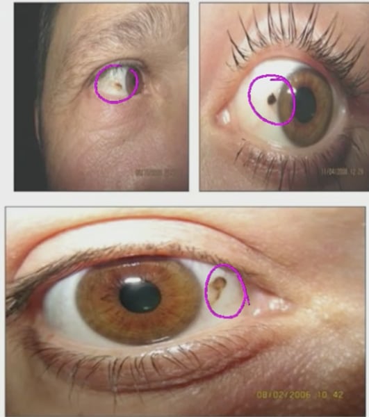
Complexion-Acquired Melanosis (CAM)
benign pigmentation of the conjunctival epithelium
What is another name for complexion acquired melanosis?
Racial melanosis (associated with darker skinned individuals more often)
What is the distribution of CAM?
OU, asymmetrical
True or False: when you move the globe, pigmentation due to CAM moves freely
True
What is the most common location to see a conjunctival nevus?
juxtalimbal region of bulbar conj
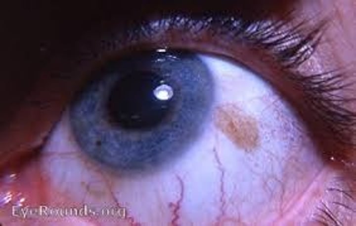
Conjunctival nevus characteristics
typically unilateral, solitary, flat (or slightly elevated), freely mobile, and are occasionally non-pigmented
What would you do if a patient comes to you with a conjunctival nevus?
Ask how long the patient has been aware of the nevus, photodocumentation to watch for growth or change over time, potentially refer for biopsy if it looks concerning
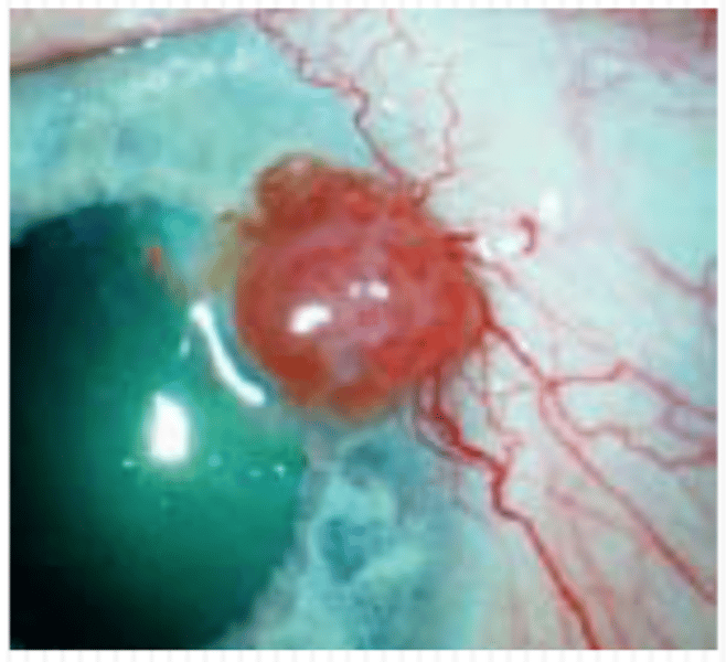
What percentage of malignant eye conditions are conjunctival melanoma?
2% (rare but deadly!)
True or False: all conjunctival melanomas are black/grey pigmented fixed nodules
False: non pigmented lesions with smooth surfaces are also possible
What is the most common cause of conjunctival melanoma?
Primary acquired melanosis (80-70%)
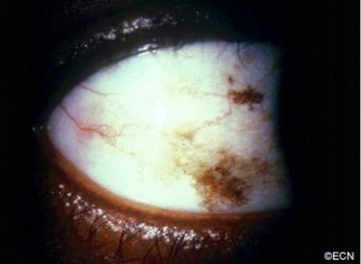
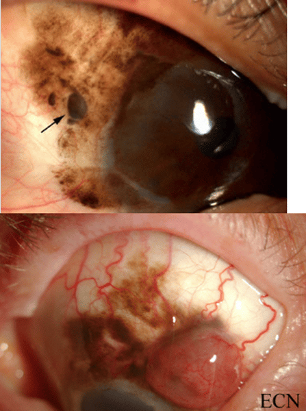
Primary acquired melanosis (PAM)
UNILATERAL pigmentation patches on conjuntiva. Has indistinct margins. Has PREMALIGNANT potential for malignant melanoma.
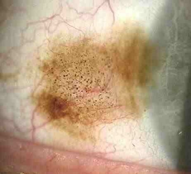
What is the main differences between CAM and PAM?
Complexion acquired melanosis is bilateral while primary acquired melanosis is unilateral. CAM is associated with dark skin individuals while PAM is associated with caucasian individuals. CAM is harmless while PAM can be premalignant.
How deadly is conjunctival melanoma?
25% mortality rate (as high as 44% if started as PAM)
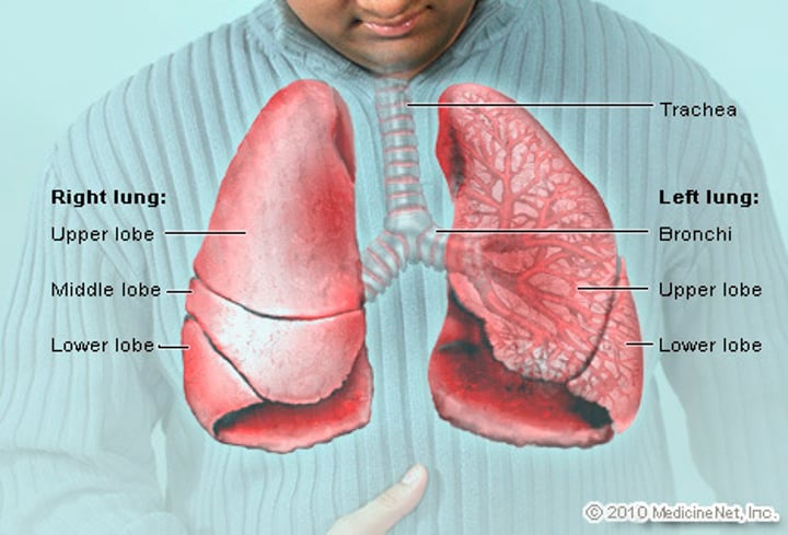
What is the most frequent metastatic site for conjunctival melanoma?
The lungs (both to the eye and from the eye)
Name some changes that can occur when a nevus evolves into a melanoma!
Size, density, increased vascular feeder vessels to the area, if it becomes fixed to the underlying sclera (except at the limbus)
True or False: Primary acquired melanosis (PAM) is fixed to the conjunctiva
False: PAM IS MOBILE! If it were fixed, you'd consider it more likely to be conj melanoma at that point
True or False: it is possible to see a patient who has PAM and melanoma occurring simultaneously in the same eye
True! Always check under the lids!
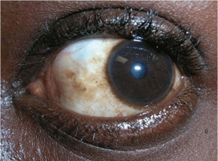
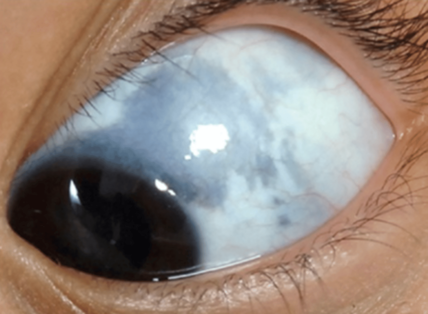
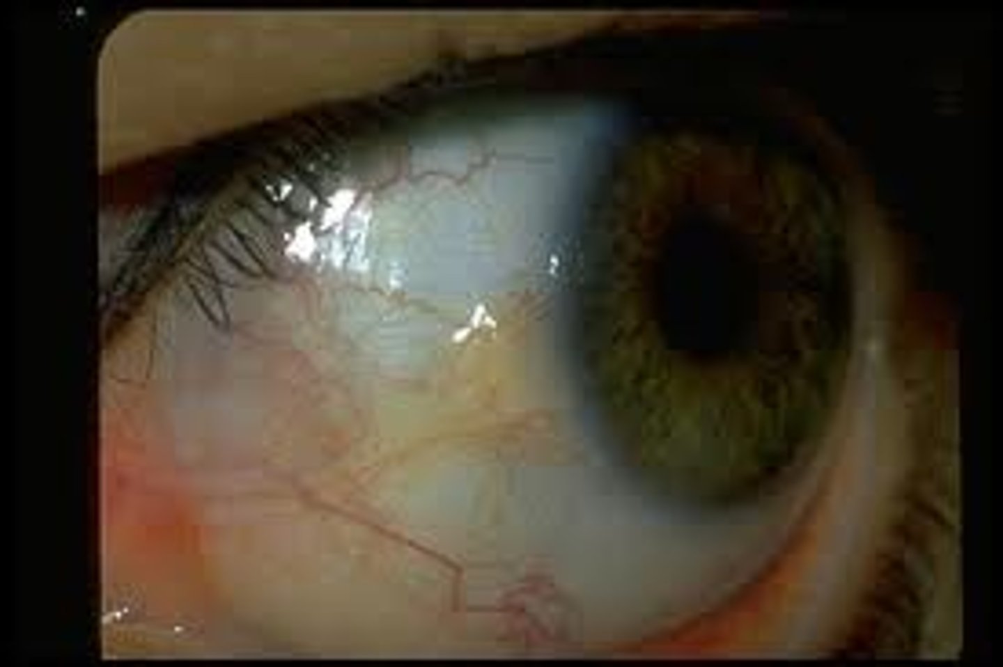
Ocular melanocytosis
-Slate gray or blue pigmentation of the sclera and episclera
-NONmobile plaque like appearance
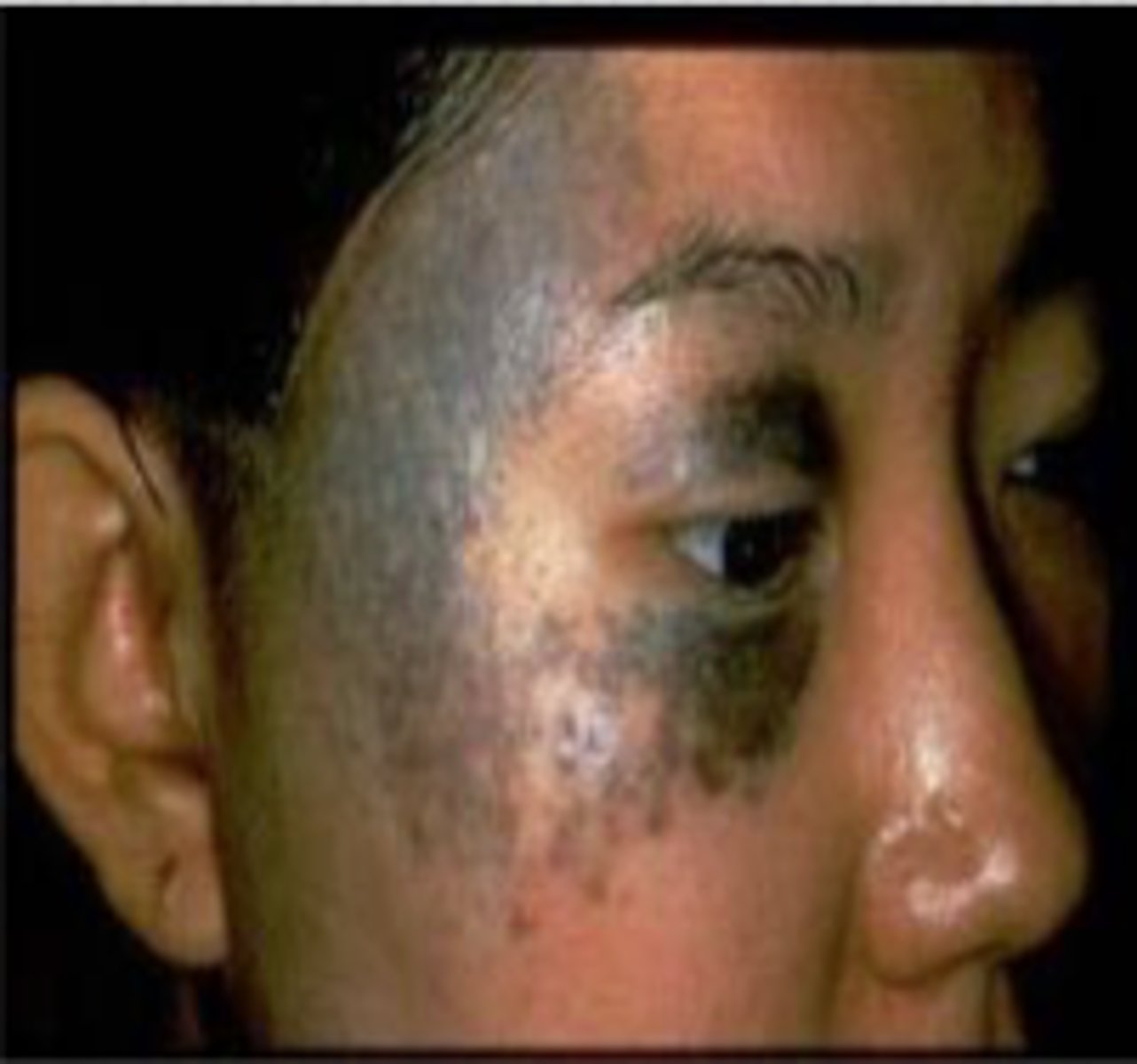
Oculodermal Melanocytosis
Ocular melanocytosis + hyperpigmentation of periorbital skin around the areas in which the 1st and 2nd divisions of the Trigeminal nerve innervate
Is oculodermal melanocytosis typically bilateral or unilateral?
unilateral
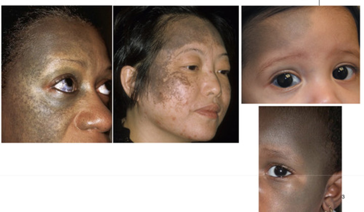
What is another name for oculodermal melanocytosis?
Nevus of Ota
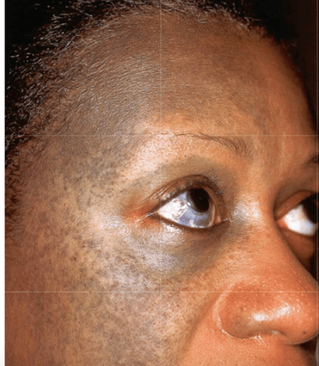
What associated features should you be concerned about with oculodermal melanocytosis?
melanoma and glaucoma
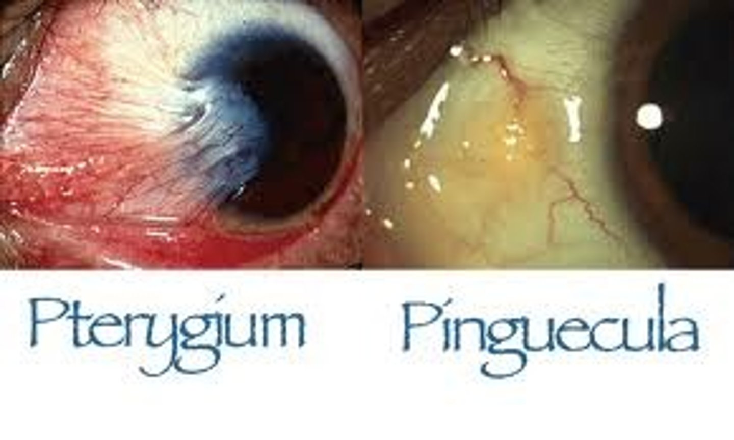
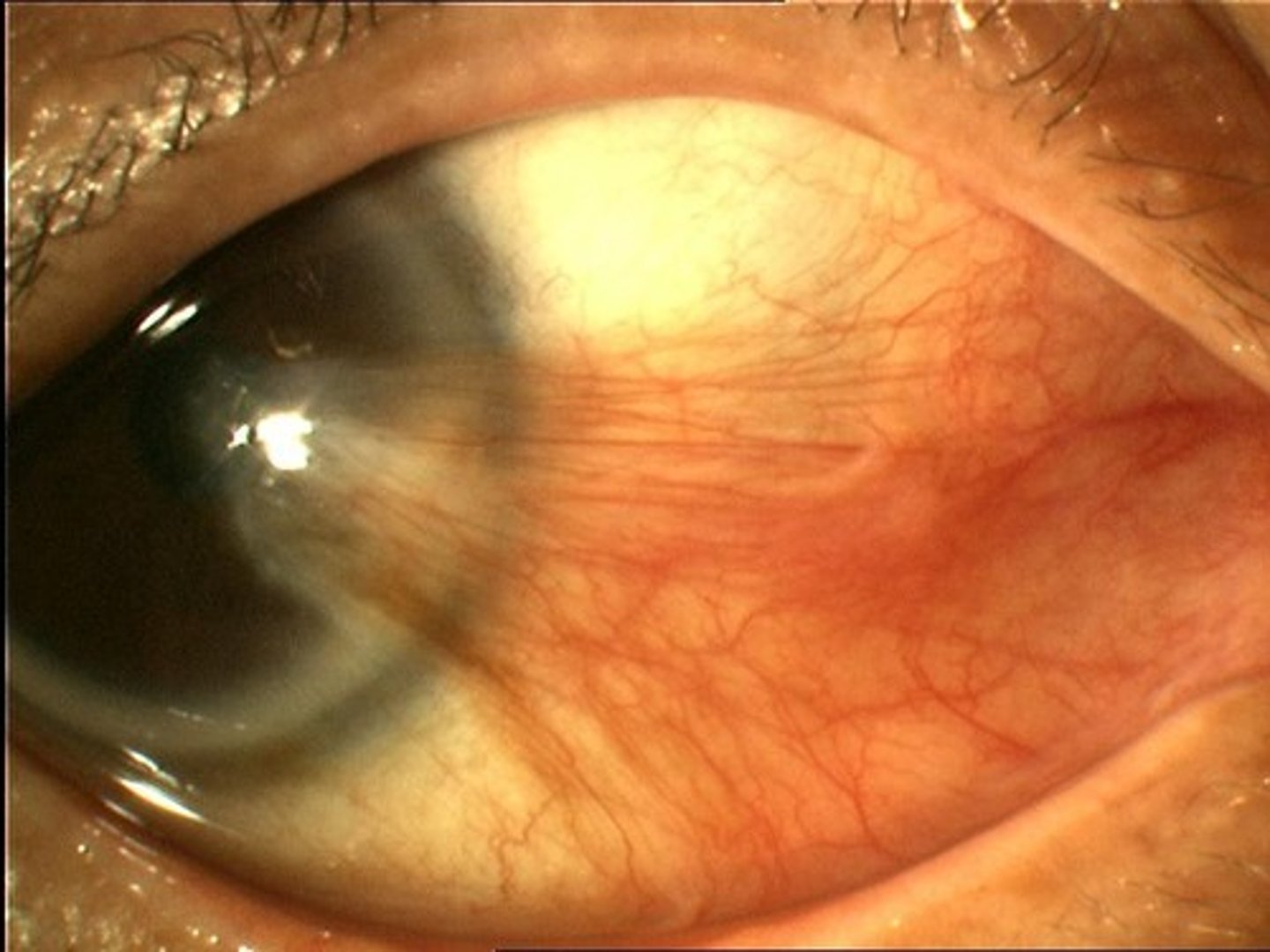
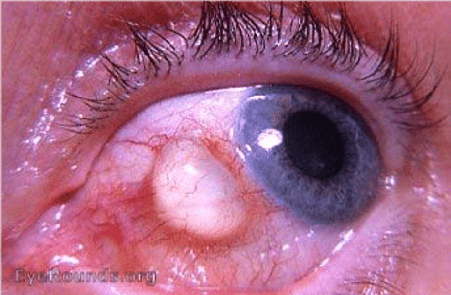
Pinguecula
yellowish mass of collagen the conjunctiva that may be related to exposure to ultraviolet light, dry climates, and dust
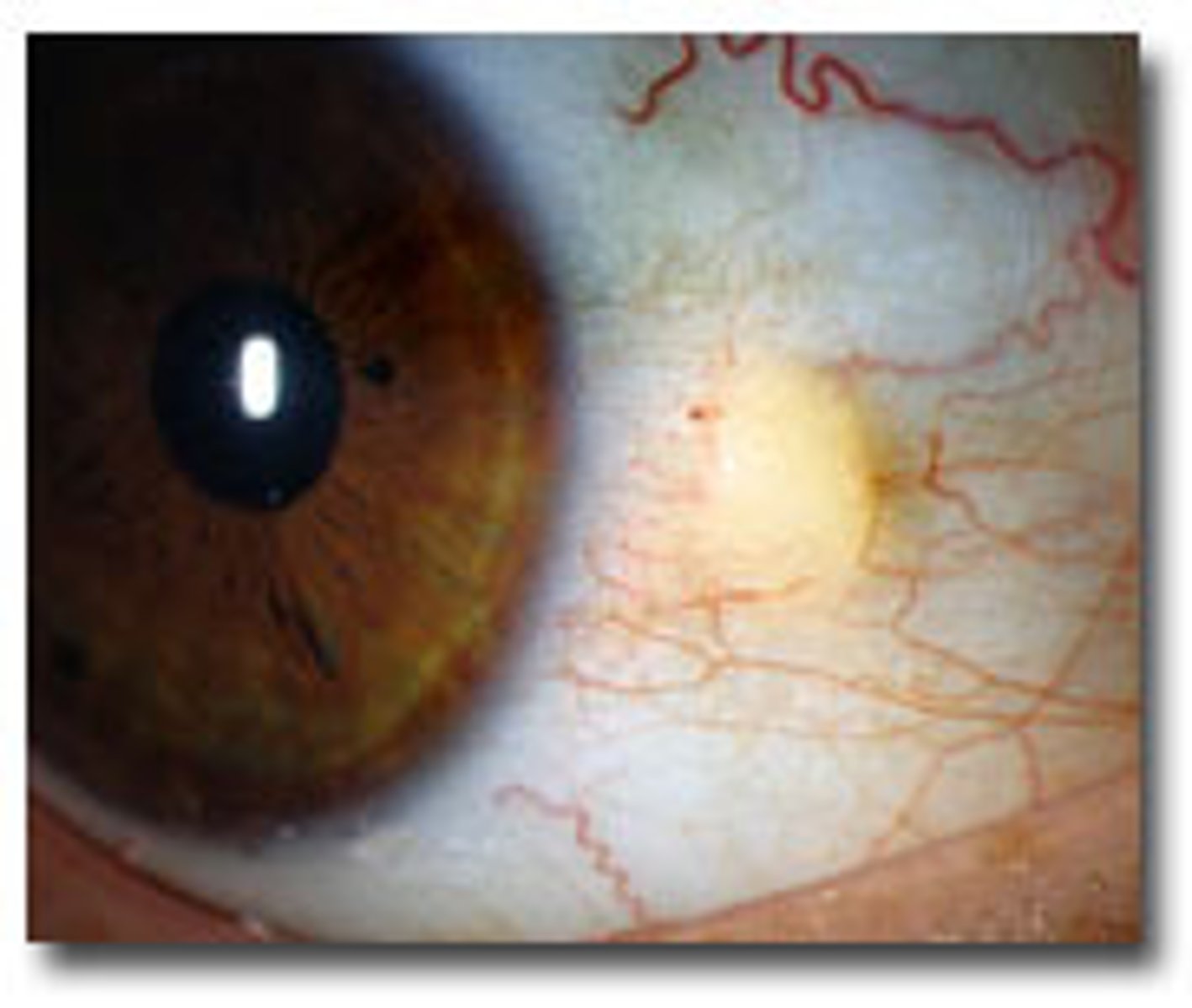
What simple solutions can you treat pinguecula with?
side shield sunglasses for UV and environmental protection as well as lubricants
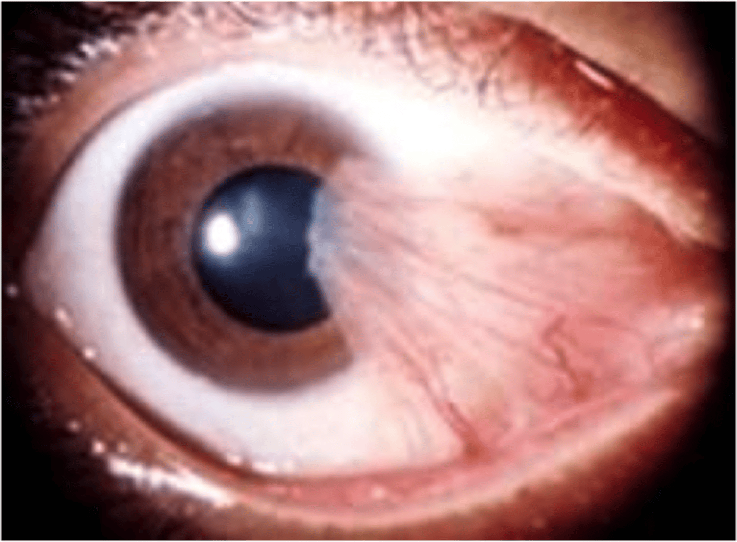
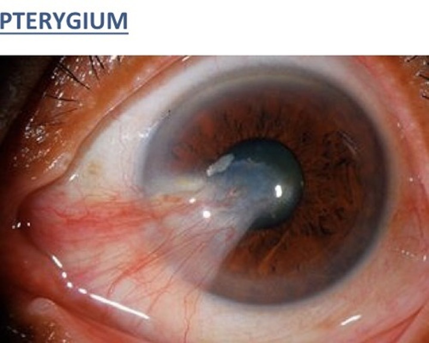
pterygium
winglike, triangular fibrovascular growth of conjunctival tissue extending to the cornea
True or false: pterygium can evolve into pinguecula
False: pinguecula can evolve into pterygium!
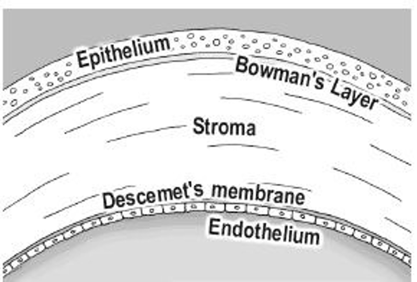
What layer of the cornea do pterygiums destroy?
Bowman's membrane (cannot regenerate)
What can pterygiums do to your vision?
Cause induced astigmatism or decrease vision
How are pterygium managed?
lubricants, sunglasses, avoid vasoconstrictors if the pterygium is inflamed
What is the issue with pterygium surgical removal?
They have a high rate of reoccurrence, so it is best to hold off on removal if the pterygium is not impacting vision
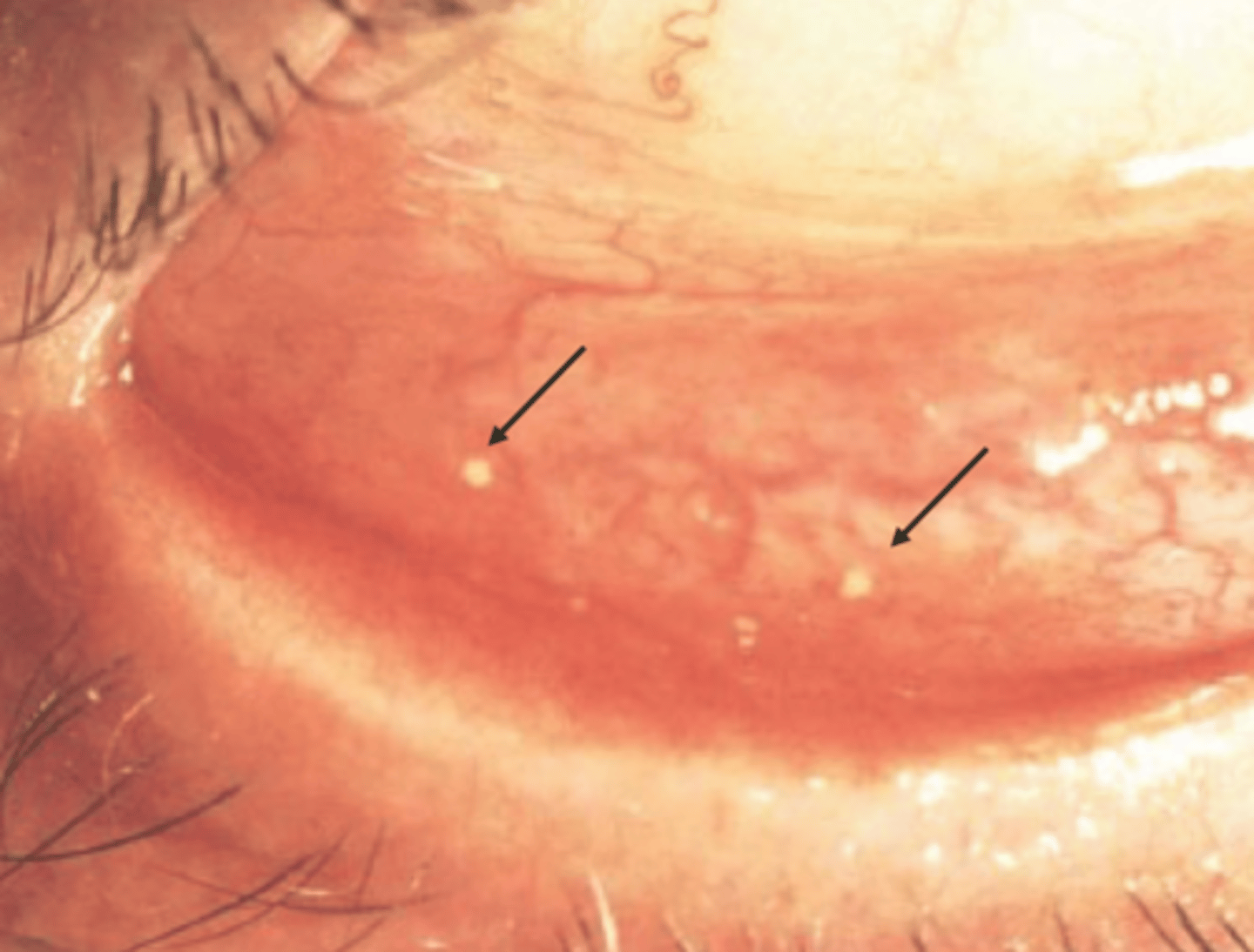
Concretions
small yellow calcified hard spots in the palpebral conjunctiva caused by cellular degeneration debris getting trapped
Conjunctival cysts
fluid filled clear cysts on the conjunctiva
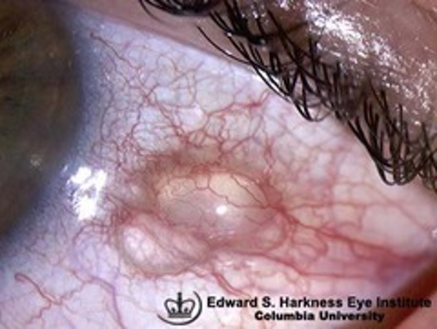
What tends to happen if you drain a conjunctival cyst?
they have a tendency to quickly refill
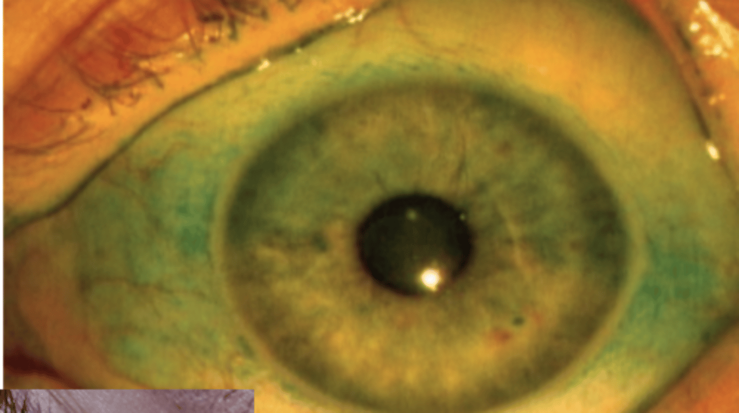
What do you use lissamine green for?
staining DEAD cells on the conjunctiva
Bitot's spot
White foamy appearing area of keratinizing squamous metaplasia of bulbar conjunctiva
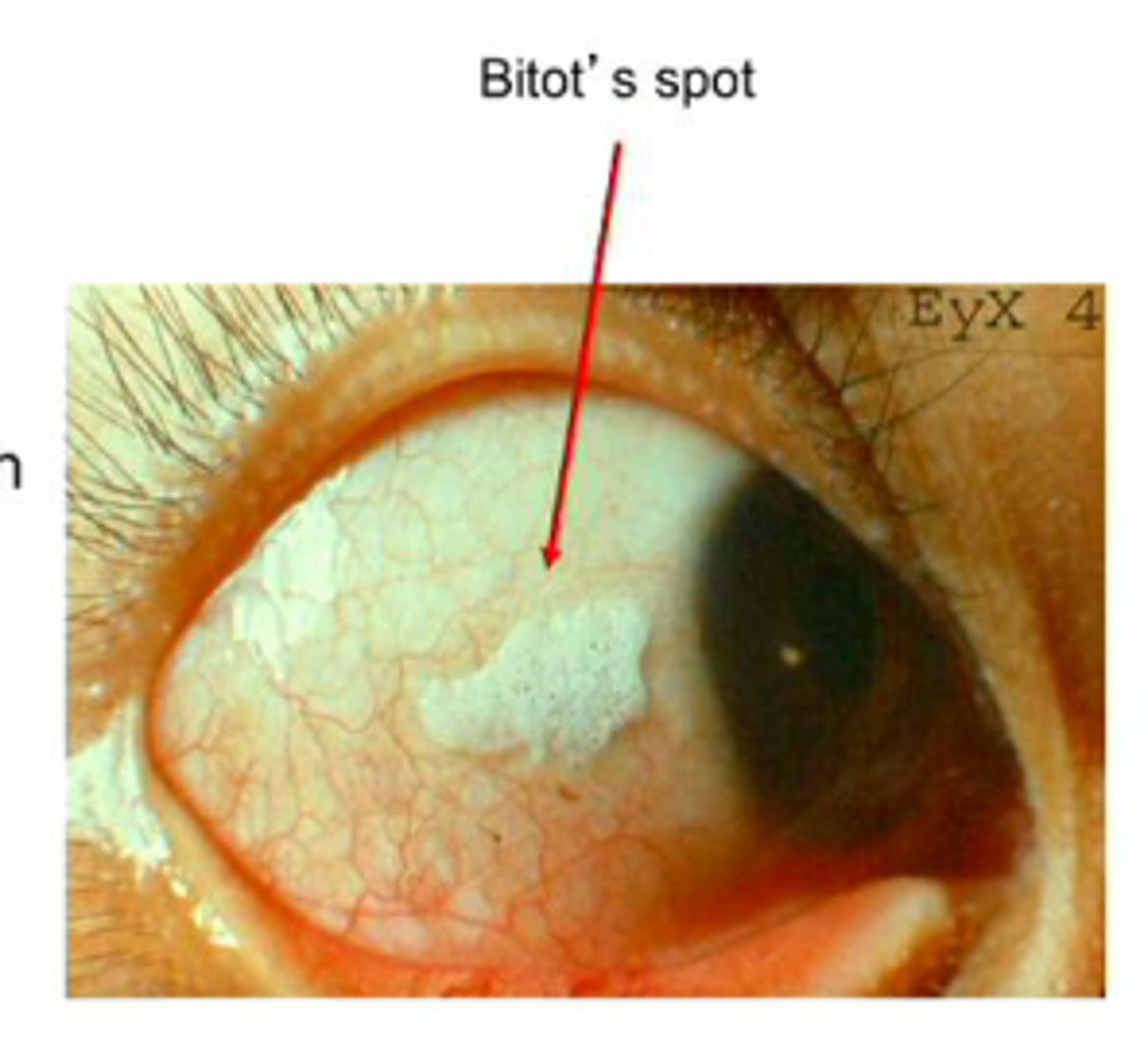
What does Bitot's Spot indicate is an issue with the patient?
Vitamin A deficiency