A&P - Neuro 1
1/64
There's no tags or description
Looks like no tags are added yet.
Name | Mastery | Learn | Test | Matching | Spaced |
|---|
No study sessions yet.
65 Terms
what is acetylcholinesterase
1 of 4 neurotransmitters (structure differs substantially from the others)
synthesized from acetate and choline and stored in synaptic vesicles
function of acetylcholinesterase
used in PNS to stimulate skeletal muscle, used in CNS to increase arousal
effect on target cell depends on receptor present (nicotinic or muscarinic)
mechanism for clearing acetylcholine from a synapse
action potential triggers its release into synaptic cleft - some briefly attaches to postsynaptic receptor - then cleared from cleft by being broken down to acetate and choline by acetylcholinesterase
acetate and choline then recycled by presynaptic cell
chronologic order of events in synaptic transmission
1. action potential arrives at axon terminal
2. voltage gated calcium channels open
3. neurotransmitter vesicles fuse w/ presynaptic membrane
4. neurotransmitter released into and diffuse across synaptic cleft
5. neurotransmitter binds to receptors on postsynaptic membrane
6. neurotransmitter diffuses across cleft
7. neurotransmitter bind to postsynaptic receptor
8. postsynaptic potential generated
effect of voltage gated calcium channel opening and neurotransmitter release at the synaptic knob
depolarization of membrane potential, neurotransmitters released into synaptic knob
what occurs when voltage gated K+ channels open in conductive segment
K+ flows out of neuron down concentration gradient causing repolarization of membrane potential (becomes more negative)
impact on resting membrane potential if there are no Na+ leak channels
if K+ only is leaked, RMP would be where K+ concentration and electrical gradients at equilibrium (-90mV)
relative concentration locations of potassium and sodium
higher concentration of K+ in cytosol vs interstitial fluid
higher concentration of Na+ in interstitial fluid vs cytosol
how repolarization and depolarization relate to Na+/K+ channel states (steps in depol and repol)
steps in depolarization:
1. reaching threshold - Na+ enters from adjacent region and voltage gated Na+ channels open
2. Na+ enters axon causing membrane to have positive potential
2b. inactivation state - Na+ channels close, becoming temporarily inactive (unable to open)
- steps repeat in adjacent regions and impulse moves toward synaptic knob -
steps in repolarization:
3. reaching threshold slowly opens K+ channels and K+ diffuses out, causing negative membrane potential
4. hyperpolarization - K+ channels stay open for a longer time so K+ exit makes cell more negative than RMP
5. reestablishing RMP - K+ channels eventually close and RMP reestablished by leak channels and Na+/K+ pumps
- steps 3-5 repeat in adjacent regions as impulse moves toward synaptic knob -
order and definition of absolute and relative refractory periods
refractory period (in general): period of time after start of action potential when difficult or impossible to fire another action potential
- absolute refractory: ab 1 ms, no stimulus can initiate another action potential, Na+ channels are open then inactivated (ensures propogation goes toward synaptic knob and doesn't reverse direction)
- relative refractory: just after absolute, another action potential is possible (Na+ channels have reset) but minimum stimulus strength is now greater (some K+ channels are still open so cell is slightly hyperpolarized and further from threshold)
all or none law of action potentials
- if threshold reached, axon potential generated and propogated down axon w/o any loss in intensity
- if threshold not reached (subthreshold), voltage gated channels stay closed - no action potential
- axon shows same intensity of response to values greater than threshold
think of a gun: sufficient pressure on trigger makes it fire, insufficient not fired; firing is the same no matter how hard you squeeze the trigger
factors allowing for the fastest nerve impulse propogation
conduction speed depends on axon diamater and myelination; thicker fibers faster than thin (less resistance) and myelinated faster than unmyelinated
function of myelin and which cells produce it in CNS/PNS
myelin: several layers of membrane of glial cells; high lipid content gives glossy white appearance and insulates axon (responsible for appearance of white matter in brain and spinal cord)
glia are neuromyelocytes in PNS and oligodendrocytes are neuromyelocytes in CNS
soma/ cell body of neuron
(aka perikaryon) spherical w/ triangular cone shaped axon hillock, plasma membrane enclosed cytoplasm, contains mucus and chromatophilic substance (nissl bodies) made of ribosomes (both free and bound)
dendrites of neuron
short unmyelinated processes branching of cell body of neuron; receive input and transfer it to cell body
axon of neuron
- long processes emanating from axon hillock (triangular region) of cell body of neuron
- cytoplasm termed axoplasm
- membrane termed axolemma
- spills into branches called axon collaterals
- tips of extensions are synaptic knobs which house synaptic vesicles containing neurotransmitter
- may be insulated by myelin sheath formed by glial cells (neurofibril nodes are the gaps btwn regions of myelin)
cytoskeleton of neuron
composed of microfilaments (actin), intermediate filaments (neurofilaments) which aggregate to form bundles called neurofibrils which provide tensile strength, and microtubules
multipolar neurons
many dendrites, one axon (mc type)
bipolar neurons
one dendrite, one axon (limited number, ex: retina)
unipolar neurons
(psedounipolar) one process extends from cell body, splits into 2 processes : peripheral process (splits into several receptive dendrites) and central process (leads to synaptic knobs in CNS)
mixed nerve
contain both sensory and motor (mc nerve)
autonomic motor division
- (visceral motor) sends involuntary commands to heart, smooth muscle, and glands
- has sympathetic and parasympathetc divisions
somatic motor division
detects stimuli we consciously perceive
somatic sensory division
detects stimuli we consciously perceive
visceral sensory division
detects stimuli we do not typically perceive
major glial cells
- astrocytes
- oligodendrocytes
- ependymal cells
- microglia
- satellite cells
- neurolemmocytes
glial cells
(neuroglia) nonexcitable support cells found in CNS and PNS, approximately same number of glial cells as neurons; accounts for about half the volume of the nervous system
astrocytes
- (star shaped) most abundant glial cell in CNS; have processes that end in vascular feet
- helps form BBB by wrapping perivascular feet around brain capillaries (controls which substances have access to brain)
- regulate tissue fluid composition (chemical environment around neurons, ex: K+ concentration forms structural support for nearby neurons, assists neuronal development, and alters synaptic cleft - add/eliminate/influence)
- occupy the space of dying neurons
ependymal cells
line cavities in brain and spinal cord; part of choroid plexus (which produces spinal fluid)
microglia
small phagocytic cells of immune system that wander CNS and replicate during infxn; remove synapses that are no longer needed
oligodendrocytes
large cells w/ slender extensions; extensions wrap around axon, forming myelin sheath
satellite cells
arranged around neuronal cell bodies in a ganglion; electrically insulate and regulate exchange of nutrients and wastes
neurelemmocytes
(schwann cells) elongated flat cells that ensheath PNS axons w/ myelin; allows for faster action potential propogation
how do glial cells differ from neurons
glial cells: nonexcitable supportive/regulatory roles - homeostasis, form myelin, schwann cells, structual support (support, protect, and nourish neurons)
neurons: excitable, transmit electrical and chemical signals - mood, muscle movement, sensory perception
what do all glial have in common
non excitable, supportive role, maintain homeostasis, do not form synapses for signal transmission (unlike neurons)
types of neuronal pools/circuits
converging, diverging, parallel-after-discharge, and reverberating
converging neuronal circuit
input converges at a single postsynaptic neuron (ex: multiple sensory input synapses on neurons in salivary nucleus - eg sight, sound, smell of cooking leads to single output of salivation)
diverging neuronal circuit
spreads info from one presynaptic neuron to several postsynaptic (ex: neurons in brain that control walking send commands to several different muscles for proper balance, posture, and motion)
reverberating neuronal circuit
uses feedback to produce repeated cyclical activity; once started, stays active until an inhibitory stimulus or synaptic fatigue (ex: circuits that keep regular breathing during sleep)
parallel after-discharge neuronal circuit
- input transmitted simultaneously along several paths to a postsynaptic cell
- since paths vary in number of synapses, signal arrives at postsynaptic cell at various times (believed to be involved in higher order thinking)
which division of the nervous system shows greater regenerative capacity
PNS
conditions for successful axon regeneration
- after traumatic injury, axon regeneration possible if neuron cell body is intact and enough neurilemma remains
- success more likely if amount of damage less extensive and distance btwn site of damage and structure it innervates is shorter
role of schwann cells/neurolemmocytes in PNS axon regrowth
steps of axon regeneration:
1. axon severed by trauma
2. proximal to the cut, axon seals off and swells
2b. distal to the cut, axon and sheath degenerate (wallerian degeneration) but neurilemma survives
3. neurilemma and endoneurium form a regeneration tube
4. axon regenerates, guided by nerve growth factors released by neurolemmocytes/schwann cells
5. axon reinnervates original effector or sensory receptor
graded potentials
small short lived changes in RMP established in receptive segment by opening chemically gated ion channels
vary in degree of change and direction of change of RMP - can be large or small, can cause depolarization (RMP becomes more positive) or hyperpolarization (RMP becomes more negative)
graded potentials in a postsynaptic neuron
postsynaptic neuron = excitatory postsynaptic potentials can be generated bc postsynaptic neuron can bind many neurotransmitters simultaneously
EPSP
excitatory post synaptic potential - post synaptic potential resuling in depolarization, depolarized by Na+ entry
IPSP
inhibitor post synaptic potential - post synaptic potential resulting in hyperpolarization, hyperpolarized by K+ entry or Cl- exit
summation of ESPS and ISPS
- occurs at axon hillock - sum may or may not reach threshold membrane potential for initiating action potential (typically ab 55 mV)
- if threshold reached, voltage gated ion channels open and action potential generated
ohm's law
current = voltage/resistance (I = v/r)
current increases w/ larger voltage and smaller resistance
- applied to neurons: current is generated when ions diffuse through channels (voltage exists across membrane d/t unequal distribution of ions)
- membrane offers resistance to ion flow which changes w/ actions of gated channels (resistance decreases when channels open)
typical resting membrane potential
-70 mV
tissue layers
3 connective tissue wrappings: epineurium, perineurium, endoneurium
epineurium
dense irregular CT enclosing entire nerve
perineurium
dense irregular CT that wraps fascicle (bundle of axons in nerve)
endoneurium
areolar CT that wraps an individual axon to separate and electrically insulate each
motor, sensory, and mixed nerves
- motor:
somatic - output to skeletal muscle
autonomic - output to cardiac muscle, smooth muscle, and glands
- sensory:
somatic - input from receptors of five senses and proprioceptors
visceral - input from receptors of internal organs eg heart and blood vessels
- mixed:
ex: Facial Nerve (CN VII) - contains both motor fibers to control facial muscles and glands, and sensory fibers responsible for taste and general sensation
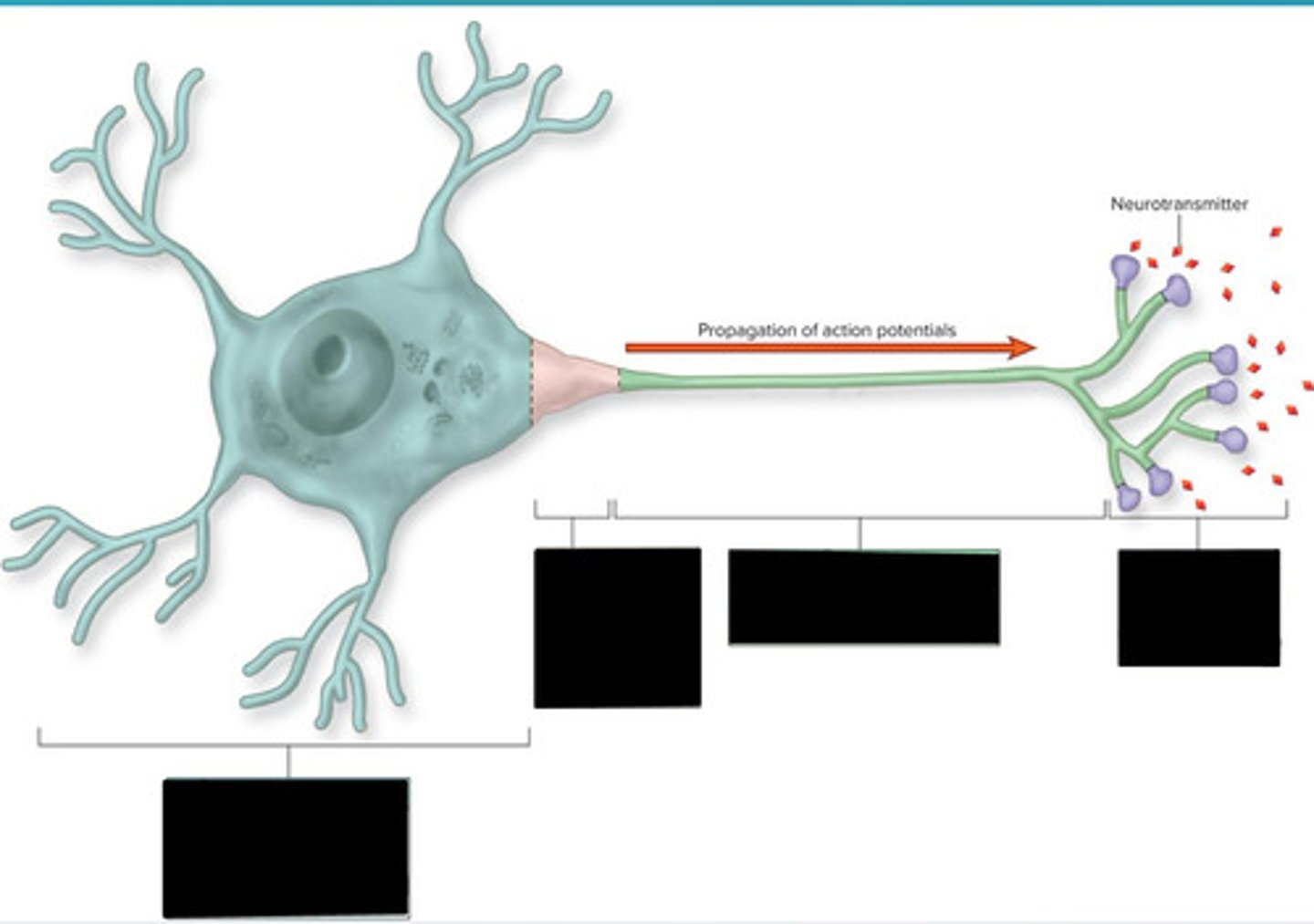
neuron segments
receptive segment, initial segment, conduction segment, transmissive segment
receptive segment of neuron
binding of neurotransmitter released from presynaptic neurons; production of graded potential
initial segment of neuron
summation of graded potentials; initiation of action potential
conduction segment of neuron
propogation of action potentials
transmissive segment of neuron
release of neurotransmitter
steps after action potentials cause opening of voltage gated Ca+ channels
occuring at synaptic knob
1. Ca+ diffuses into knob (Ca+ pumps have established gradient)
2. Ca+ binds to proteins associated w/ synaptic vesicles and triggers vesicles to fuse w/ membrane
3. neurotransmitter is release via exocytosis
4. neurotransmitter binds to ligand receptors
5. only one type is released at a time, depending on frequency of action potentials that reach synaptic knob
synaptic knob
tips of axon branching extensions containing synaptic vesicles which contain neurotransmitters
interneurons
(aka association neurons)
- receive, process, and integrate info from many other neurons
- communicate btwn sensory and motor neurons
- usually multipolar, located w/in CNS, and make up 99% of our neurons
peripheral axon regeneration is possible if what remains
neuron cell body is intact and enough neurolemma remains
label image; click on image first, test yourself, then check definition
left to right:
receptive segment
initial segment
conductive segment
transmissive segment