Biology - A2.2 Cell Structure
1/61
There's no tags or description
Looks like no tags are added yet.
Name | Mastery | Learn | Test | Matching | Spaced | Call with Kai |
|---|
No analytics yet
Send a link to your students to track their progress
62 Terms
Cells definition
Cells are the fundamental units of life and the smallest unit of self-sustaining life
Cell theory, three points to summarise
Cells are the basic structural unit of life
Living organisms consist of cells
Cells come from preexisting cells
Unicellular vs. Multicellular
Organsims that are unicellular only have one cell
Larger organisms are usually multicellular and have differentiated and specialised cells
Two categories of living organisms
Prokaryotes and eukaryotes. (viruses aren’t living)
Structures common to cells in all living organisms
plasma membrane enclosing the cell composed of lipids, cytoplasm composed mainly of water for metabolism, DNA for genetic material (and ribosomes)
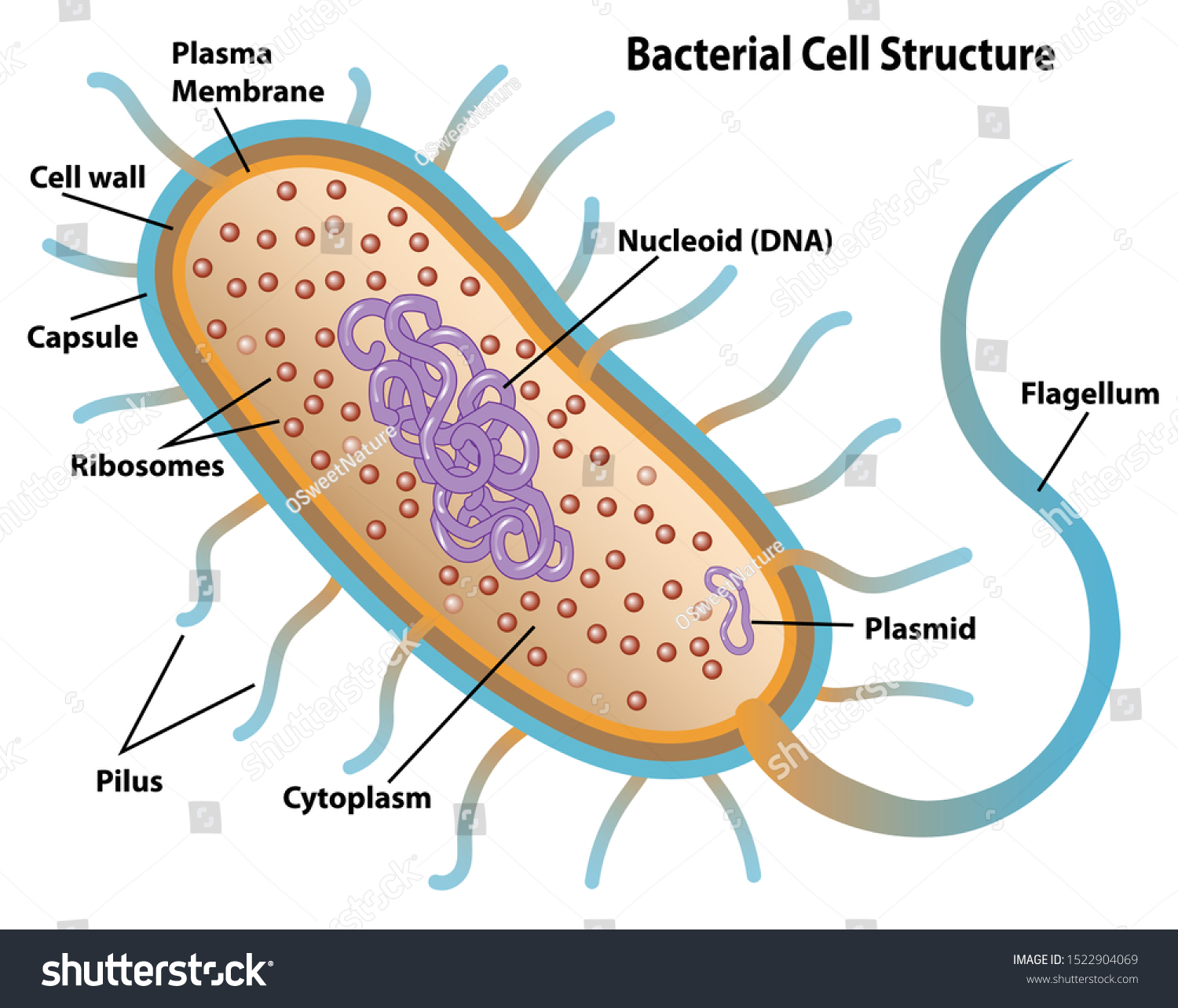
Prokaryote 3 basic structures
Rods (bacili), spheres (cocci), spirals (spirilli)
Prokaryote cell components
Cytoplasm,
nucleoid (lighter part of cytoplasm where single naked polymer/molecule of DNA/chromosome is located),
70S ribosomes,
plasma membrane,
cell wall made from peptidoglycans (sugars and proteins)
pili are exterior extensions/appendages made from proteins that can transfer genetic material and attach the bacteria to another surface
flagella are appendages for movement
plasmids are small circle DNA that is often transfered
meosome are the powerhouse of the prokaryotes, produce energy/ATP
capsule is an outer layer that protects the bacteria from different environments and resists it from bursting
Cytoplasm
Contains water and many substances dissolved or suspended in it. There are also enzymes bc the cytoplasm is a key role in metabolism.
Plasma membrane
Membrane on the outside that encloses the contents of the cell and controls the entry and exit of substances.
The plasma membrane ensures favorable inner environment that might be different from exterior (e.g. solute concentrations and pressure).
Made from phospholipids
Fragile and can burst if interior pressure is too great, lysis
Some animal cells have projections called microvilli
DNA
DNA is the genetic material for all cells. Can be located either inside the nucleus or in the cytoplasm (nucleoid)
Prokaryote nucleoid
lighter part of cytoplasm where single naked polymer/molecule of DNA/chromosome is located
Pili from prokaryotes
Exterior extensions/appendages made from proteins that can transfer genetic material and attach the bacteria to another surface
Prokaryote flagella
appendages for movement
Prokaryote plasmid
small circle DNA that is often transfered
Prokaryote mesosome
Folded invaginations of the plasma membrane are the powerhouse of the cell. Produce energy/ATP
Prokaryote capsule
Outer layer that protects the prokaryote from exterior environments and form bursting
Compartmentalization of cells
Eukaryotic cells are compartmentalized so some areas are seperated from the rest of the cell/cytoplasm with one or two membranes, organelles such as nucleus are an example
Nucleus
In eukaryotic cells the DNA is associated with histone protein and found in the nucleus.
There is a double membrane with nuclear pores that allows communication with the rest of the cell
There is a nucleolus (dense structure involved in ribosome synthesis)
Common structures for all eukaryotic cells
Nucleus, 80S ribosomes, mitochondria, cytoplasm, plasma membrane, endoplasmic reticulum and Golgi apparatus
Ribosomes
Small structures in all cells that are either free, or associated with the endoplasmic reticulum and synthesise proteins
Vacuoles
In eukaryotic cells (smaller in animal cells), they are structures that contain nutrients
Eukaryotic centrioles
Associated with nuclear division, they are composed of microtubes that can divide the nucleus in cell division
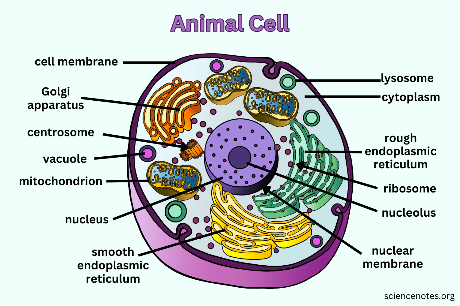
Components of a generalised animal cell
Ribosomes (80S)
Vacuoles
Cytoplasm
Nucleus
Mitochondria
Plasma membrane
Golgi apparatus
Endoplasmic reticulum (smooth or rough)
Centrioles
Lysosomes
Eukaryotic lysosomes
Sacs bound by a single membrane that contain enzymes and transport them
Not usually present in plant cells
Function Golgi apparatus
Structures that can store, modify and package proteins (included in the last stages of making a protein)
Endoplasmic reticulum
structure within the cytoplasm
Rough ER has ribosomes which are for protein synthesis
Smooth ER (without ribosomes) is part of lipid synthesis
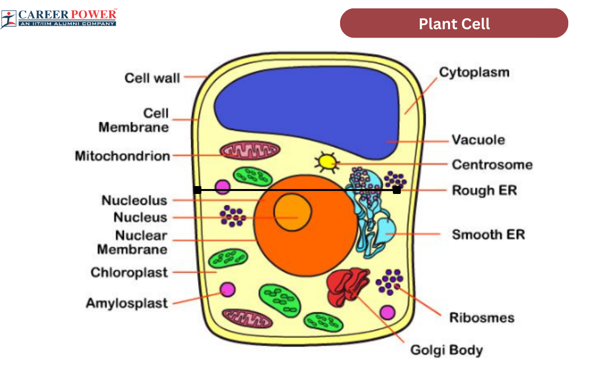
Components of a generalised plant cell
Chloroplasts
Cytoplasm
Golgi apparatus
Mitochondria
Plasma membrane
Endoplasmic reticulum
Cell wall
Large central vacuole
Starch granules - amyloplasts
Nucleus
Amyloplasts/Starch granules
Carbohydrates stored in amyloplasts which store and synthesise carbs
Cell wall
A semi-rigid structure composed of mainly cellulose, provides structural support
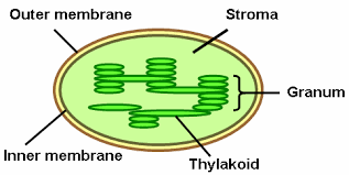
Chloroplasts
Specialised plastids containing the green pigment chlorophyll. Function: photosynthesis
Have a double membrane and thylakoids
Eukaryotic cytoskeleton
Microtubules and microfilaments that guide the movement of organelles around the cell
Differences in eukaryotic cells (between animal, plant and fungi)
composition of cell walls, the size and function of vacuoles, presence of chloroplasts/plastids, general size, presence of centrioles, cilia and flagella
Differences in eukaryotic cells (between animal, plant and fungi) in terms of cell wall
Aren’t present for animals, made out of cellulose for plant cells and made of chitin for fungi
Size and function of vacuoles differing for eukaryotic cells
Small and temporary for animal cells, large and permanent for plant and fungi cells.
Function is to store substances and pressurise the cells for both plant cells and fungi cells.
Presence of plastids/chloroplasts differs for animal, plant and fungi eukaryotic cells
In both fungi and animal they aren’t present whilst plant cells have both chloroplasts and amylosplasts
Presence of centrioles differs for animal, plant and fungi eukaryotic cells
Absent for plant and fungi cells
Used in animal cells for cell reproduction
Compare and contrast size of different eukaryotic cells
Animal cells: 10-30 micrometres
Plant cells: 40-100 micrometres
Fungi cells: 2-10 micrometres
Presence of cilia/flagella (undulipodia) for animal, plant and fungi cells
Can be present for animal cells (e.g. sperm cells)
Usually absent for plant and fungi cells
Name four examples of atypical eukaryotic cells
Red blood cells, phloem sieve tube elements, skeletal muscles, aseptate fungal hyphae
Why are red blood cells atypical eukaryotic cells?
They have no nucleus so that they can carry more haemoglobin but they’re lifespan is shorter because they cannot repair themselves
Why are phloem sieve tube elements atypical eukaryotic cells?
These cells only have plasma membrane and a smaller amount of cytoplasm so that the liquid can flow easily.
An adjacent cell (a companion cell) is associated with the sieve tubes so that they remain alive.
Why are skeletal muscle cells atypical eukaryotic cells?
They are multinucleate and very long because groups of cells fuse together to form the skeletal muscle cells.
Why are aseptate fungal hyphae atypical eukaryotic cells?
They are multinucleate (coenocytes), as when the cells grow and the nucleus divides, there is no septa created (hence aseptate)
Tips for drawing from electron micrographs
Label
Use straight lines for labels
Draw with a well-sharpened pencil
No sketching
The cell wall is the inside layer between the two lines whilst the plasma membrane is the inner-most line (for plant cells and fungi cells)
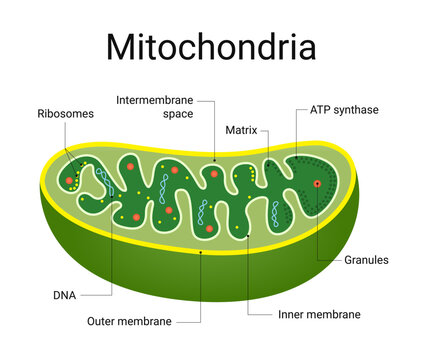
Mitochondria
Organelles that carry out respiration to produce ATP
Have a double membrane and the inner membrane goes in towards the center
Formula to calculate magnification
Size of image (what it measures with a ruler, usually in mm) divided by actual size of structure (usually given)
remember, both should be in the same units (micrometres or millimetres)
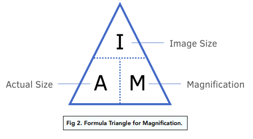
Definition magnification
How much an object is enlarged from its true size
written: x 400 e.g.
Stage micrometer + Eyepiece graticule
A special microscope slide that has divisions on it that are usually 100 micrometres apart.
An eyepiece graticule is a graticule with a ruler
You line them up and find out how many units of the eyepiece are equivalent to 100 micrometres.
Then divide the 100 by the units and you get what 1 eyepiece granule unit is equivalent to.
What are the 8 essential life processes that all (most) living organisms carry out?
Nutrition (supplying nutrients for energy, growth and repair)
Metabolism (all biochemical reactions including catabolic and anabolic)
Growth (increase the number of cells)
Response to stimuli (appropriate action after perceiving stimuli)
Excretion (removal of waste products)
Homeostasis (maintaining a constant internal environment)
Reproduction (producing offspring)
Movement (ability to change position)
Unicellular organisms
The cell has to carry out all life processes. E.g. Chlamydomonas and Paramecium
(In multicellular organisms, life processes can be done by different cells)
Chlamydomonas and all life processes in one cell
Excretion through freely permeable cell wall
Metabolic reactions occur in cytoplasm
Nutrition through storing starch in chloroplasts and food vacuoles
Reproduction and growth as the nucleus can divide to produce genetically identical nuclei
Movement with two flagella
Response to stimuli as the light sensitive ‘eyespot’ can sense brightest light and swim towards it
Homeostasis as the water level is kept constant with the contractile vacuoles
Paramecium and unicellular life processes
Movement with whip-like cillia
Response to stimulia as the cilia can move the cell in direction of changes in environment
Nutrition through food vacuoles
Metabolic reactions take place in the cytoplasm
Homeostasis by regulating water contents with contractile vacuoles
Excretion happens by waste products diffusing out through the plasma membrane
Reproduction and growth as the nucleus can divide
Electron microscope
Instead of using light, they have a beam of electrons. Can be transmission (goes through the specimen) or scanning (bounces of specimen)
+higher resolution
-no living tissue
-only black and white images
Freeze fracture electron microscopy
1) Sample is frozen
2) Sample is fractured at weakest point
3) Some ice is removed through etching
4) Vapour of carbon fired to obtain a replica (the replica is then observed with an electron microscope)
+can observe surfaces inside cells
Cryogenic electron microscopy (cryo-EM)
Once a sample is flash frozen the electron beams can go through computer software to get a 3D image of the structure of proteins
+3D image
+Less intense electron beams than traditional electron microscopy
Fluorescent stains
Stains used that under UV or violet/blue light re-emit light
the chemicals bind to some to highlight those areas
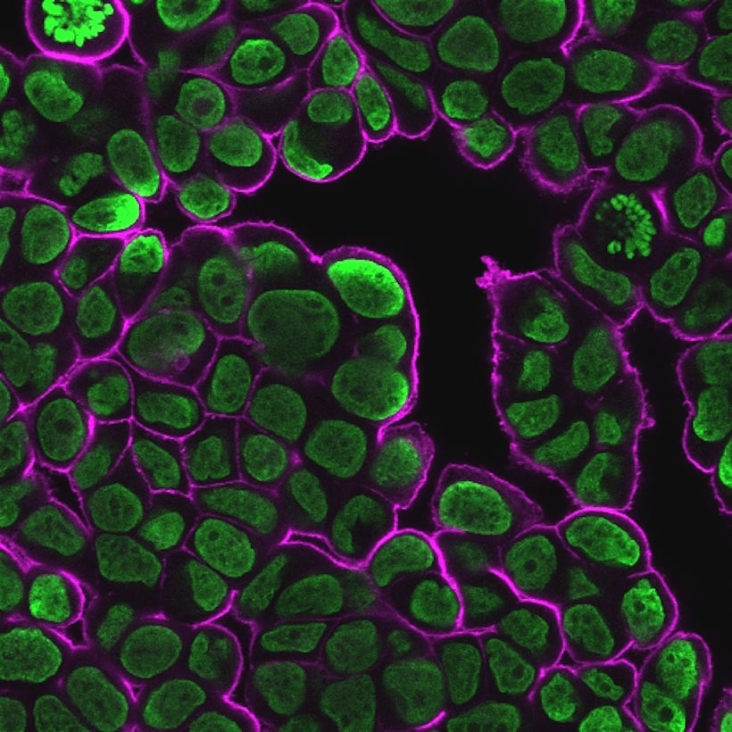
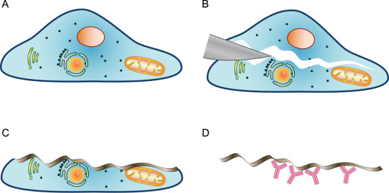
Immunofluorescent stains
The stains attach to some antibodies to attach to proteins and antigens
If the right proteins are targeted, the amount of infected cells can be visually monitored through immunofluorescent staining.
Endosymbiotic theory
Evidence suggests all eukaryotic cells originated from a common unicellular ancestor (prokaryotic cell)
1) 2 billion years ago a large prokaryote with a nucleus and that was sexually reproducing engulfed (through endocytosis) a smaller prokaryote that did aerobic respiration
2) They had a symbiotic (mutually beneficial) relationship and after evolving the smaller prokaryote became mitochondria.
3) The cell could then engulf a prokaryotic cell that could photosynthesise which could evolve into a chloroplast.
Evidence for endosymbiotic theory
Both chloroplasts and mitochondria have:
double membranes (one from the outside of the larger prokaryote and one from the smaller one itself)
Their own naked and circular DNA (hence prokaryote bc of plasmid)
70S ribosomes (as prokaryotes do)
Similar size to prokaryotes
Independent division
Multicellular organisms main advantage
Cells can be specialised for specific and unique functions
Therefore, the organisms become larger, more complex, and have greater diversity.
Cell differentiation definition and process
The process for developing specialized tissues in multicellular organisms
Done by switching genes on or off according to the function of that cell
Gene expression changes if there are changes in the environment
Evolution of multicellularity
All plants and animals are multicellular and so are many fungi and eukaryotic algae
Multicellularity evolves repeatedly