Avian Diagnostic Imaging
1/48
Earn XP
Description and Tags
Flashcards covering avian diagnostic imaging techniques, anatomy, and common radiographic lesions by organ system.
Name | Mastery | Learn | Test | Matching | Spaced | Call with Kai |
|---|
No analytics yet
Send a link to your students to track their progress
49 Terms
What is the most frequent imaging modality and diagnostic test used in birds?
Radiography
Why is general anesthesia usually needed for avian radiographs?
Due to high respiratory rate and small size, which makes it challenging to get stable images.
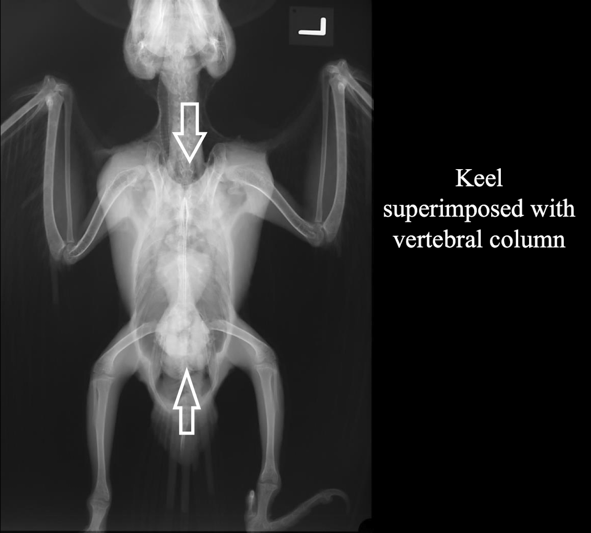
What is important to consider regarding avian radiographic anatomy?
Knowledge of avian radiographic anatomy is important as it cannot be extrapolated from mammals due to anatomic diversity.
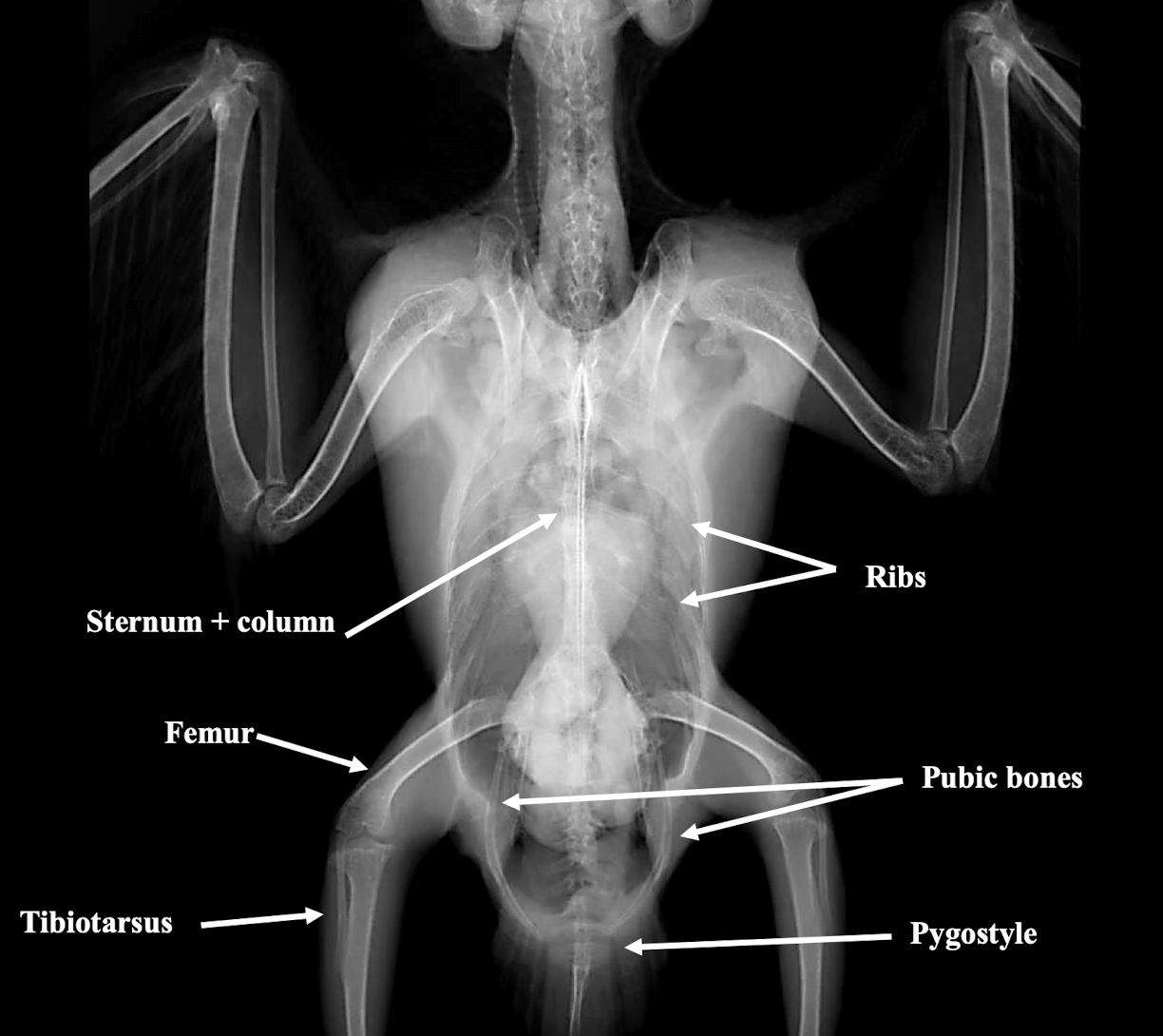
Correct radiograph positioning VD
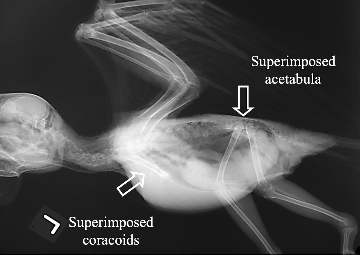
Correct radiograph positioning Lateral
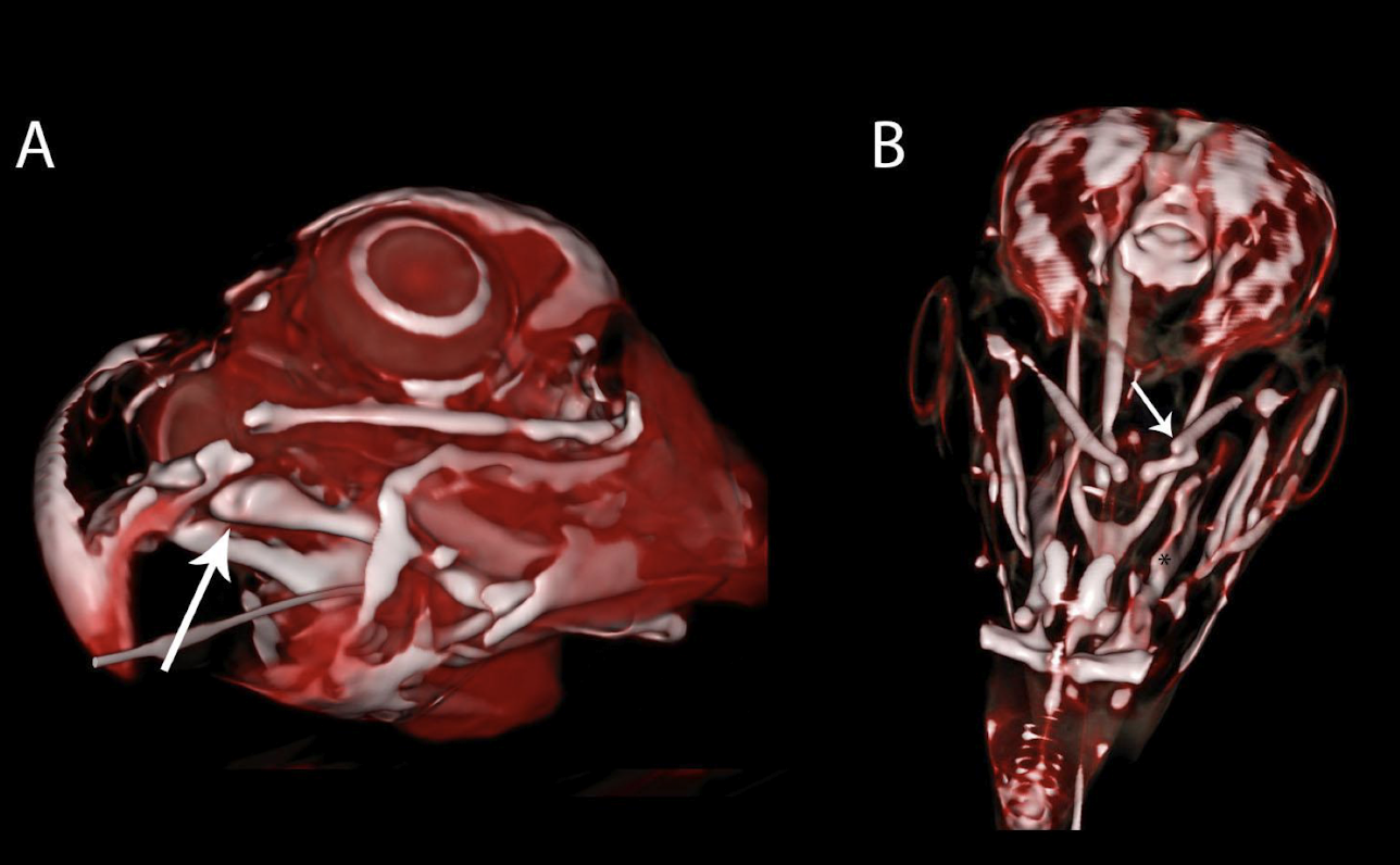
What advanced imaging modality is better for visualizing the avian skull than radiography?
Computed Tomography (CT)
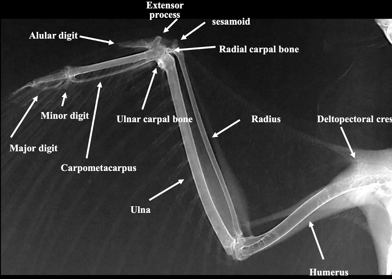
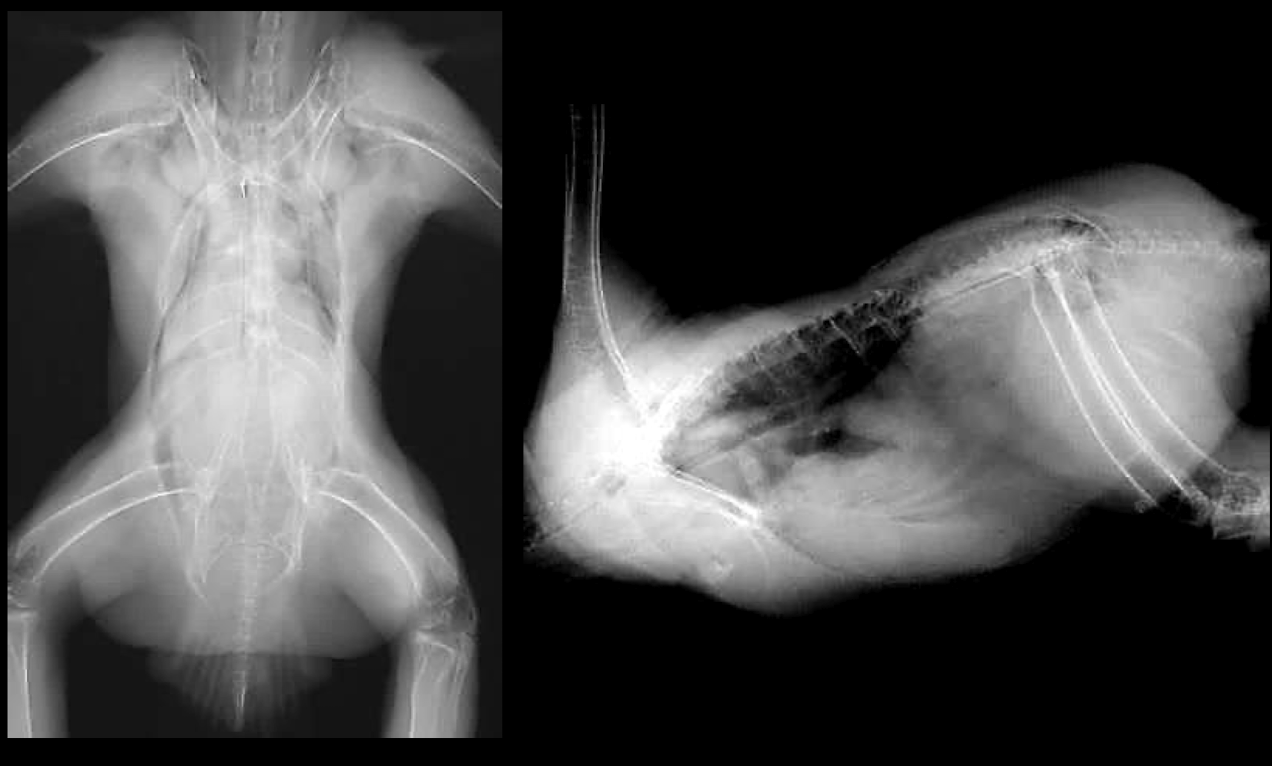
What are the standard radiographic views for avian wings?
VD (ventrodorsal) and caudo-cranial views.
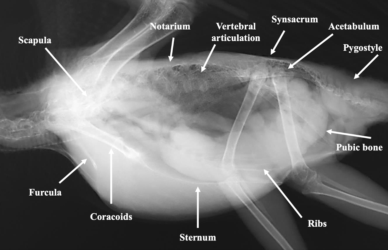
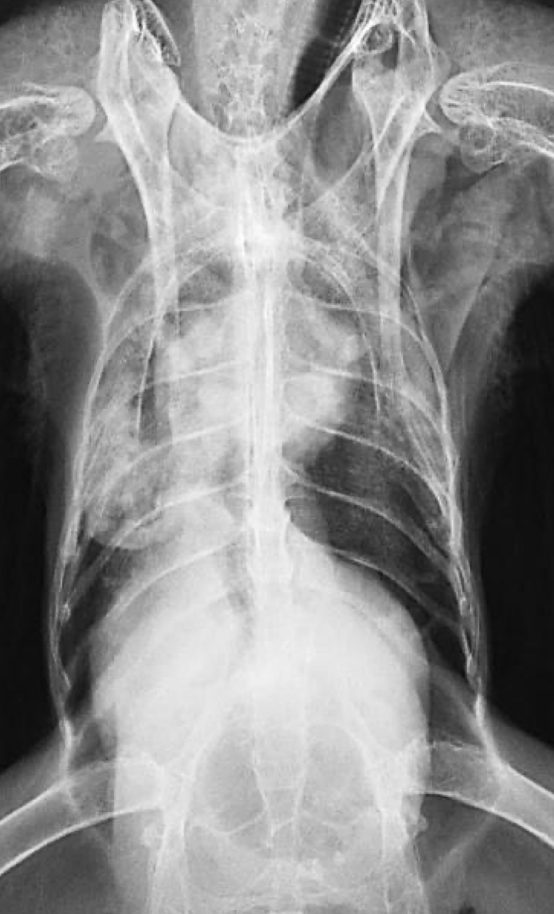
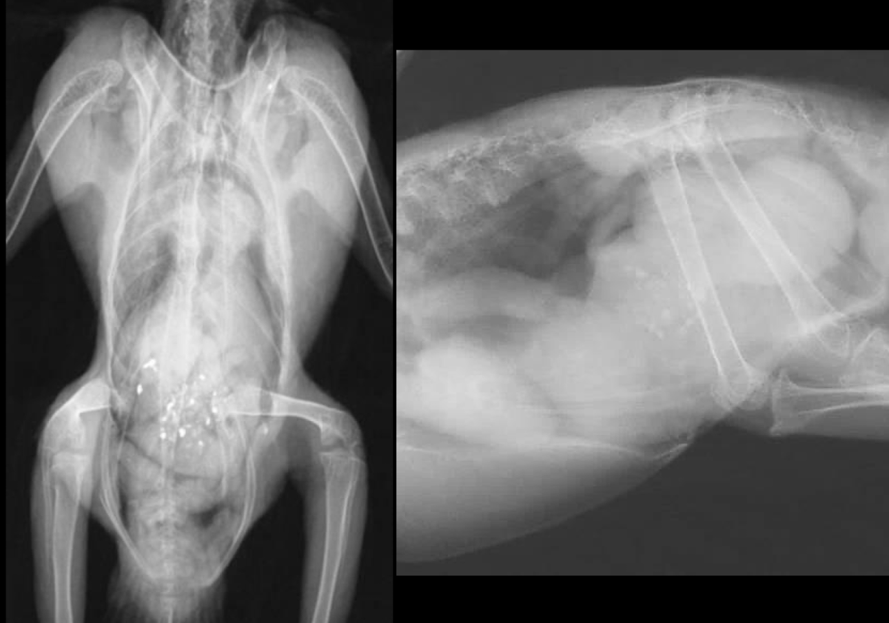
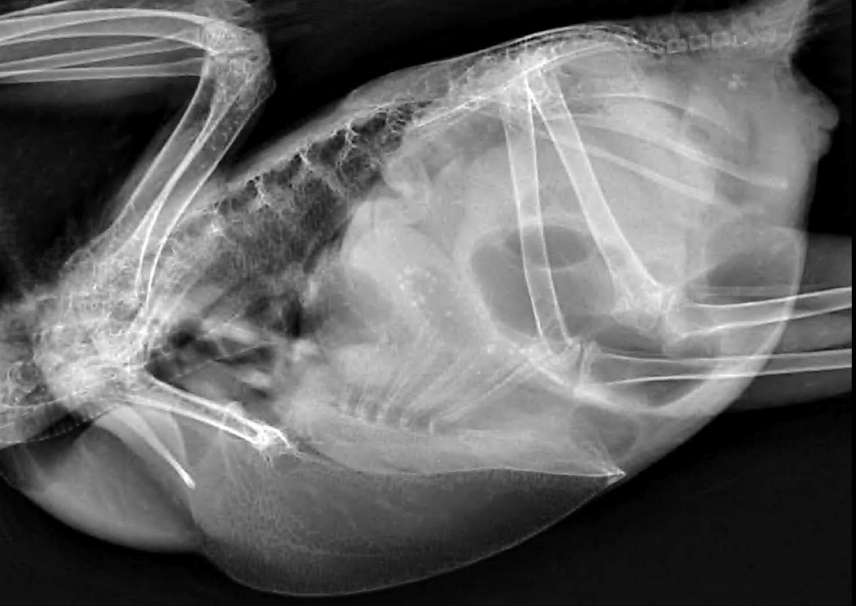
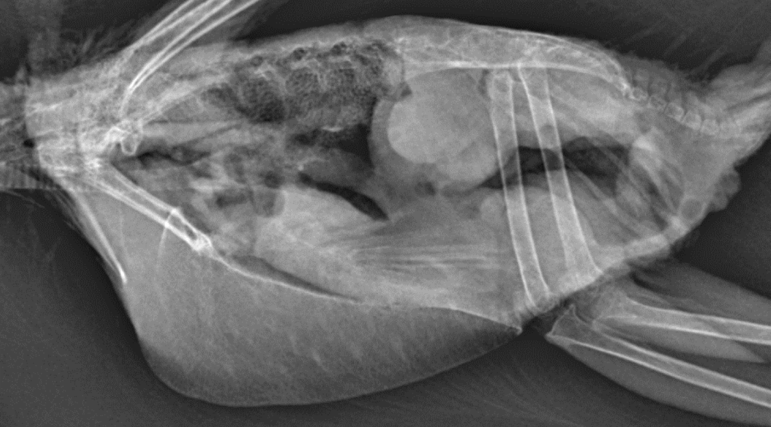
Normal avian body xray
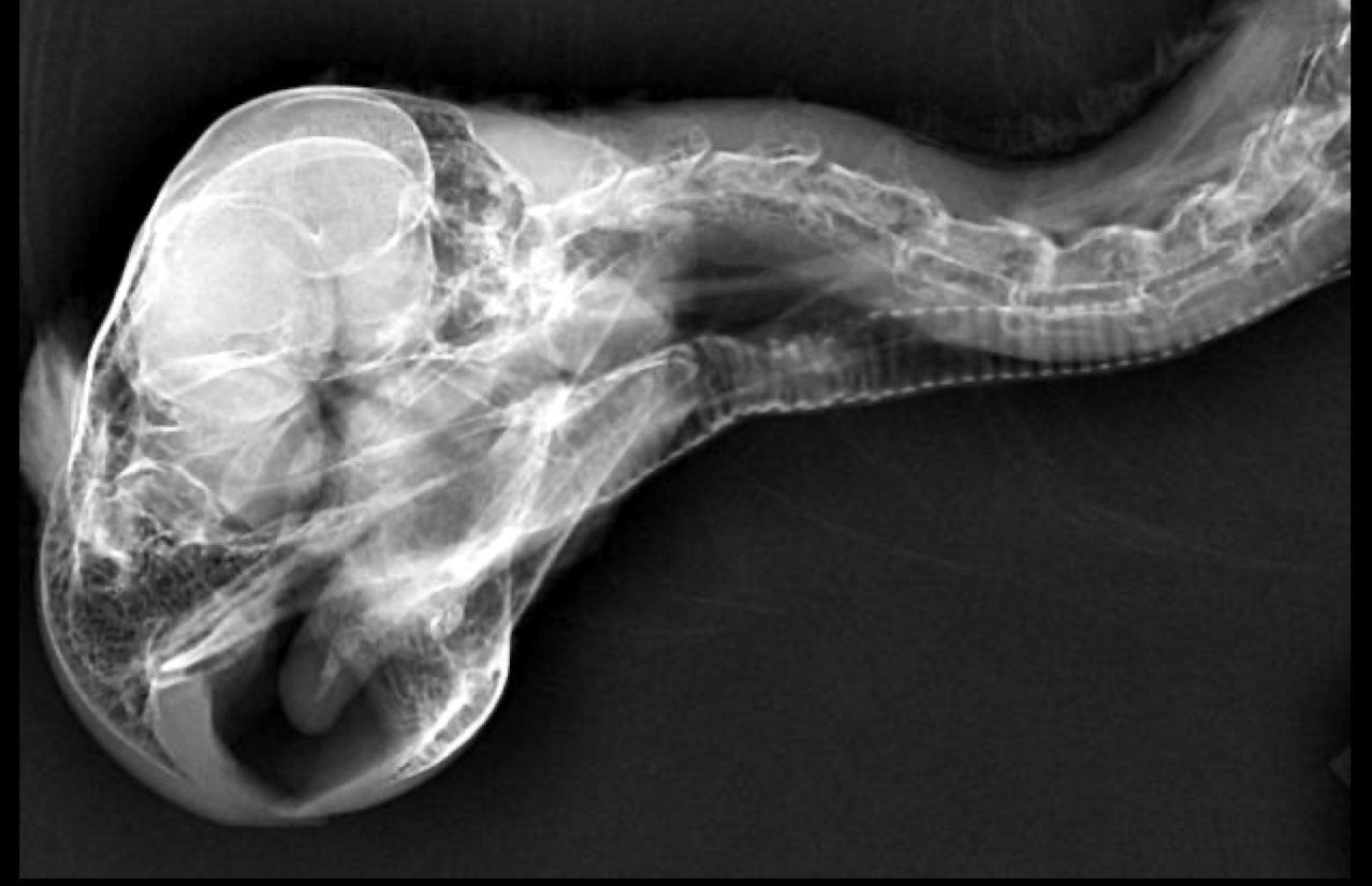
Normal avian wing xray
Normal avian lateral xray
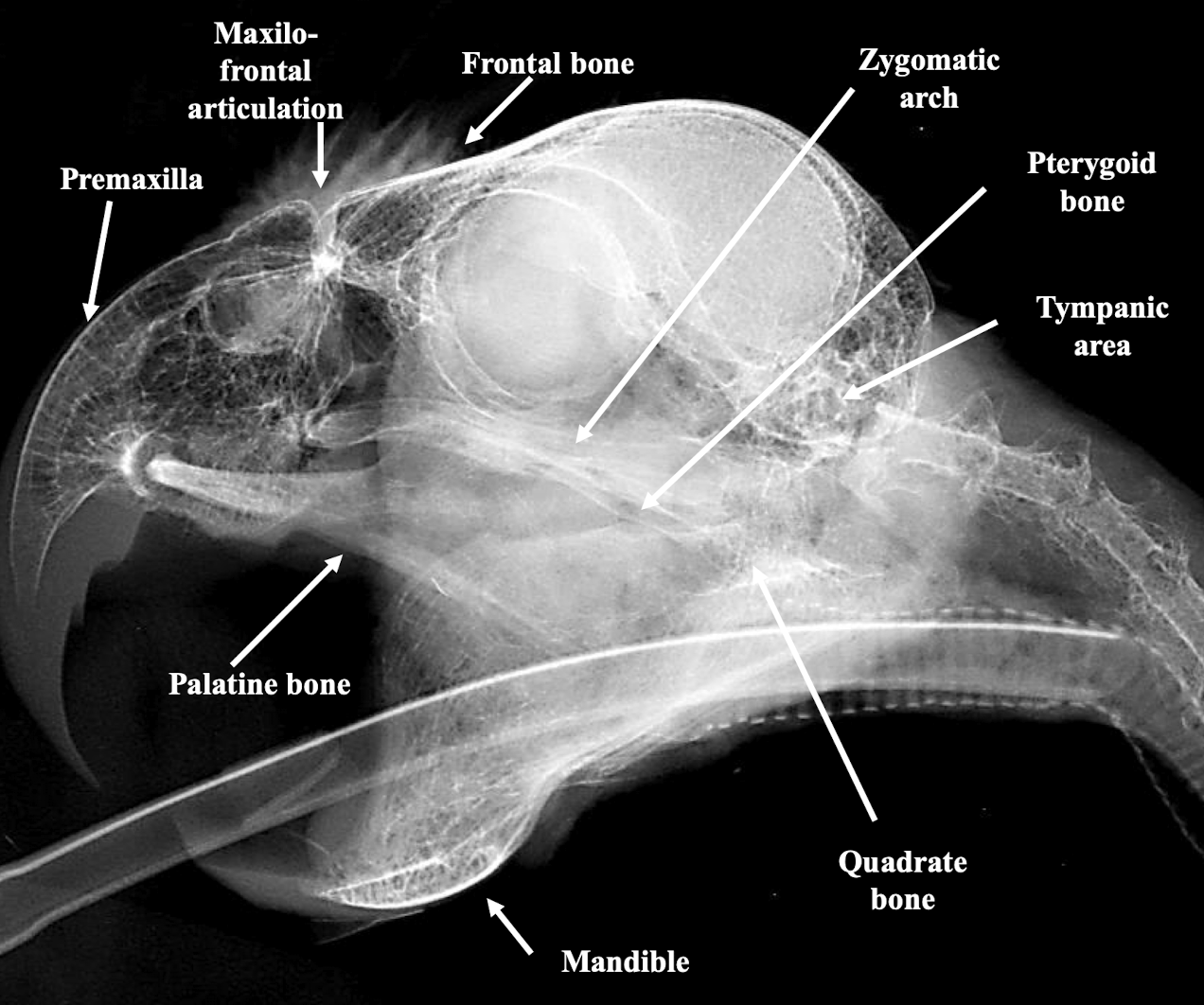
Normal avian skull xray
Name two common skeletal lesions identifiable on avian radiographs.
Fracture and Luxation.
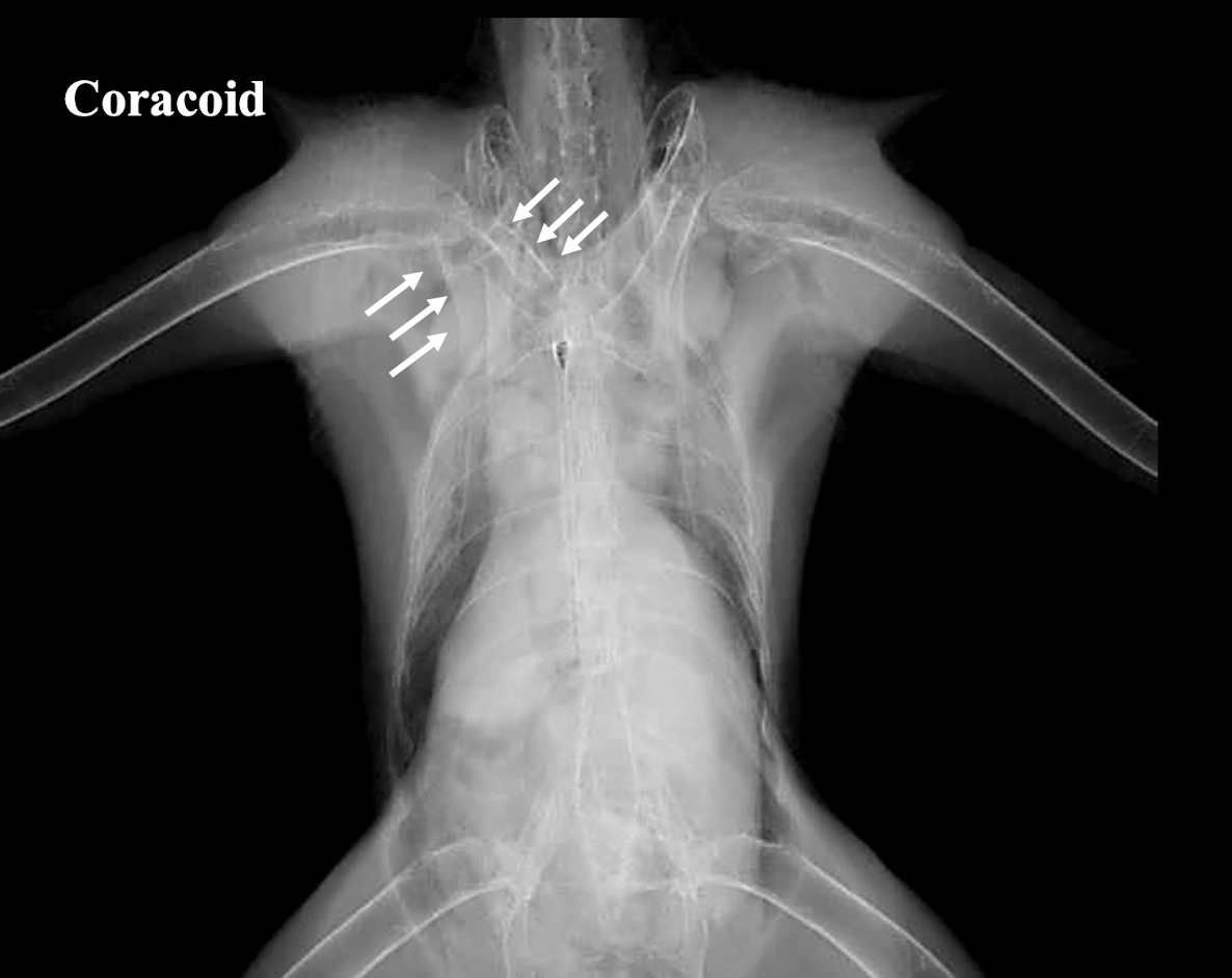
Coracoid fracture
What is Polyostotic Hyperostosis?
A common radiographic lesion of the skeleton in birds, indicating abnormal bone growth. Normal for egg laying hen due to inc level of calcium

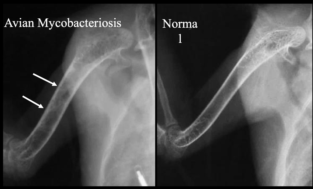
What two specific avian diseases can cause pathologic hyperostosis?
Avian Mycobacteriosis and Avian Leukosis.
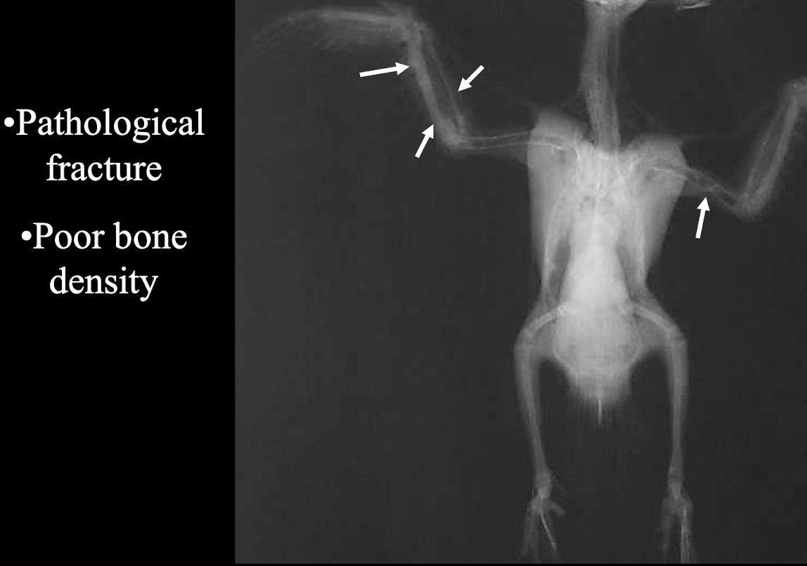
What radiographic signs are indicative of Metabolic Bone Disease in birds?
Pathological fracture and poor bone density.
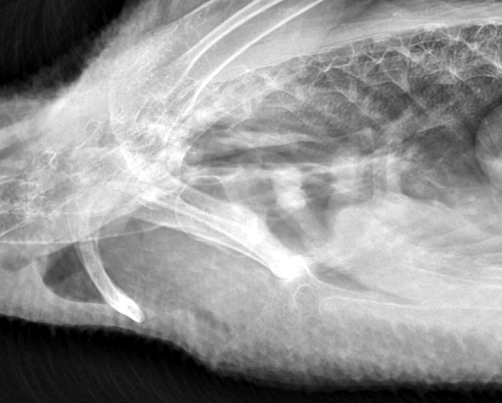
Name one unique anatomical feature of the respiratory system often visualized in ducks.
Syringeal Bulla.
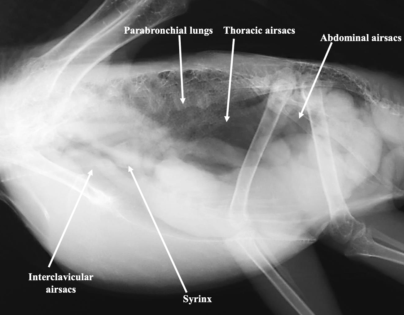
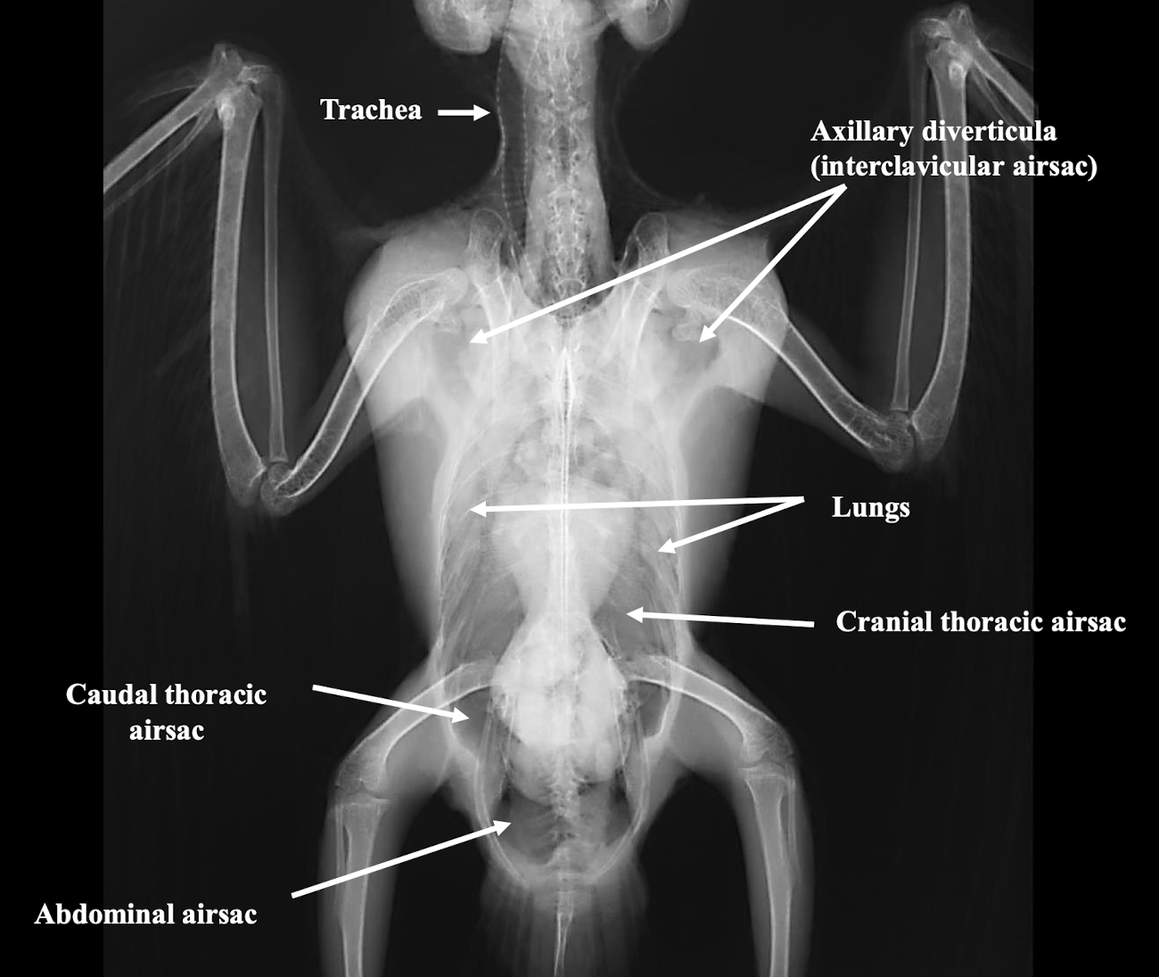
Which major air sacs are typically identified in avian respiratory imaging?
Interclavicular, cranial thoracic, caudal thoracic, and abdominal airsacs.
What radiographic finding in older parrots may be a normal variant?
Prominent Syrinx.
Name three common radiographic lesions of the avian respiratory system.
Pneumonia, Bacterial Airsacculitis, and Fungal Airsacculitis.
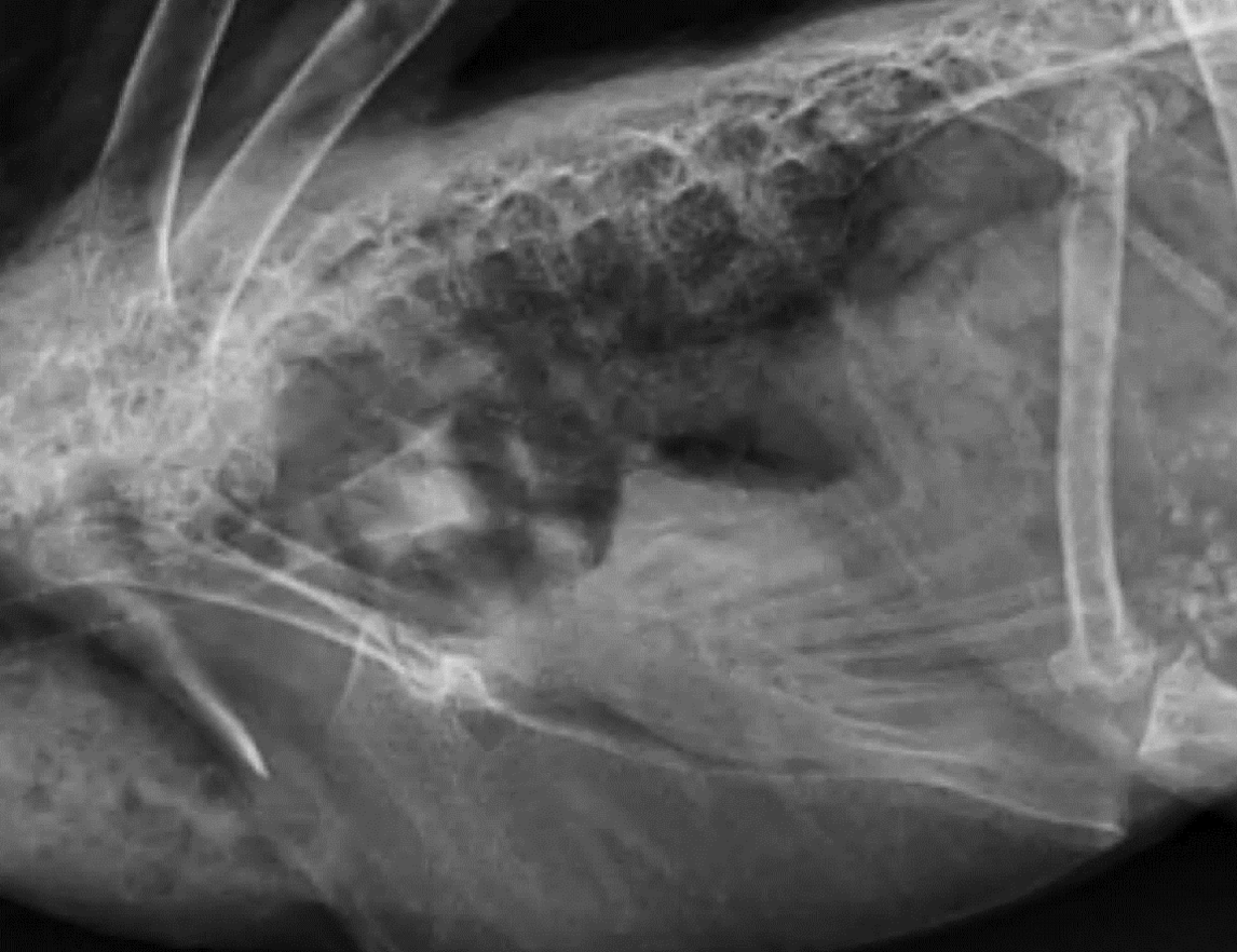
Whats wrong?
pneumonia
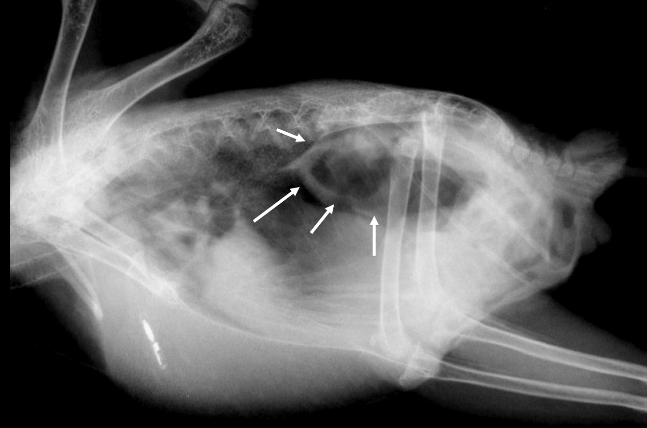
Whats wrong?
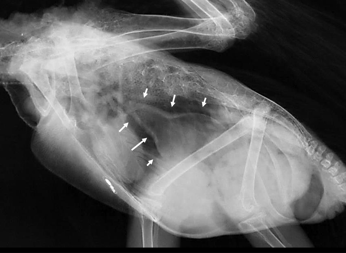
Bacterial airsacculitis → thickening of airsac wall
Whats wrong?
Fungal airsacculitis
What is wrong here?
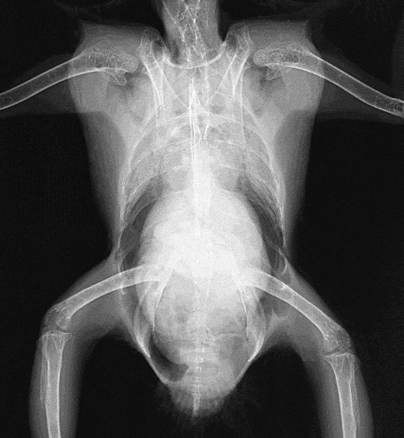
obesity
What is wrong here?
tracheal stenosis
How does an avian heart's size compare to that of mammals, especially in falcons?
The heart is larger than in mammals, especially in falcons.
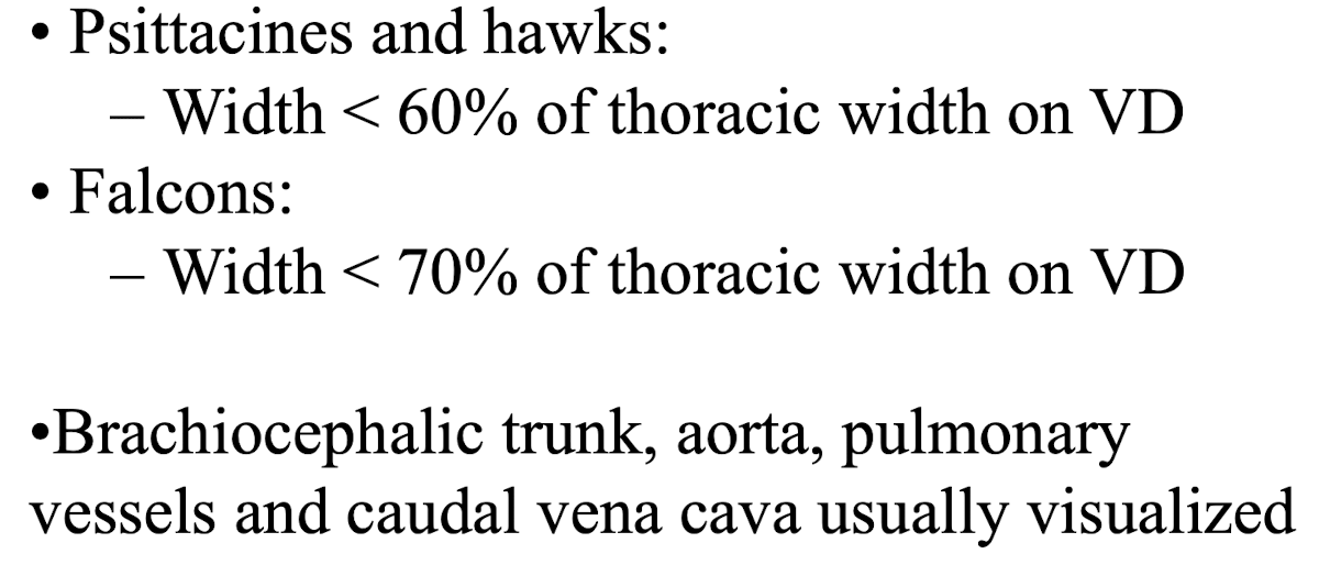
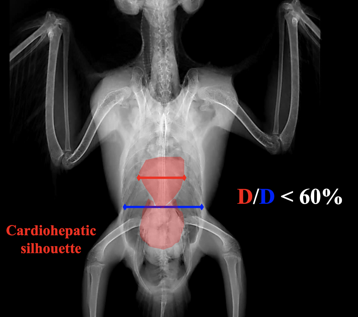
What is the normal heart width on a VD radiograph for psittacines and hawks?
Less than 60% of the thoracic width.
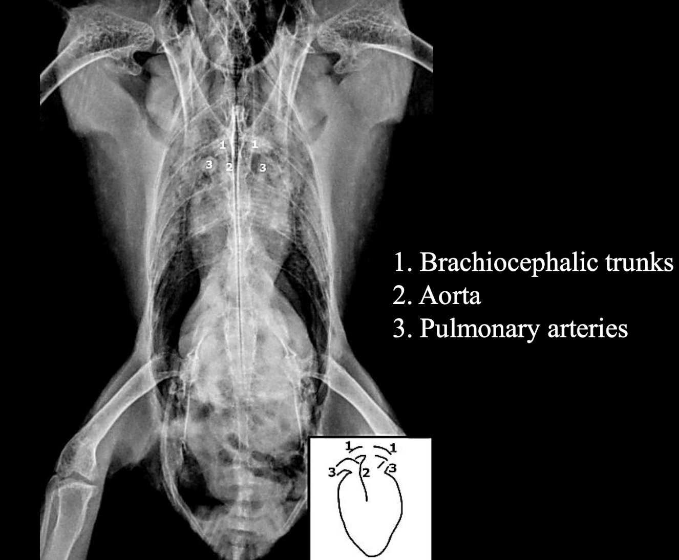
Great vessels in the heart VD
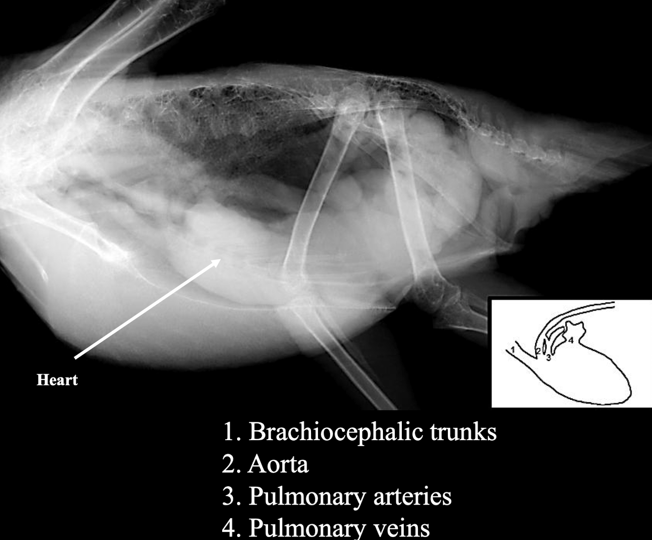
Great vessels in the heart lateral
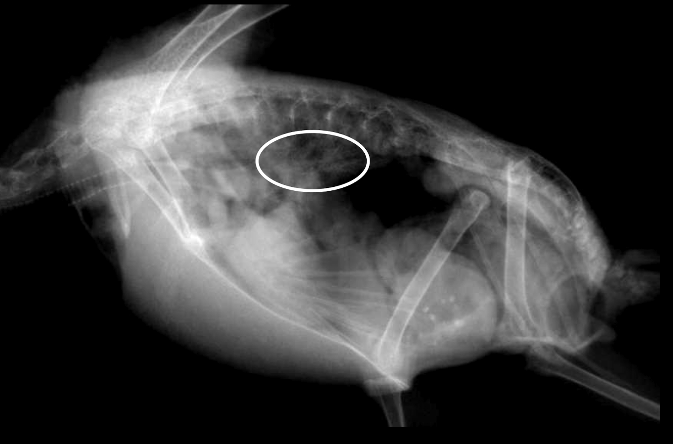
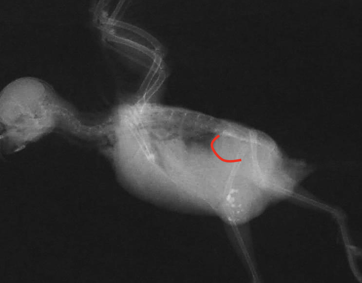
Whats wrong?
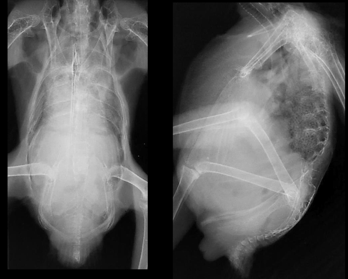
congestive heart failure
What is a common radiographic lesion of the avian cardiovascular system that presents as fluid accumulation?
Pericardial Effusion.
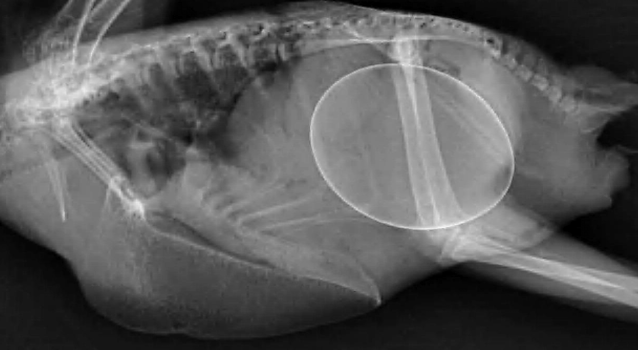
What is wrong?
arterial calcification
What is the typical range for the proventriculus to clavicular length ratio (d/D) in avian radiographs?
d/D < 0.5.
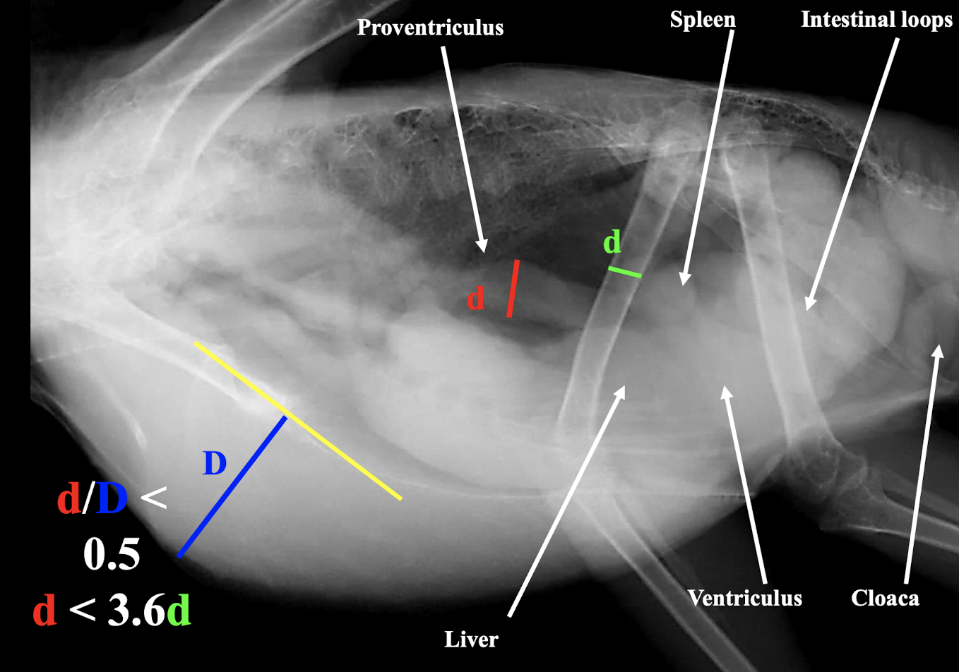
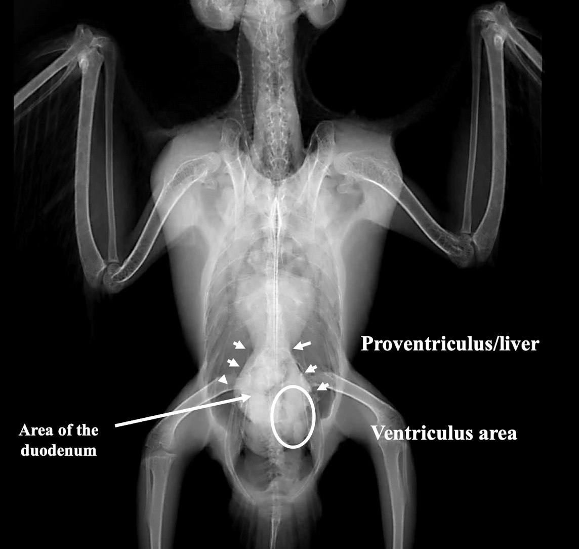
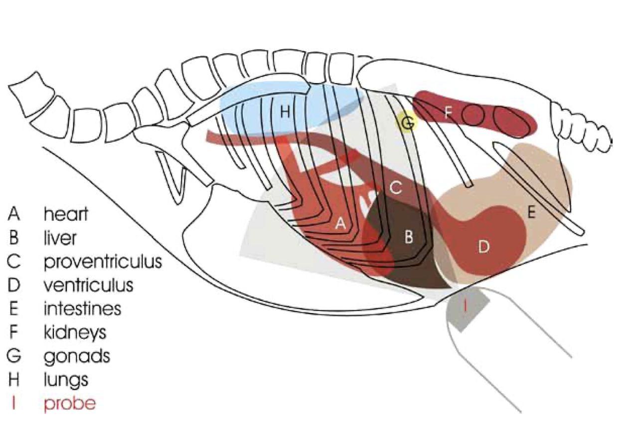
GI tract
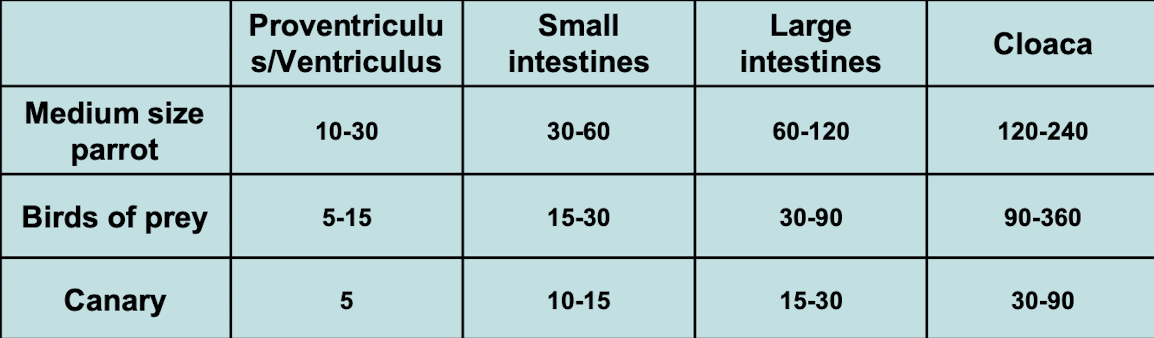
When are barium studies indicated in birds?
To localize the GI tract, assess hypomotility, identify obstruction/occlusion, or evaluate changes in location/shape of other organs.
Name two common radiographic lesions of the avian gastrointestinal system.
Proventricular Dilation and metal/grit in the ventriculus.
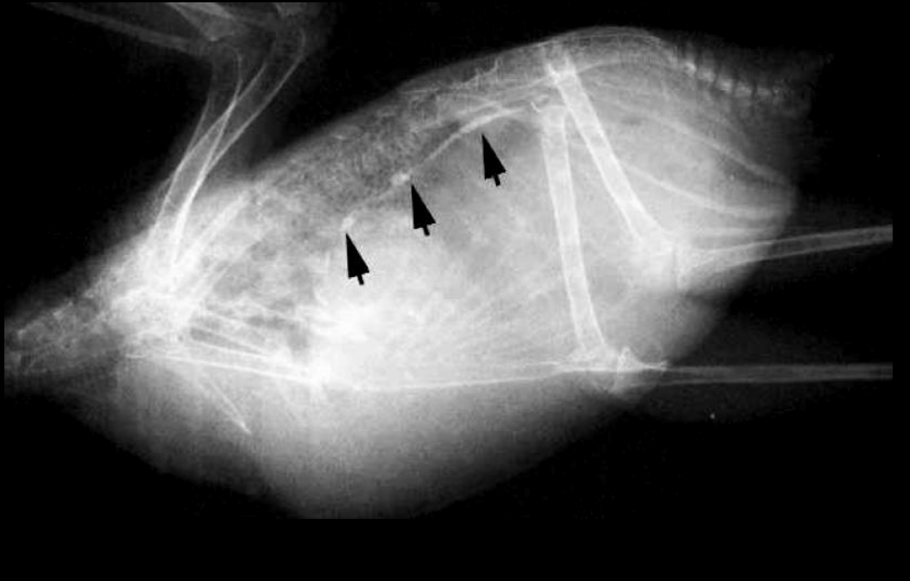
What is wrong?
Proventricular dilation
What is wrong?
metal/grit in ventriculus
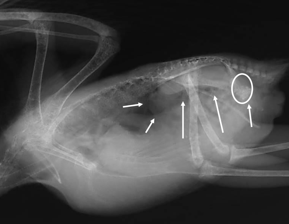
What is wrong?
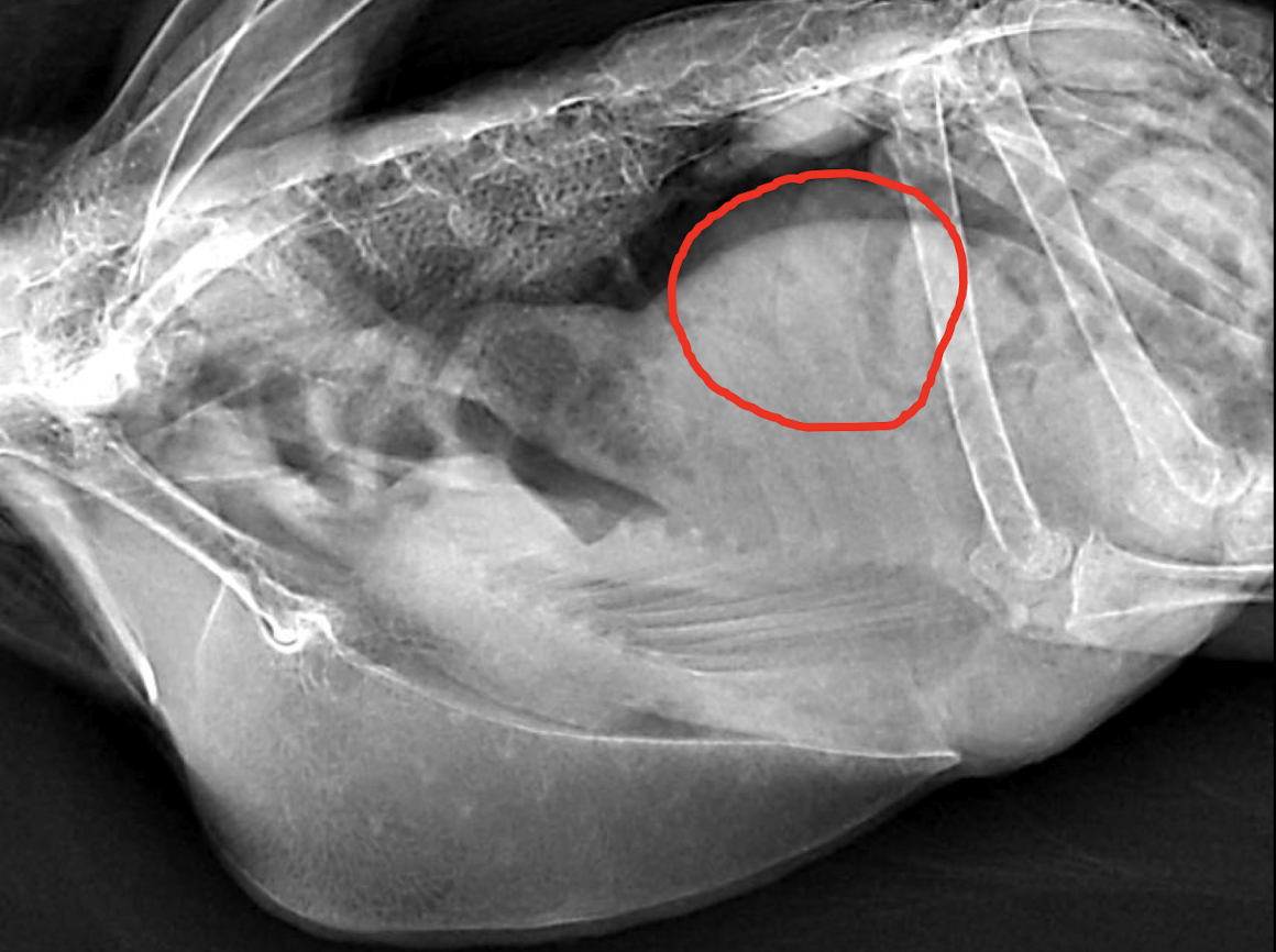
splenomegaly
What is wrong?
hepatomegaly
What is wrong?
mechanical ileus → neoplasia
What is a relatively common radiographic lesion of the avian urinary system?
Renal Mass.
Name three common radiographic lesions of the avian reproductive system.
Dystocia, Egg Binding, and Oviductal Impaction.
What is wrong?
oviductal impaction
What is wrong?
gonadal mass
What are the primary indications for using CT in avian diagnostics?
When radiographs are insufficient, for skull fractures, upper respiratory disease, and arterial calcification.
What makes ultrasonography difficult in birds?
Lack of ultrasound windows due to the keel and air sacs → mostly just for echocardiography
Which organs are rarely visualized with ultrasonography in birds?
Kidneys, gonads (unless large), and spleen.
What is a key advantage of endoscopy in birds for diagnostic purposes?
It allows direct visualization of all abdominal and thoracic organs without the need for insufflation and enables targeted biopsies. use a 2.7mm 30o endoscope!