Looks like no one added any tags here yet for you.
Anterior rami of T1-T11
These nervous structures form the intercostal nerves
- Bonus: "" T12 forms the subcostal nerve
Posterior rami of T1-T12
These nervous structures pass posteriorly to supply the joints, muscles, and skin of the back in the thoracic region
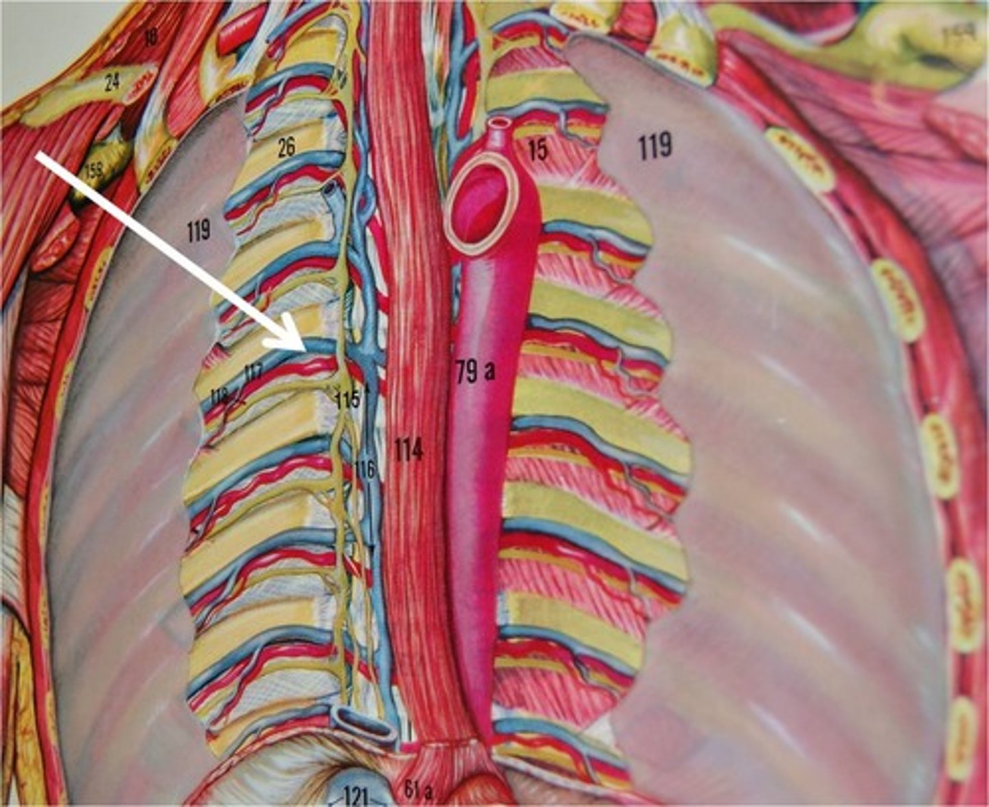
Between the pleura
In the posterior thorax, the intercostal nerve lies here instead of between the intercostalis intimi and internal intercostal muscle
- Top to bottom: VAN
myotome
Group of muscles innervated by a single spinal nerve
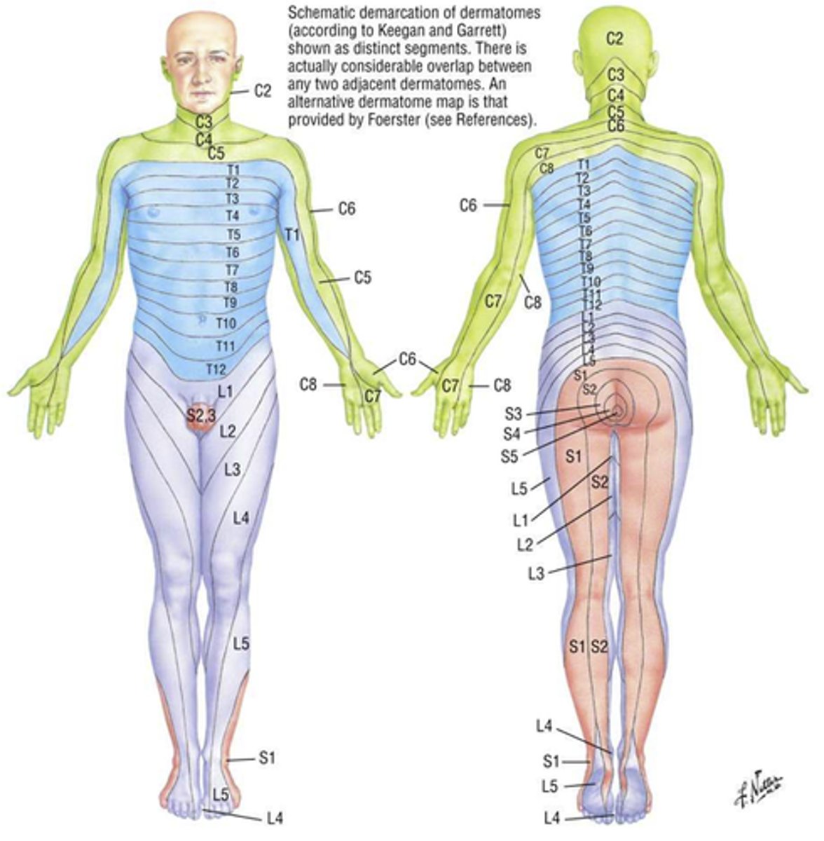
dermatome
Area of skin supplied by a single spinal nerve
- T4 includes the nipple
- T10 includes the umbilicus
First intercostal nerve (T1)
This spinal nerve joints the ventral ramus of C8 to form the lower trunk of the brachial plexus
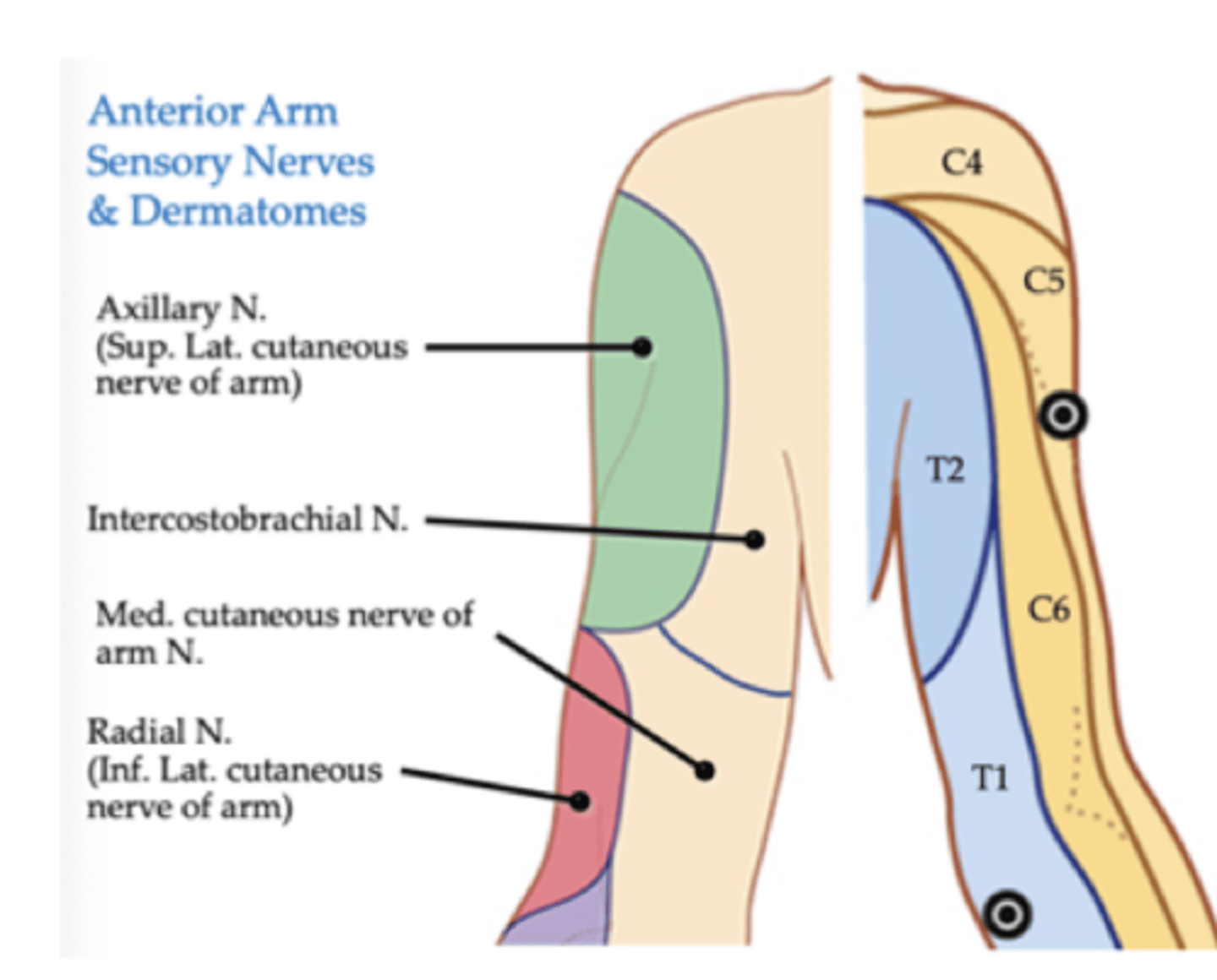
Second intercostal nerve (T2)
This spinal nerve gives a branch that joins the medial cutaneous nerve of arm, called intercostobrachial nerve
- In CAD, pain is referred along this nerve to the medial arm
mediastinitis
Due to:
- Deep infection of the neck
- Penetrating chest wound
- Esophageal perforation
Air can escape to CT spaces and produce subcutaneous emphysema (air bubbles below the skin)
mediastinoscopy
Diagnostic procedure to obtain tracheobronchial lymph specimens
- Small incision above suprasternal notch
- Superior mediastinum is explored
- Tuberculosis, sarcoidosis, lymphoma, lung cancer
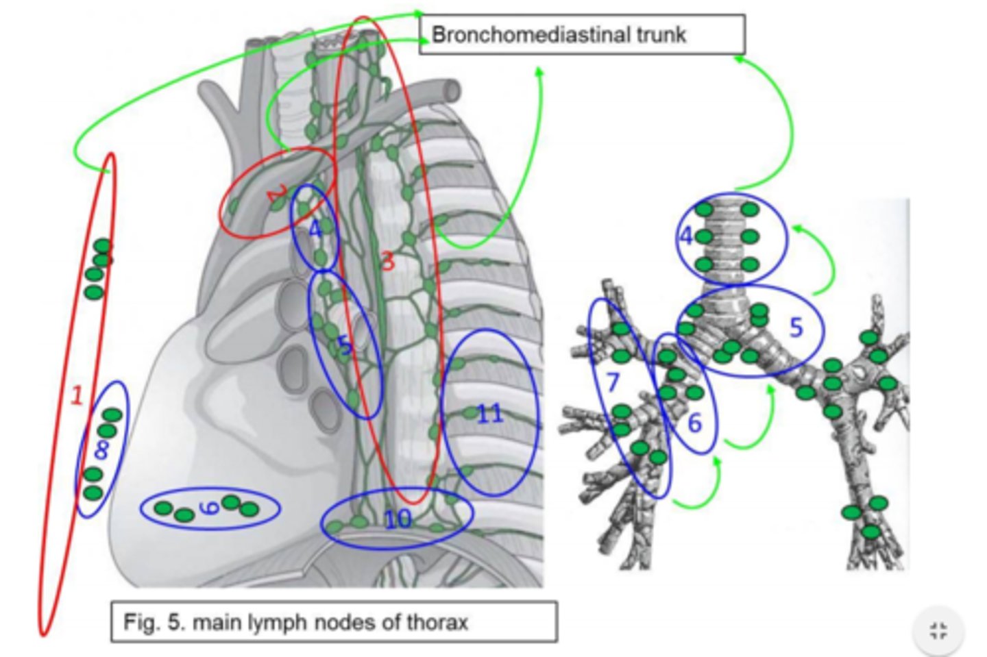
mediastinal lymph nodes
Lung cancer typically involves these.
Enlargement can compress:
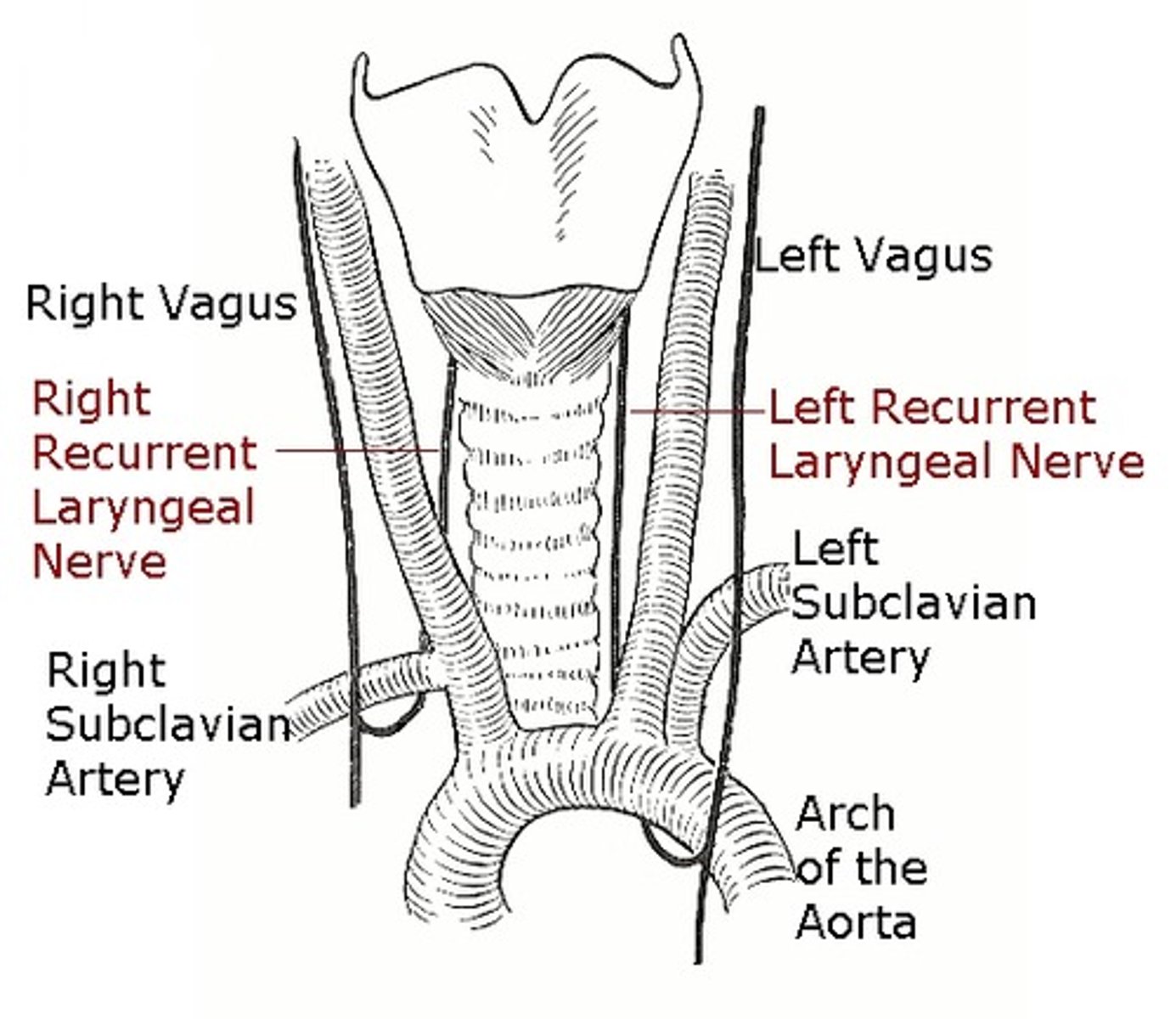
- left recurrent laryngeal nerve (hoarseness)
- Superior vena cava (congestion of veins of upper body)
- Sympathetic trunk, phrenic nerves, trachea, bronchi, esophagus
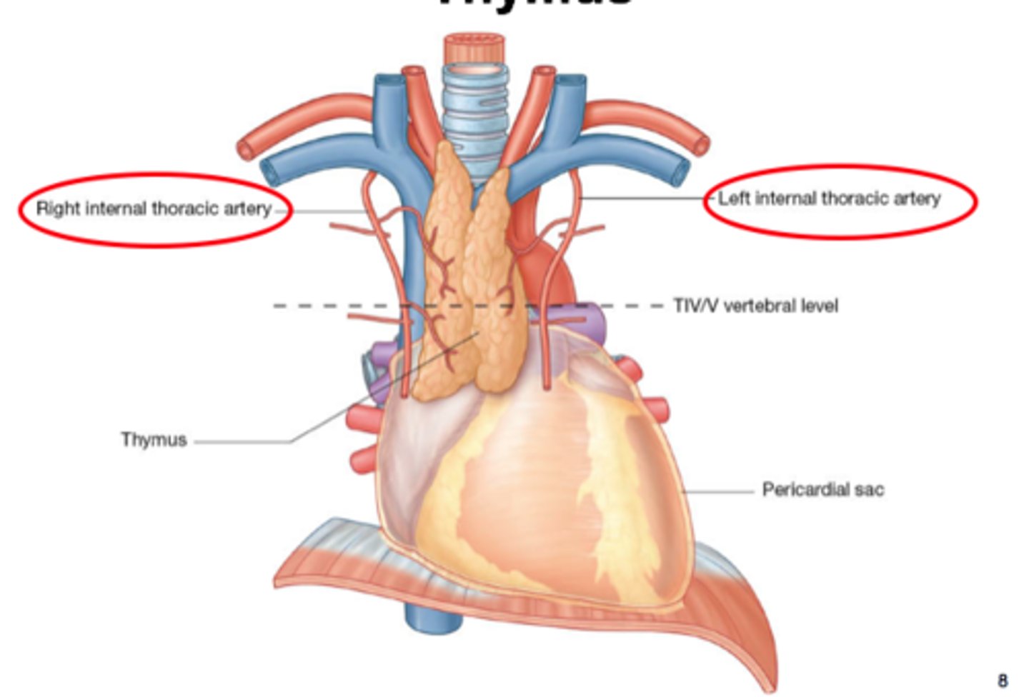
thymus
This organ is supplied as follows:
- Artery: Inferior thyroid, internal thoracic, mediastinal arteries
- Nerve: Small branches of the vagus, recurrent laryngeal, phrenic
- Venous: Drains into left brachiocephalic and internal thoracic veins
DiGeorge Syndrome
Most common cause of thymic aplasia - thymus fails to develop > T cells do not mature - non-functional
- Immunodeficiency
thymic hyperplasia
Idiopathic inflammation of the thymus often associated with steroids, rebound to chemotherapy, radiation, burns, or other systemic stress
- Lymphoid hyperplasia
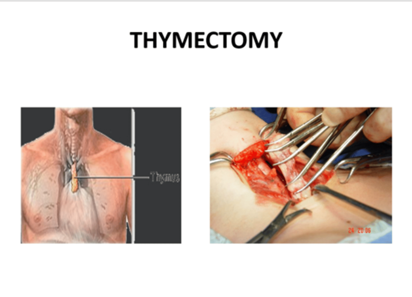
thymoma
Tumor of the thymus, found in 10-15% of patients with myasthenia gravis (progressive muscular weakness at NMJ)
- Compression of the recurrent laryngeal nerve
- Treatment by complete thymectomy
3rd/4th ICS
Where should the sternotomy be carried in a thymectomy procedure?
- For patients with myasthenia gravis and thymoma
Cervical esophagus (upper)
Artery: Inferior thyroid artery
Vein: Inferior thyroid vein
Lymph: Deep Cervical nodes
- This structure has skeletal muscle
Thoracic esophagus (middle)
Artery: Esophageal branches of the aorta and bronchial arteries
Vein: Azygos and hemiazygos vein
Lymph: Posterior mediastinal nodes
- This structure has a mix of skeletal and smooth muscle
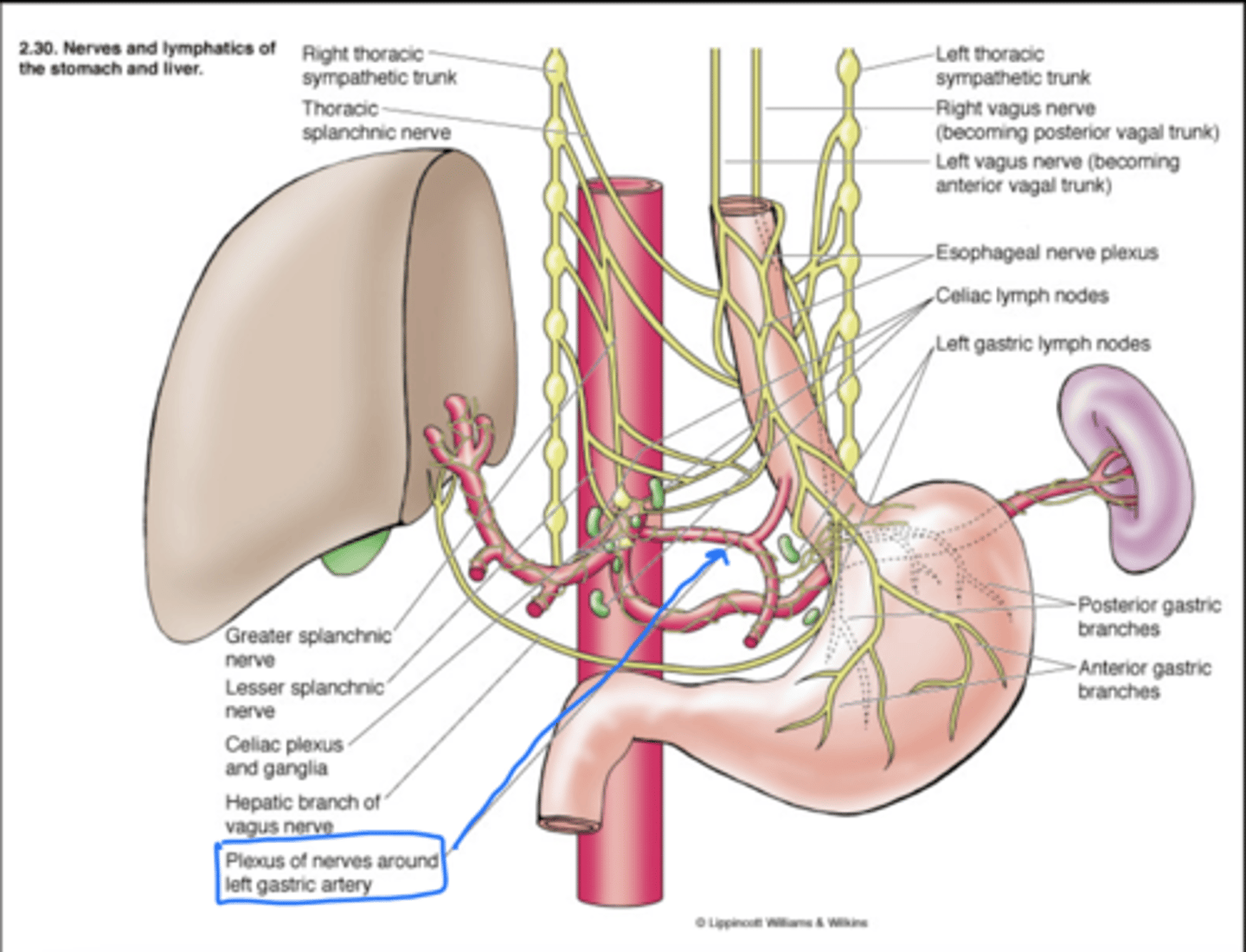
Abdominal esophagus (lower)
Artery: Esophageal branches of left gastric and left inferior phrenic arteries
Vein: Hemiazygos vein, tributary of interior vena cava + left gastric vein, tributary of portal vein
Lymph: Left gastric nodes
- This structure has only smooth muscle
Recurrent laryngeal and esophageal plexus (vagus nerves)
The parasympathetic nerve supply to the esophagus is supplied by these nerves
- 1st supplies the upper half, 2nd supplies the lower half
- These nerves are sensory, motor, and secretomotor in function
Middle cervical and upper 4 thoracic ganglia
These ganglia supply the sympathetic innervation for the esophagus.
- 1st supplies the upper half, 2nd supplies the lower half as the esophageal plexus
- These nerves are sensory and vasomotor in function
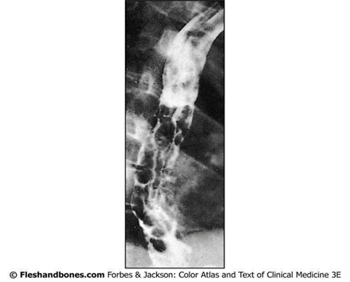
Portal hypertension
What condition leads to esophageal varices?
- The lower esophagus is drained by these - dilation of esophageal veins > rupture
- On barium swallow - worm-like shadows
Left atrial enlargement, aortic arch aneurysm, lymph node enlargement, abnormal right subclavian artery
What 4 conditions leads to dysphagia due to compression of the esophagus from the outside?
Barrett's esophagus
Lower esophageal squamous epithelium > gastric columnar epithelium with goblet cells due to chronic acid exposure
- Typically due to malfunction in LES
- Associated with esophageal adenocarcinoma
Lower 1/3
Malignant tumors of the esophagus typically affect this portion
- This drains to the celiac lymph nodes past the diaphragm, spreading to these nodes
Esophagojejunostomy
If there is a tumor of the esophagus, often the esophagus, stomach, spleen, and omenta will all need to be resected due to the communication with celiac lymph nodes.
How is the continuity of the gut restored?
Trachea
This structure is supplied by:
- Artery: Inferior thyroid arteries
- Vein: Left brachiocephalic vein
- Lymph: Pretracheal and paratracheal nodes
Vagus and recurrent laryngeal
These nerves supply parasympathetic innervation to the trachea
- Sensory, secretomotor (to the mucus membrane), and motor (to the trachealis) in function
Middle cervical ganglion
This ganglion supplies sympathetic innervation to the trachea
- Travels along thyroid arteries
- Vasomotor
Mediastinal shift
The trachea is normally midline in position, any shift of the trachea indicates a _
- Palpated in the suprasternal notch
Thyroid, thymus, lymph nodes, aortic arch
The trachea may be compressed by enlargement of these 4 structures, leading to dyspnea, cough, and hoarseness
Between 2nd/3rd tracheal rings
Where is a tracheostomy typically done?
- In the case of blockage in nose/larynx