Cardiovascular Pathology
1/122
There's no tags or description
Looks like no tags are added yet.
Name | Mastery | Learn | Test | Matching | Spaced |
|---|
No study sessions yet.
123 Terms
T
T or F: Cardiac hypertrophy and dilation are beneficial to a point
Eccentric
Chronic volume overload leads to what type of hypertrophy?
Exercise and pregnancy
What are stimuli for physiological cardiac hypertrophy?
F
T or F: Physiological hypertrophy causes depressed cardiac function
Aortic stenois, mitral regurgitation, etc.
What are stimuli for pathological cardiac hypertrophy?
Primary cardiac hypertrophy
Absence of known cause and other cardiac conditions,
inherited non-sex-linked genetic trait
Secondary cardiac hypertrophy
Due to sustained increase in cardiac workload (volume or pressure overload)
Concentric
Chronic pressure overload leads to what type of hypertrophy?
Preload
Valvular insufficiency increases the ___________ on the ventricles
Afterload
Semilunar valvular stenosis, outflow tract stenosis and hypertension increase the ___________ on the ventricles
Decrease
AV valvular stenosis and pericardial diseases __________ the preload on the ventricles
A. Eccentric
B. Concentric
ID type of cardiac hypertrophy for:
A. Left
B. Right
1. Initiation
2. Compensation
3. Deterioration
What are the cellular stages of cardiac hypertrophy?
Initiation
Cellular stage of cardiac hypertrophy characterized by increase in cell size (sarcomeres/mitochondria)
Compensation
Cellular stage of cardiac hypertrophy characterized by stable hyperfunction (no clinical signs)
Deterioration
Cellular stage of cardiac hypertrophy characterized by degeneration of hypertrophied cardiomyocytes (loss of ventricular contractility or compliance)
Blood vessels (which deliver oxygen to cardiomyocytes)
During initiation of cardiac hypertrophy, the cell size increases; however, what does not increase leading to hypoxia/necrosis?
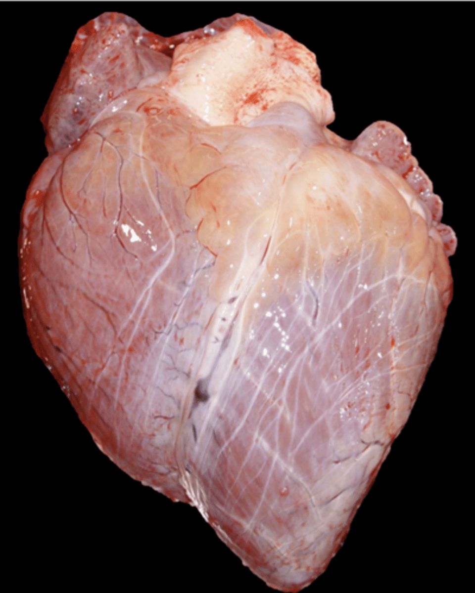
Broad base
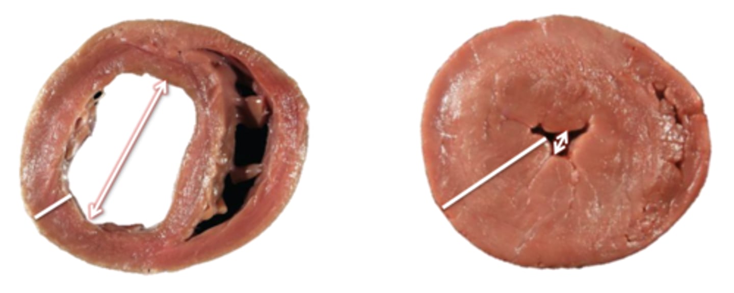
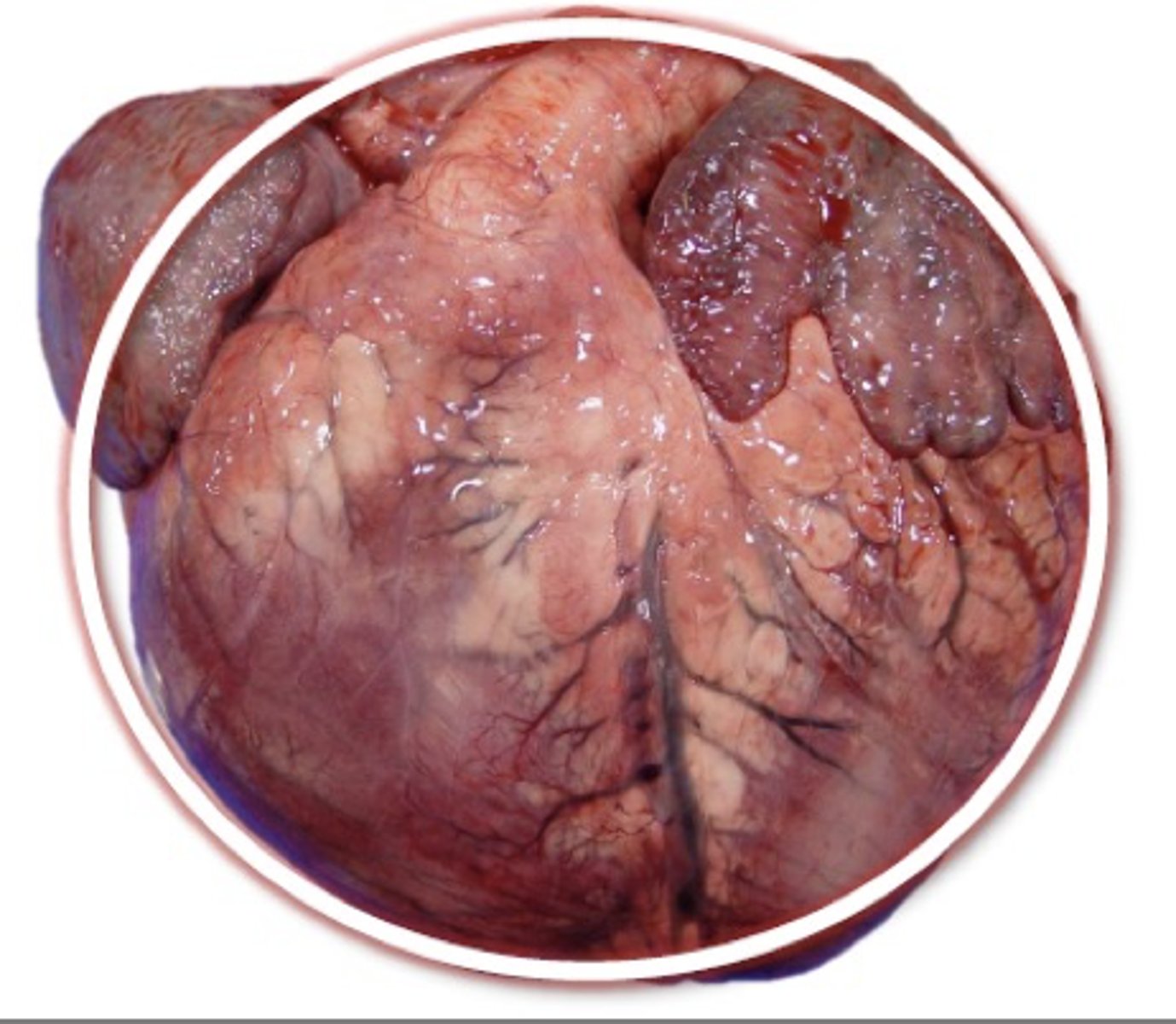
What gross change is apparent due to right cardiac hypertrophy?

Increased length
What gross change is apparent due to left cardiac hypertrophy?
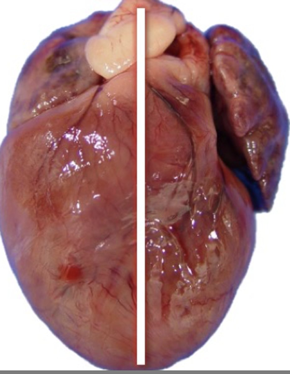
Globose (rounded)
What gross change is apparent due to bi-ventricular hypertrophy?

D, G
If a patient has a heart that is gross in appearance as shown in the picture, what are some underlying causes? (Multiple answers apply)
A. Mitral valve disease
B. DCM
C. Aortic stenosis
D. Pulmonary hypertension
E. HCM
F. VSD
G. Pulmonic stenosis
A, C, E
If a patient has a heart that is gross in appearance as shown in the picture, what are some underlying causes? (Multiple answers apply)
A. Mitral valve disease
B. DCM
C. Aortic stenosis
D. Pulmonary hypertension
E. HCM
F. VSD
G. Pulmonic stenosis
B, F
If a patient has a heart that is gross in appearance as shown in the picture, what are some underlying causes? (Multiple answers apply)
A. Mitral valve disease
B. DCM
C. Aortic stenosis
D. Pulmonary hypertension
E. HCM
F. VSD
G. Pulmonic stenosis
0.5-1.0
Hearts should be ___________% of the body weight
18 grams
In cats, if the heart weighs more than ____________ it is hypertrophied, irrespective of body weight
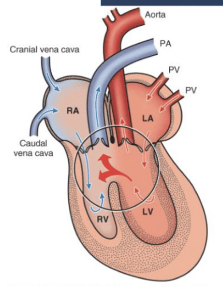
Ductus venosus
Connects the umbilical vein to the inferior vena cava, bypassing the liver
Foramen ovale
Connects the two atria in the fetal heart, bypassing the lungs
Ductus arteriosus
Connects the pulmonary artery to the aorta, bypassing the lungs
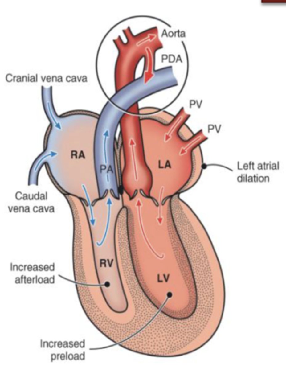
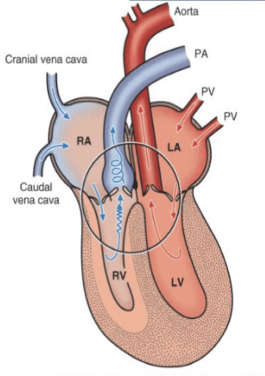
1. Patent ductus arteriosus (PDA)
2. Atrial septal defect (ASD)
3. Ventricular septal defect (VSD)
What are the three examples of left to right shunts?
Patent ductus arteriosus
Failure of the ductus arteriosus to close after birth, resulting in an abnormal opening between the pulmonary artery and the aorta
1. Blood shunts from left to right via PDA
2. Increases blood flow to the lung
3. Increases venous return to the LA & LV
4. Volume overload of LV
5. LV eccentric hypertrophy
Describe the hemodynamics of PDA
1. Blood shunts left to right via PDA
2. Increases blood flow to the lung
3. Pulmonary hypertension
4. Pressure overload of RV
5. RV concentric hypertrophy
Describe the hemodynamics of rare cases of PDA

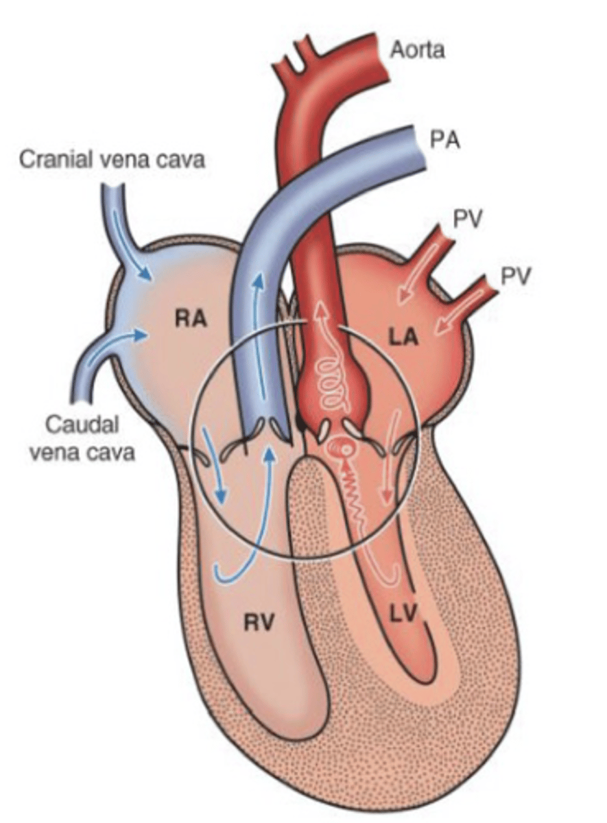
Atrial septal defect
Congenital defect in the septum dividing the left and right atria
Dogs and cattle
ASD is most common in what species?
1. Blood shunt from LA to RA
2. Increase in blood volume in the right ventricle
3. Volume overload of RV
4. RV eccentric hypertrophy
Describe the hemodynamics of ASD
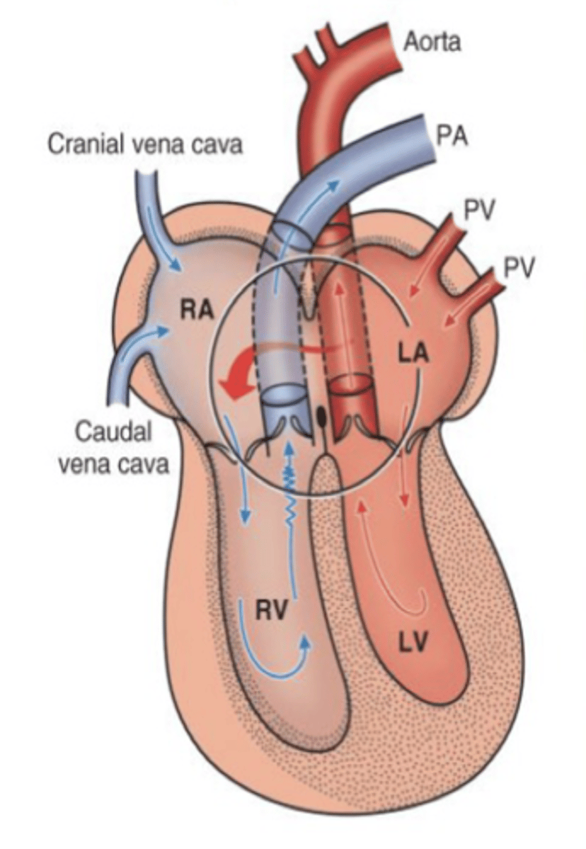
Ventricular septal defect
Congenital opening in the septum separating the ventricles
Horses and cattle
VSD is most common in what species?
High VSD
Defect in the membranous portion of the septum
Low VSD
Defect in the muscular portion of the septum
High
Is a high or low VSD more common?
1. Blood shunt from LV to RV
2. Increase in blood volume in the RV
3. Equalization of pressure across the ventricles
4. LV hypertrophy and RV hypertrophy
Describe the hemodynamics of VSD
Eisenmenger complex
Shunt reversal
Stenosis
Narrowing of the valve orifice
Valvular
What type of pulmonic stenosis is most common in dogs?
1. Stenotic valve restricts outflow
2. Pressure overload of RV
3. RV concentric hypertrophy
4. Right heart failure
Describe the hemodynamics of pulmonic stenosis
1. Stenotic valve restricts outflow
2. Pressure overload of LV
3. LV concentric hypertrophy
(If severe can lead to arrhythmias and sudden cardiac death, left heart failure)
Describe the hemodynamics of subaortic stenosis (SAS)
Cats
Cardiomyopathies are most common in what species?
1. Hypertrophic cardiomyopathy
2. Dilated (congestive) cardiomyopathy
3. Restrictive (infiltrative) cardiomyopathy
What are the three classifications of primary cardiomyopathies?
Hypertrophic cardiomyopathy
What is the most common form of feline cardiomyopathy?
1. Left side heart failure
2. Pulmonary edema
3. Dyspnea
Describe how hypertrophic cardiomyopathy lead to dyspnea?
Myosin binding protein c
Mutations in the cardiac protein called ____________________ can lead to feline HCM
Symmetrical
If both the septum and free wall are affected in feline HCM, it is considered what?
Diastolic (ventricle cannot fill properly)
Feline HCM causes impaired (systolic or diastolic) function
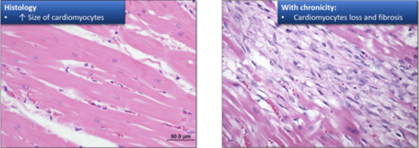
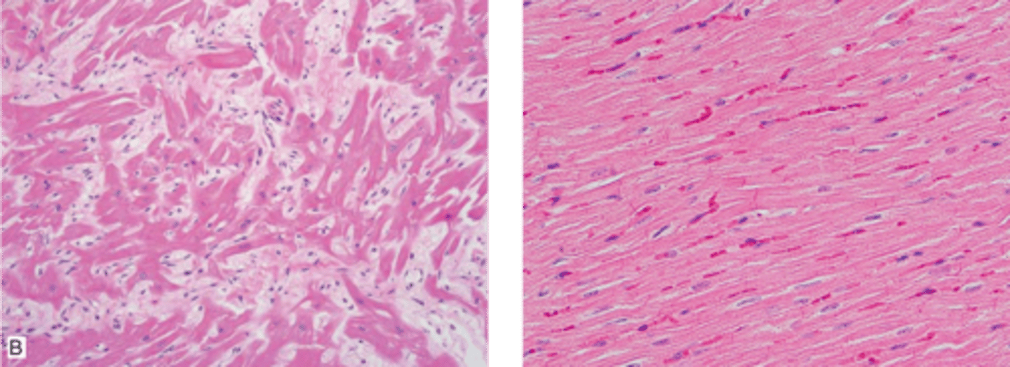
Myofibers are in disarray with interstitial fibrosis present
Why is the image on the left not normal myocardium?
1. Severe atrial enlargement (because blood cannot flow to left ventricle)
2. Caudal abdominal aorta (saddle thrombus)
What are some sequelae of feline HCM?
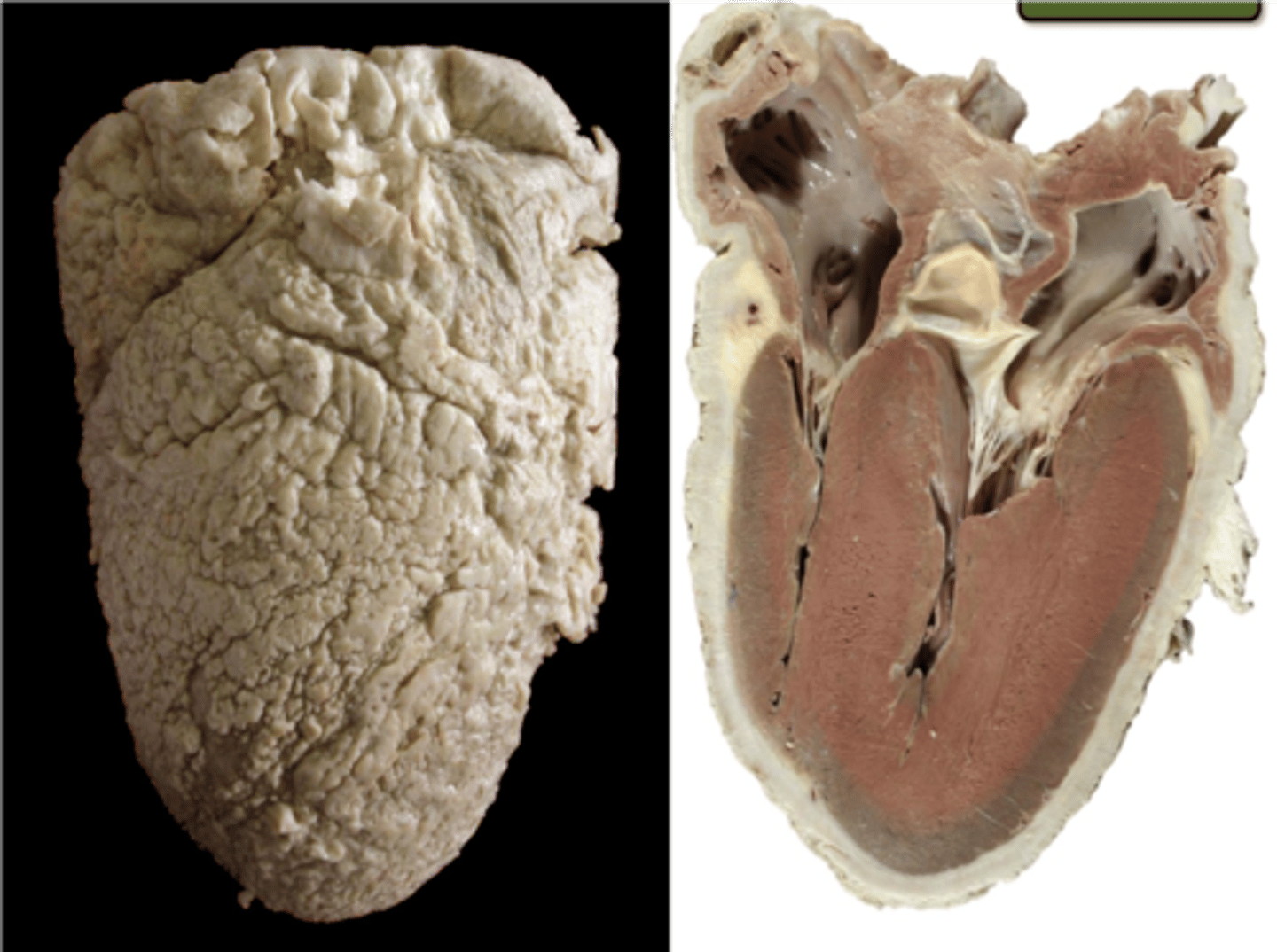
Dilated cardiomyopathy
All chambers enlarged, ventricles are dilated, flabby with thin walls
Taurine
A _____________ deficiency leads to myocardial failure (dilated cardiomyopathy)
Systolic
Dilated cardiomyopathy causes impaired (systolic or diastolic) function
Diastolic
Restrictive cardiomyopathy causes impaired (systolic or diastolic) function
Young to middle aged, large breed dogs (St. Bernard, Great Dane)
Dilated cardiomyopathies are common in what canine breeds?
Striatin
Arrhythmogenic right ventricular cardiomyopathy (ARVC) is common in Boxers and caused by a mutation in what gene?
Right ventricle
ARVC can lead to ventricular arrhythmia of _________ origin
Pericardium
A double-layered serous membrane that surrounds the heart
Hydropericardium
Transudate in the pericardial sac
1. Increases in hydrostatic pressure: right heart failure
2. Hypoproteinemia: emaciation, protein losing enteropathy/nephropathy
What is the pathogenesis of hydropericardium?
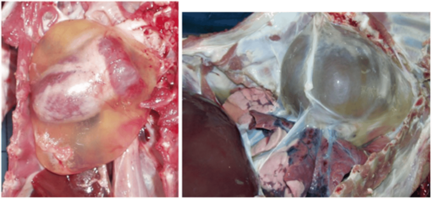
Hemopericardium
Blood in the pericardial cavity
1. Cardiac puncture
2. Aortic rupture
3. Ulcerative atrial endocarditis
4. Atrial hemangiosarcoma in dogs
What are causes for hemopericardium?
Horses and turkeys
Aortic rupture leading to hemopericardium is most common in what species?
Atrial hemangiosarcoma
What is the most common cause for hemopericardium in dogs?
Cardiac tamponade
Acute compression of the heart caused by fluid accumulation in the pericardial cavity
Diastolic filling
Cardiac tamponade may prevent what?
Serous atrophy of pericardial fat
Epicardial fat appears gelatinous
Emaciation
Serous atrophy of pericardial fat occurs due to what?
BCS
Serous atrophy of pericardial fat is indicative of poor what?
Fibrinous pericarditis
Pericardial sac contains effusion rich in fibrin that clots soon after exposure to air
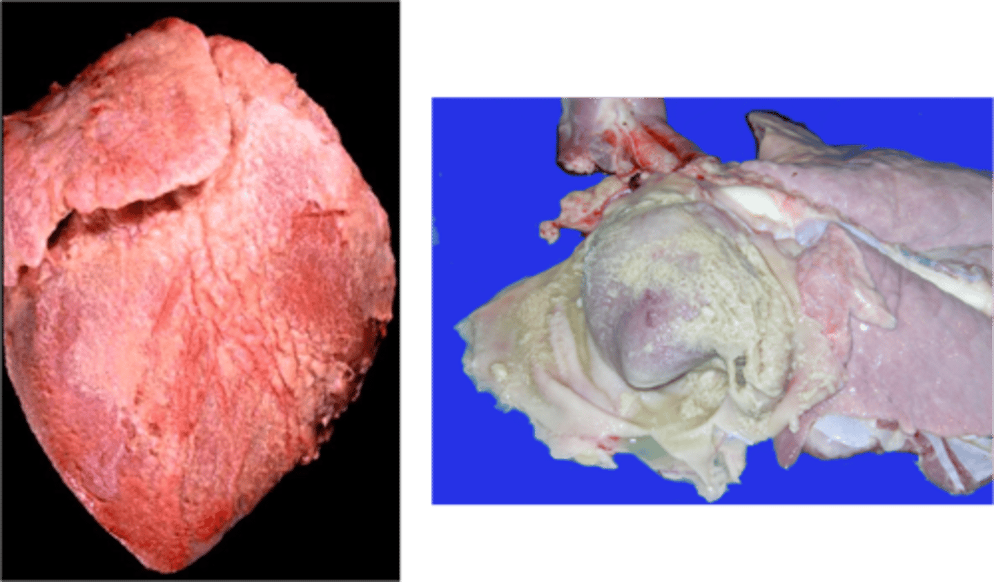
Vascular
Fibrinous pericarditis is reflective of ____________ disease
Fibrinous pericarditis
FIP in cats leads to what pericardial disease?
"Shaggy heart" or "bread and butter heart"
What are names given for fibrinous pericarditis?
Purulent pericarditis
Pericarditis where pericardial space is invaded by infection that enters due to neighboring inflammation
Traumatic reticulopericarditis
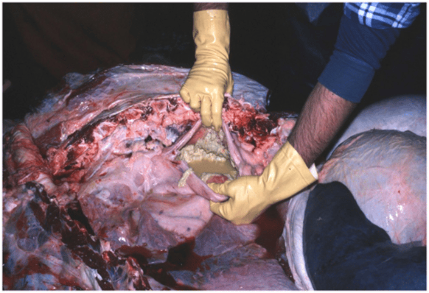
Uncommon consequence of hardware disease due to penetration of a sharp metal object from the reticulum into the pericardium
Chronic constrictive pericarditis
What is the ultimate fate of purulent and fibrinous pericarditis?
Chronic constrictive pericarditis
A fibrous thickening of the pericardium that prevents adequate filling of the ventricles (diastolic dysfunction) and eventually results in cardiac failure
1. Ischemic
2. Toxic
3. Nutritional
4. Neurogenic
Myocardial necrosis is due to what?
Brain-heart syndrome
Injury to brain can cause myocardial necrosis due to catecholamine surge
Vitamin E and selenium
Nutritional myocardial necrosis is due to deficiencies in what?
F (common in humans)
T or F: Myocardial necrosis due to ischemia is common in animals
1. Coagulative necrosis
2. Infiltration of leukocytes
3. "Healing" begins (fibroblast proliferation)
What are the steps of myocardial necrosis?
T
T or F: There is no regenerative power of myocardium in adults
1. Loss of cross striations
2. Karyolysis
Acute myocardial necrosis (12-24 hours) is characterized by what microscopically?
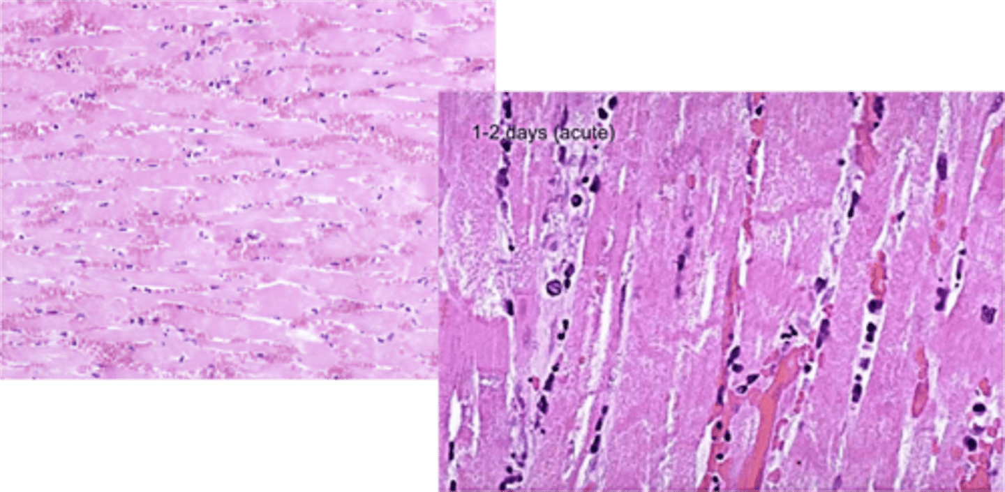
Macrophages and neutrophils infiltrate and phagocytize necrotic debris
Acute myocardial necrosis (2-3 days) is characterized by what microscopically?
1. Fibrosis
2. Mineralization
Myocardial fibrosis (> 12-20 days) is characterized by what microscopically?
Monensin toxicity
Incredibly toxic in horses causes myocardial degeneration and necrosis in left ventricle
Ionophores and Doxorubicin
What are some important toxins that cause toxic myocardial necrosis?
White muscle disease
Nutritional myopathy due to vitamin E or selenium deficiency
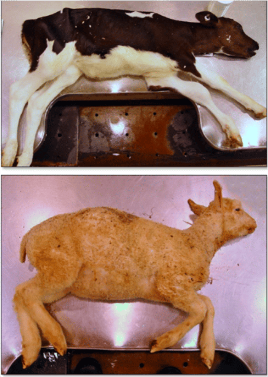
Young farm animals
White muscle disease typically affects what group of animals?
T
T or F: White muscle disease can affect skeletal muscle, or cardiac muscle, and sometimes both
1. Selenium and vitamin E deficiency
2. Free radicals are produced during normal cardiac metabolism
3. Decreased scavenging of free radicals
4. Peroxidation of cell membranes
5. Cardiac and skeletal muscle necrosis and mineralization
Describe the mechanism of white muscle disease
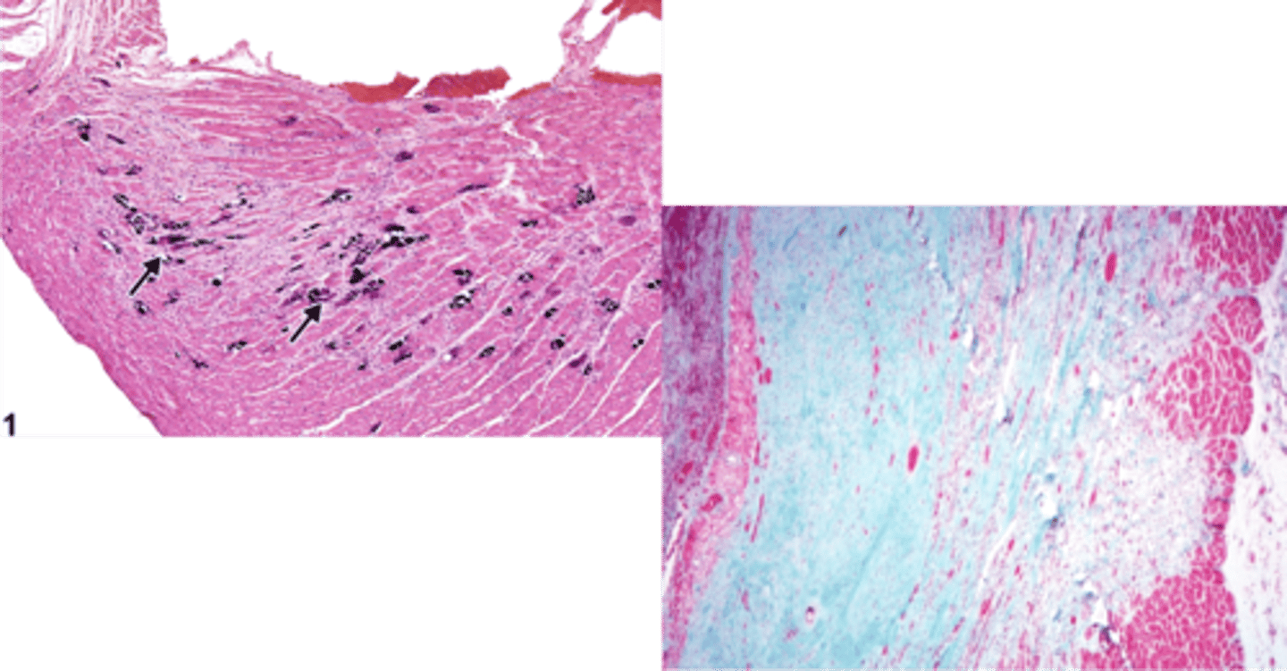
Left ventricle
White muscle disease of calves typically affects what?
Right ventricle
White muscle disease of lambs typically affects what?
T
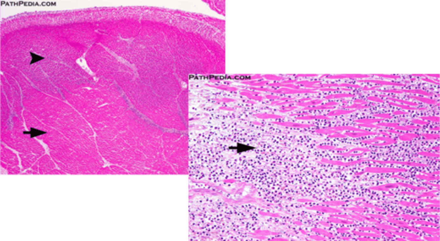
T or F: Myocarditis rarely occurs alone, it is typically part of a systemic disease