A&P - 6.4 Bone Formation and Development
1/21
There's no tags or description
Looks like no tags are added yet.
Name | Mastery | Learn | Test | Matching | Spaced |
|---|
No study sessions yet.
22 Terms
ossification
(also, osteogenesis) bone formation or development
osteogenic pathways
intramembranous ossification
endochondral ossification
intramembranous ossification
process by which bone forms directly from mesenchymal tissue
compact and spongy bone develops directly from sheets of mesenchymal (undifferentiated) connective tissue
begins in utero during fetal development and continues on into adolescence
examples of bones formed via intramembranous ossification: flat bones of the face, most of the cranial bones, and the clavicles (collarbones)

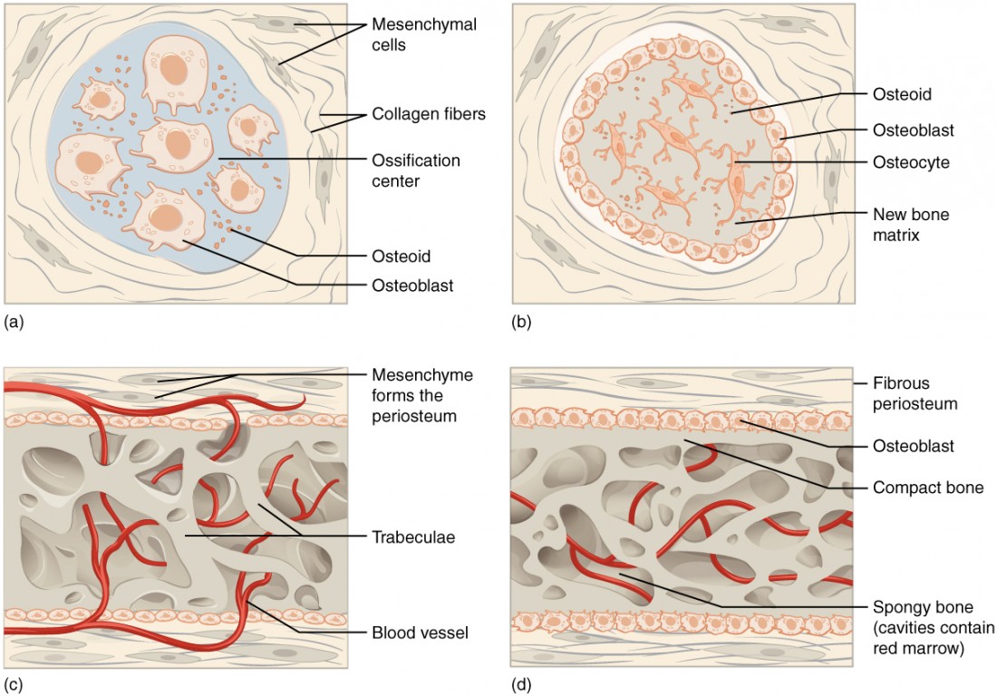
process of intramembranous ossification
begins when mesenchymal cells in the embryonic skeleton gather together and begin to differentiate into specialized cells
some of these cells will differentiate into capillaries while others will become osteogenic cells and then osteoblasts
although they will ultimately be spread out by the formation of bone tissue, early osteoblasts appear in a cluster called an ossification center
when osteoid calcifies (hardens) within a few days as mineral salts are deposited on it, thereby entrapping the osteoblast within
once entrapped, the osteoblasts become osteocytes
as osteoblasts transform into osteocytes, osteogenic cells in the surrounding connective tissue differentiate into new osteoblasts
osteoid secreted around the capillaries results in a trabecular matrix, while osteoblasts on the surface of the spongy bone become the periosteum
the periosteum then creates a protective layer of compact bone superficial to the trabecular bone
the trabecular bone crowds nearby blood vessels, which eventually condense into red marrow
ossification center
clusters of osteoblasts found in the early stages of intramembranous ossification


osteoid
uncalcified bone matrix
osteoblasts secrete osteoid
endochondral ossification
process in which bone forms by replacing hyaline cartilage
cartilage does not become bone
instead, cartilage serves as a template to be completely replaced by new bone
cartilage remains on the epiphyseal (growth) plate and at join surface as articular cartilage
takes much longer than intramembranous ossification
examples of bones formed via endochondral ossification: bones are the base of the skull and long bones

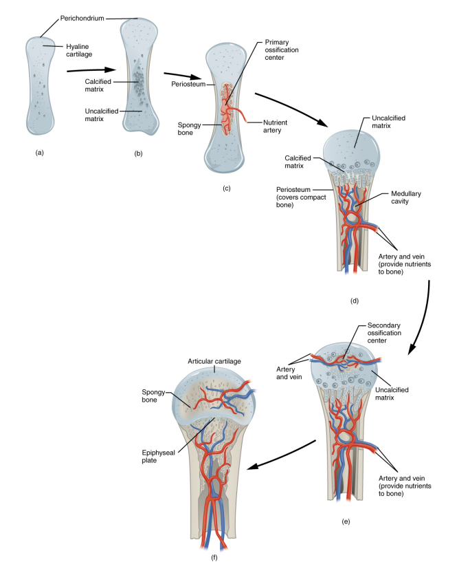
process of endochondral ossification
mesenchymal cells differentiate into chondrocytes (cartilage cells)
the cartilage model of the future bony skeleton and the perichondrium form
because as the matrix calcifies nutrients can no longer reach the chondrocytes which results in death and disintegration of the surrounding cartilage
the blood vessels invade the space carrying osteogenic cells with them resulting in enlarging cavities which eventually combine to become the medullary cavity
as the cartilage grows capillaries penetrate cartilage which initiates the transformation of the perichondrium into the bone-producing periosteum
osteoblasts form a periosteal collar of compact bone around the cartilage of the diaphysis
by the second or third month of fetal life, bon cell development and ossification ramps up and creates the primary ossification center
cartilage and chondrocytes continue to grow at ends of the bone
secondary ossification centers develop
cartilage remains at epiphyseal (growth) plate and at joint surface as articular cartilage

perichondrium
membrane that cover cartilage
medullary cavity
hollow region of the diaphysis; filled with yellow marrow

primary ossification center
region, deep in the periosteal collar, where bone development starts during endochondral ossification

secondary ossification center
region of bone development in the epiphyses
after birth, this same sequence of event; matrix mineralization, death of chondrocytes, invasion of blood vessels from the periosteum, and seeing with osteogenic cells that become osteoblast, occurs in the epiphyseal regions, and each of these centers of activity is referred to as a secondary ossification center

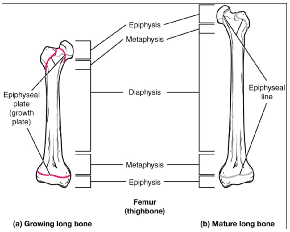
epiphyseal plate
(growth plate) - area of longitudinal growth in a long bone
epiphyseal plates are visible in a growing bone
area of hyaline cartilage that separates epiphyses and diaphysis of children’s bones
epiphyseal lines are the remnants of epiphyseal plates in a mature bone
composed of four zones of cells and activity
zones of cells and activity in epiphyseal plate
reserve zone
proliferative zone
zone of maturation and hypertrophy
zone of calcified matrix

reserve zone
region of the epiphyseal plate that anchors the plate to the osseous tissue of the epiphysis
contains small chondrocytes within the matrix

proliferative zone
region of the epiphyseal plate that makes new chondrocytes to replace those that die at the diaphyseal end of the plate and contributes to longitudinal growth of the epiphyseal plate
next layer toward the diaphysis and contains stacks of slightly larger chondrocytes
longitudinal growth of bone is a result of cellular division in the proliferative zone
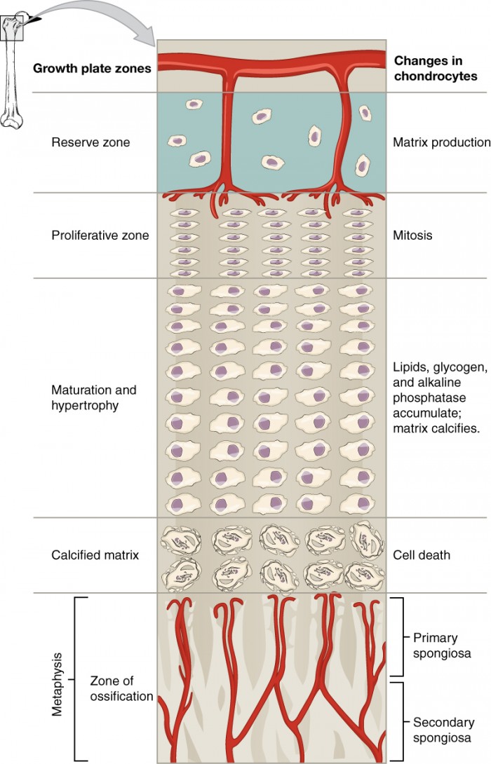
zone of maturation and hypertrophy
region of the epiphyseal plate where chondrocytes from the proliferative zone grow and mature and contribute to the longitudinal growth of the epiphyseal plate
chondrocytes here are older and larger than those in the proliferative zone
the most mature cells are situated closer to the diaphyseal end of the plate
longitudinal growth of bone is a result of maturation of cells in the zone of maturation and hypertrophy

zone of calcified matrix
region of the epiphyseal plate closest to the diaphyseal end; functions to connect the epiphyseal plate to the diaphysis
most of the chondrocytes are dead because the matrix around them has calcified
capillaries and osteoblasts from the diaphysis penetrate this zone and the osteoblasts secrete bone tissue on the remaining calcified cartilage
connects the epiphyseal plate to the diaphysis
a bone grows in length when osseous tissue is added to the diaphysis

epiphyseal line
completely ossified remnant of the epiphyseal plate
when the chondrocytes in the epiphyseal plate cease their proliferation and bone replaces the cartilage, longitudinal growth stops, all that remains of the epiphyseal plate is the epiphyseal line

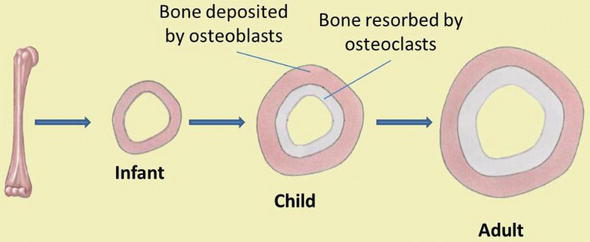
appositional bone growth
increase diameter of existing bones
does not form original bones
osteogenic cells differentiate into osteoblasts that add bone matrix under periosteum via intramembranous ossification
adds successive layers of circumferential lamellae
trapped osteoblasts become osteocytes
deeper lamellae recycled and replaced by osteons
osteoclasts remove matrix at inner surface to enlarge medullary cavity
osteoclasts resorb old bone that lines the medullary cavity

modeling
process, during bone growth, by which bone (matrix) is resorbed on one surface of a bone and deposited on another
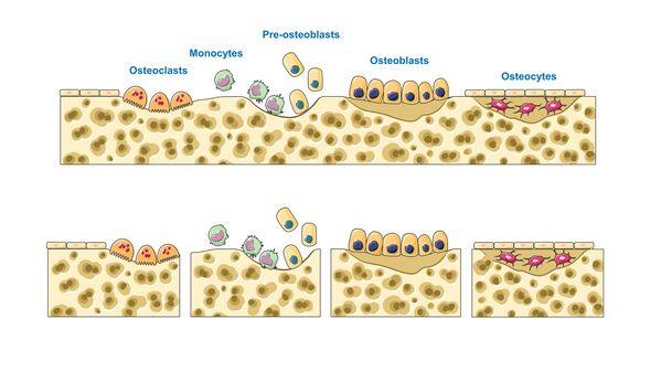
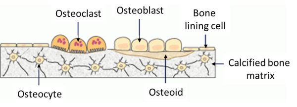
bone remodeling
process by which osteoclasts resorb old or damaged bon at the same time as and on the same surface where osteoblasts form new bone to replace that which is resorbed
during adult life
injury, exercise, and other activities lead to remodeling