Lecture 4: Alterations in Chromosomes and Genes
1/31
There's no tags or description
Looks like no tags are added yet.
Name | Mastery | Learn | Test | Matching | Spaced | Call with Kai |
|---|
No analytics yet
Send a link to your students to track their progress
32 Terms
How common are cytogenetic disorders?
Cytogenetic disorders: diseases caused by abnormal chromosomes (number or structure).
Nearly 1% of live births have chromosomal abnormalities.
2% of pregnancies in women >35 (older eggs have been in meiosis longer → more errors).
10% of stillbirths.
50% of spontaneous abortions (miscarriages).
Many cancers involve chromosomal changes.
How are cytogenetic studies performed?
Take a blood sample (use white blood cells).
Culture cells and stimulate them to divide.
Arrest at metaphase with colcemid (chromosomes are easiest to see).
Colcemid a drug that halts cells in metaphase so chromosomes can be seen.
Use a hypotonic solution to swell cells and spread chromosomes.
Hypotonic: Lower solute concentrations outside; higher solute concentrations inside.
Fix and stain chromosomes on a slide for viewing.
Resolution: detects large changes (~1-10 Mb).
Big chromosomal abnormalities.
Can also use fetal cells from amniotic fluid, chorionic villis, or bone marrow.
What is G banding?
Treat cells with trypsin and Giemsa stain.
Creates pattern of light and dark bands on chromosomes.
Pattern correlates with:
% GC vs. AT content.
Distribution of repetitive elements.
Each chromosome has unique pattern → can identify all 24 human chromosomes.
What is a karyotype?
Output of g-banding.
Standard chromosomal set of an individual.
Chromosomes arranged by size: 1-22 (autosomes) + gender chromosomes (XX or XY).
Used to identify chromosomal abnormalities.
Karyotype vs.kkaryogram:
Karyotype: the full set of chromosomes in a cell; includes number, size, shape; also refers to the analysis.
Karyogram: the arranged picture of chromosomes after analysis.
What is FISH (Fluorescence In Situ Hybridization)?
Uses fluorescent probes to detect specific DNA sequences on chromosomes.
Process:
Fix chromosomes, denature DNA.
The chromosomes are in interphase or metaphase.
Interphase = gene location or copy number.
Metaphase = structure.
Labeled probe hybridizes to target sequence.
View under fluorescence microscope.
Can work on dividing (metaphase) or non-dividing (interphase) cells.
Resolution: several megabases.
What is spectral karyotyping (SKY)?
"Chromosome paint" - colors each chromosome differently.
Uses combinatorial labeling (different ratios of fluorophores).'
Different ratios are assigned pseudocolors.
Pseudocolors = artificial colors added to visualize different DNA probes.
Makes it easy to see translocations and large rearrangements.
Useful for studying cancer (complex chromosomal changes).
To view:
Epifluorescence microscopy: view fluorescent probes.
CCD camera: captures bright, clear images.
Fourier spectroscopy: sees all emission colors at once.
What is array CGH (comparative genome hybridization)?
Detects DNA copy number changes (gains/losses) across genome.
Method:
Label patient DNA and control DNA with different colors.
Green = control DNA; red = sample DNA.
Hybridize to chip with 100,000+ oligonucleotides.
Oligonucleotide: synthetic, short DNA/RNA sequences used to target specific genetic sequences.
Measure fluorescence ratio.
Red = Green (ratio ≈ 1): Normal / balanced signal. Nothing unusual.
Red ≠ Green (ratio ≠ 1): Indicates a difference.
Shows duplications and deletions.
Cannot detect translocations, inversions, small mutations, or rearrangements.
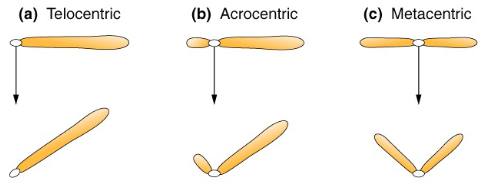
What are the chromosome classifications by centromere position?
Metacentric: centromere in middle (equal arms).
Acrocentric: centromere near end (one very short arm).
Telocentric: centromere at end (does NOT occur in humans).
What is triploidy and tetraploidy?
Triploidy: 3 complete chromosome sets (69 chromosomes).
Caused by fertilization by 2 sperm OR failure in meiotic division.
Almost always lethal.
Tetraploidy: 4 complete chromosome sets (92 chromosomes).
Usually arises from mitotic error in the zygote.
Always lethal.
What is aneuploidy?
Abnormal number of chromosomes (not a multiple of 23).
About 5% of pregnancies.
Types:
Monosomy: missing one chromosome (45 total).
Trisomy: extra chromosome (47 total).
Caused by nondisjunction (chromosomes fail to separate properly in meiosis; before fertilization).
Affects all of the cells in the body.
What is mosaicism?
Two or more cell populations with different chromosome numbers in same person.
Example: 47,XXX/46,XX (some cells have extra X, some normal).
Caused by nondisjunction during early embryonic development (not in egg/sperm).
Occurs in mitosis; after fertilization.
Only affects some cells.
Usually causes milder symptoms than if all cells affected.
What are the main structural chromosomal abnormalities?
Deletion.
Duplication.
Inversion.
Translocation.
Ring chromosome.
Isochromosome.
Deletion
Piece of the chromosome missing.
Duplication
Piece present in extra copies.
Inversion
Segment flipped 180°.
Translocation
Segment moves to another chromosome.
Reciprocal:
Two non-homologous chromosomes exchange segments.
Can be balanced (no net gain/loss of material) or unbalanced.
Balanced carriers usually healthy but at risk for unbalanced offspring.
Robertsonian: two acrocentric chromosomes join.
Acrocentric: centromere near the end, tiny p arm, long q arm.
Ring chromosome
Ends join together forming a circle.
Isochromosome
Chromosome with two identical arms.
Frameshift
The second image highlights how a single base shift causes a "domino effect," altering the entire protein downstream from the mutation.
Point mutations (missense and nonsense)
Missense: One nucleotide changes → different amino acid in the protein (e.g., His → Pro). Can alter protein function.
Nonsense mutation: Nucleotide change creates a premature STOP codon → protein is truncated and usually non-functional.
Large scale & structural mutations
Duplication Mutation: A section of DNA is copied twice. Can cause an “overdose” of protein instructions → may affect traits like lip shape or size.
Repeat Expansion: A short DNA sequence (e.g., “CAG”) repeats too many times. Can cause neurological or developmental disorders.
What are other factors affecting inheritance?
Reduced penetrance: not everyone with mutation shows symptoms.
Variable expressivity: same mutation causes different severity in different people.
Modifying loci: other genes affect how mutation is expressed.
Anticipation: disease gets worse/earlier in successive generations.
Genomic imprinting: parent of origin matters for gene expression.
Uniparental disomy: both copies of chromosome from one parent.
What are the three types of mutations by origin?
Inherited (germline): passed from parents to offspring.
De novo: occur in egg, sperm, or just after fertilization (new mutation, not in parents).
Acquired (somatic): occur during person's life in body cells.
NOT passed to offspring (unless in germline cells).
What are loss of function mutations?
Reduce or eliminate normal protein function.
Mechanisms:
Nucleotide substitutions, deletions, rearrangements.
Premature stop codons.
Impaired protein folding.
Examples:
Turner syndrome (missing X chromosome).
Thalassemias (reduced hemoglobin).
What are gain of function mutations?
Enhance or alter normal protein function.
Types:
Increase amount of protein.
Increase protein's normal function.
Novel property in new protein.
Wrong timing/location of production.
Illustrates importance of gene regulation.
What mutations can affect RNA?
Splice junction mutations: affect where introns are removed.
Intron mutations: affect splicing signals.
Coding sequence mutations: can affect splicing even in exons.
Defects in capping and tailing: affect mRNA stability and translation.
What are housekeeping proteins vs. specialty proteins?
Housekeeping proteins:
Present in every cell.
Maintain basic cell structure and function.
90% of expressed genes.
Specialty proteins:
Tissue-specific.
Produced in limited cell types.
Have unique functions.
10% of expressed genes.
What is genetic heterogeneity?
Allelic heterogeneity: different mutations in SAME gene cause similar disease.
Example: many different CFTR mutations all cause cystic fibrosis.
Locus heterogeneity: mutations in DIFFERENT genes cause similar disease.
Example: hearing loss can be caused by mutations in many different genes.
Modifier genes: genes unrelated to main defect can alter how disease manifests.
Autosomal disorder
Definition: Mutation on chromosomes 1–22.
Note: Can be dominant or recessive; affects males & females equally.
X-linked disorder
Definition: Mutation on the X chromosome.
Note: Males more affected; females may be carriers if recessive.
Codominant disorder
Definition: Both alleles are fully expressed.
Example: AB blood type, sickle cell trait.
Mitochondrial disorder
Definition: Mutation in mtDNA.
Note: Maternal inheritance only, often affects energy-demanding tissues.