ALL LC - vinter + summer
1/61
Earn XP
Description and Tags
for exam, combined from credit, Includes all from document (parasite life cycles to know).
Name | Mastery | Learn | Test | Matching | Spaced | Call with Kai |
|---|
No analytics yet
Send a link to your students to track their progress
62 Terms
What parasite has this type of lifecycle?
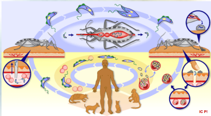
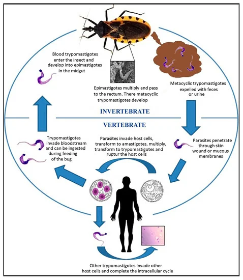
Subdivision of trypanosoma: STERCORARIA (T. Cruzi)
Trypomastigotes: The cycle starts with trypomastigotes present in the blood of an infected host. These are the infectious form of the parasite.
Reduviid bugs: The Trypomastigotes are ingested by Reduviid bugs (the vector) during a blood meal.
Epimastigotes: Inside the bug, the Trypomastigotes transform into Epimastigotes and reproduce through binary fission over 8-10 days.
Trypomastigotes in feces: The Epimastigotes eventually transform back into Trypomastigotes and are excreted in the bug's feces.
Skin lesion: The Trypomastigotes can enter a new host through a skin, wound, during scratching of the bite site.
invasion of cells: Once inside the host, Trypomastigotes invade host cells. (macrophages)
Amastigotes: Inside the macrophages, they transform into Amastigotes, where they multiply.
Lymph nodes (lnn.): From the macrophages, the Amastigotes can spread to the lymph nodes.
Blood: Eventually, they return to the bloodstream in their Trypomastigote form, continuing the cycle → epimastigote → trypomastigote etc.
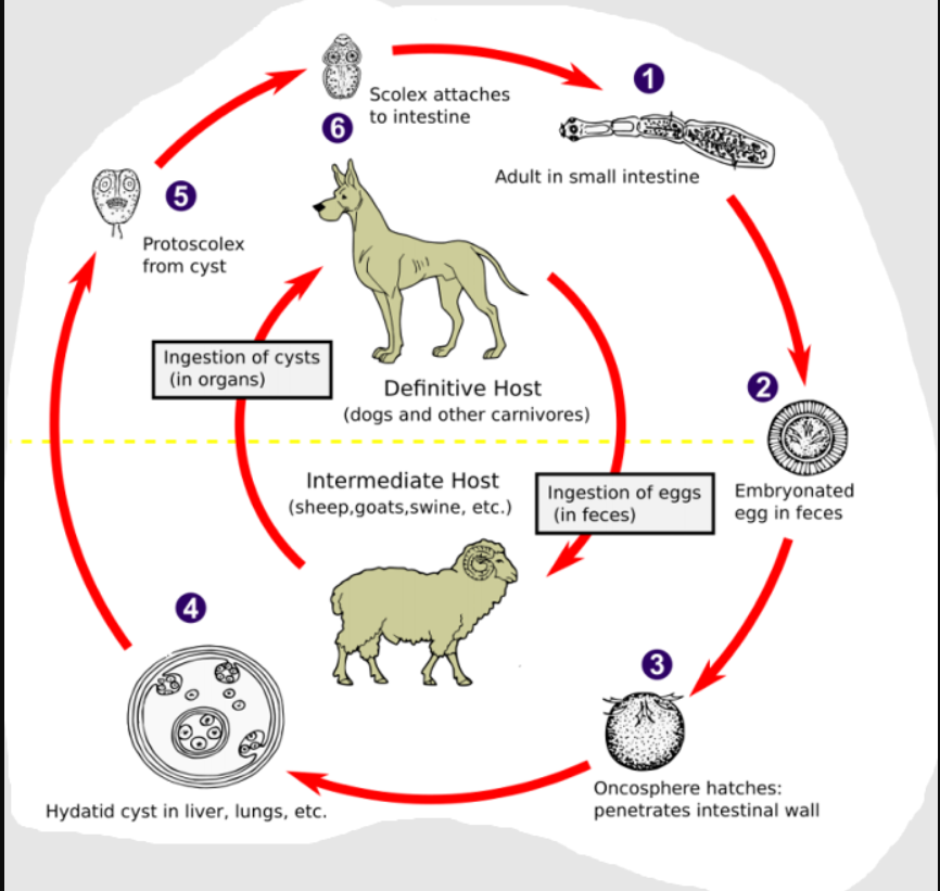
To which parasite does this lifecycle belong to?
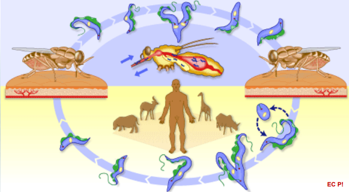
Subdivision of Trypanosoma: SALIVARIA (T.bruzei)
Trypomastigotes in Blood of infectected host
Tse Tse Fly: The tse tse fly (the vector) ingests blood during a meal, getting the trypomastigotes.
Binary Fission in the Fly: Inside the tse tse fly, the Trypomastigotes undergo binary fission, multiplying in number.
Salivary Gland: The parasites migrate to the salivary glands of the tse tse fly.
Epimastigotes: In the salivary glands, they transform into Epimastigotes. This form continues to reproduce through binary fission.
Trypomastigotes Formation: They revert to the Trypomastigote form, ready to infect a new host.
Inoculation to the Host: During a blood meal, the tse tse fly inoculates/injects Trypomastigotes into the host's bloodstream.
Binary Fission in Blood and CNS: Once in the host, Trypomastigotes multiply through binary fission in the bloodstream and can invade the central nervous system (CNS).
Which parasite does this life cycle belong to?
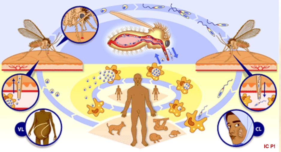
Leishmania.
complex life cycle, involving two main hosts, the sandfly vector and the mammalian host (ex. humans)
begins when sandfly vector feeds on the blood of an infected mammal, it will ingest the amastigotes from it.
These amastigotes will transform into promastigotes in the midgut of sandfly, and multiply by binary fission. These will then migrate to the sandfly`s salivary glands (pharynx of vector)
During another blood meal, the sandfly will inoculate/inject the promastigotes into the skin of a new mammalian host, where they invade macrophages and transform back into amastigotes.
Inside the macrophages, they multiply by binary fission → rupture of the affected cell and subsequent infection of new macrophages.
The infection can cause agglomeration → spread to visceral organs, skin and mucosa.
This is the life cycle of?
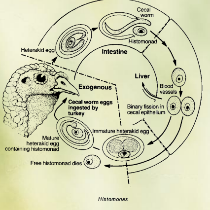
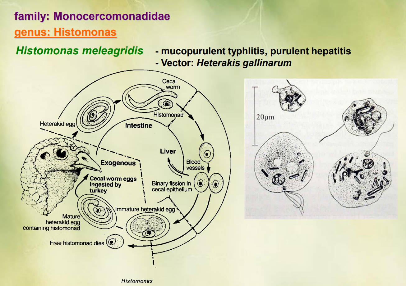
Genus Histomonas of the family monocercomonadidae.
Histomonas meleagridis
cycle begins with heterakid eggs containing histomonas, which are ingested by turkeys.
Inside the turkey`s intestine, the eggs hatch, releasing immature heterakid worms (cecal worms)
the histomonas enter the cecal epithelium, multiplying by binary fission
as they multiply, the invade blood vessels → spreads
histomonads continue to grow, eventually leading to death of free histomonads.
mature heterakid eggs containing the histomonas can now be released in the turkey`s feces.
These cecal worm eggs can then be ingested by another turkey → continuation of cycle.
This is the life cycle of?
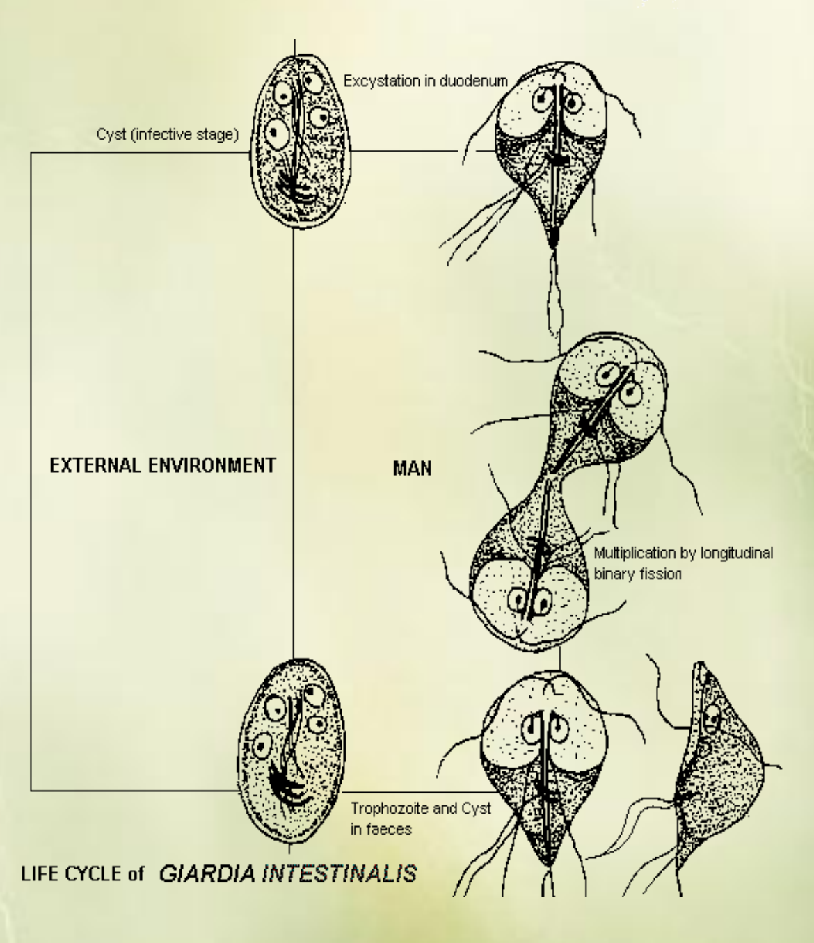
Life cycle of Giardia Intestinalis
ingestion: cysts are ingested by the host through contaminated water or food
excystation: in the intestines, cysts are exposed to stomach acid → activating the release of trophozoites.
trophozoite stage: multiplication by binary fission, attaching to intestinal wall, GIT symptoms.
encystation: trophozoites transform back into cysts
excretion: cysts are excreted in the host`s feces, where they can contaminate water or soil, completing the life cycle.
Way of transmission: water, food (with cysts)
occurrence: spring autumn.
This is the life cycle of?
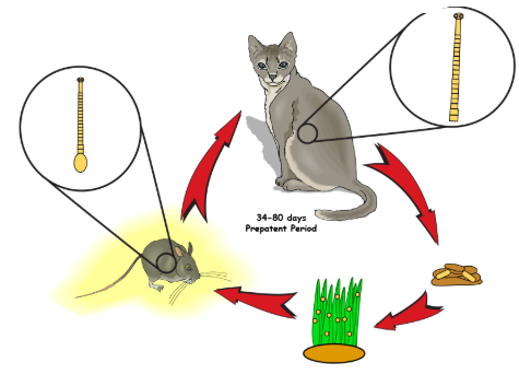
Eimeria
In rabbits, E. intestinalis, FH: rabbits, Small intestine
Exogenous sporogony: Formation of sporozoites within the oocyst from a single organism outside the host environment.
Infection: Sporulated oocysts are ingested by a new host, releasing sporozoites.
Endogenous merogony I: Sporozoites invade host cells and undergo asexual replication to produce merozoites.
Endogenous merogony II: More rounds of asexual replication occur, producing additional merozoites.
Endogenous merogony III: The final round of asexual replication occurs, leading to the production of many merozoites ready to infect new cells.
Endogenous gamogony: Some merozoites differentiate into microgametes and macrogametes, resulting in sexual reproduction within the host → zygote formed into oocyst → unsporulated oocysts are passed in the feces.
Sporulation occurs in the environment.
This is the life cycle of?
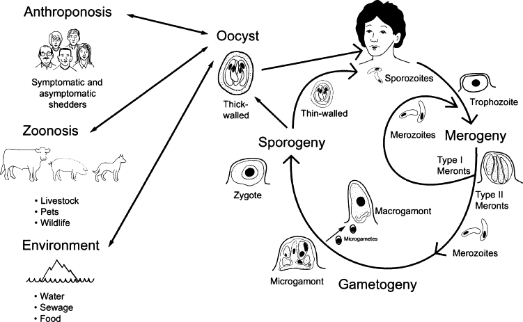
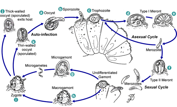
Cryptosporidium (genus) - of family cryptosporidiidae.
Incl. C. parvum (cattle, man, mammals), canis (dog), felis (cat), hominis (man).
Direct cycle, occurs in one host, in small intestine.
Ingestion: The cycle begins when a host ingests oocysts containing sporozoites from contaminated water or food.
Excystation: Inside the gastrointestinal tract, the oocysts are exposed to stomach acids, activating them to release sporozoites.
Sporozoite: The released sporozoites then invade the epithelial cells of the intestines.
Trophozoite formation: Within these cells, the sporozoites develop into trophozoites, which are the active feeding forms.
Merogony: The trophozoites then undergo asexual reproduction known as merogony, resulting in the production of merozoites.
Merozoite release: These merozoites are released back into the intestinal lumen, where they can infect new epithelial cells or develop into gametes.
Gamogony: Some merozoites differentiate into male and female gametes, ready for sexual reproduction.
Fertilization: Male gametes (microgametes) fertilize female gametes (macrogametes), forming a zygote.
Oocyst formation: The zygote develops into an oocyst, which can either sporulate inside the host to become infectious or be excreted in the feces of the host, thus reinitiating the cycle when ingested by another host.
which parasite does this life cycle belong to?
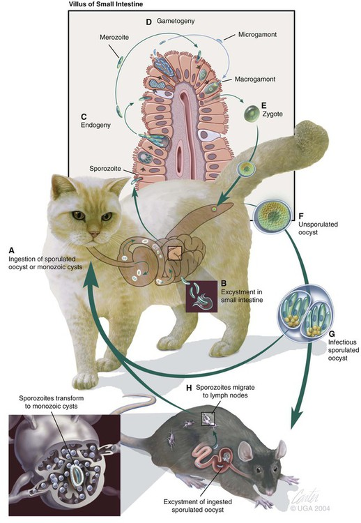
Cystoisospora (genus), of family sarcocystidae of order Eimerida.
intermediate host: dormozoite/hypnozoite, DH: extraintestinal stage, Endogeny = schizogony/merogony. IP/PH: dormozoite/hypnozoite
Life cycle of Isospora felis/CYSTOISOSPORA FELIS (new name), which is typical of the Isospora spp.
A) Ingestion: The host ingests sporulated oocysts or monozoic tissue cysts from contaminated food or water.
B) Excystation: In the gastrointestinal tract, the oocysts release sporozoites.
C) Sporozoite Stage: Sporozoites invade the epithelial cells of the intestine. Asexual reproduction happens (endodyogony - replicating once to make 2 daughter cells)
D) Merogony (Schizogony): Inside host cells, sporozoites replicate asexually in several rounds to produce multiple merozoites.
E) Gametogony: Some merozoites differentiate into male and female gametes, leading to sexual reproduction in the intestine. With the formation of a zygote.
F) Oocyst Formation: The fertilized gametes develop into unsporulated oocysts that are shed in feces.
G) Sporulation: The oocyst matures into an infectious sporulated oocyst, which can be ingested by the definitive or intermediate host.
H) Intermediate Host: excysted sporozoites migrate to tissues and form cysts.
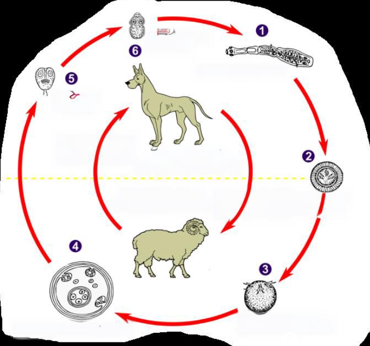
This is the life cycle of?
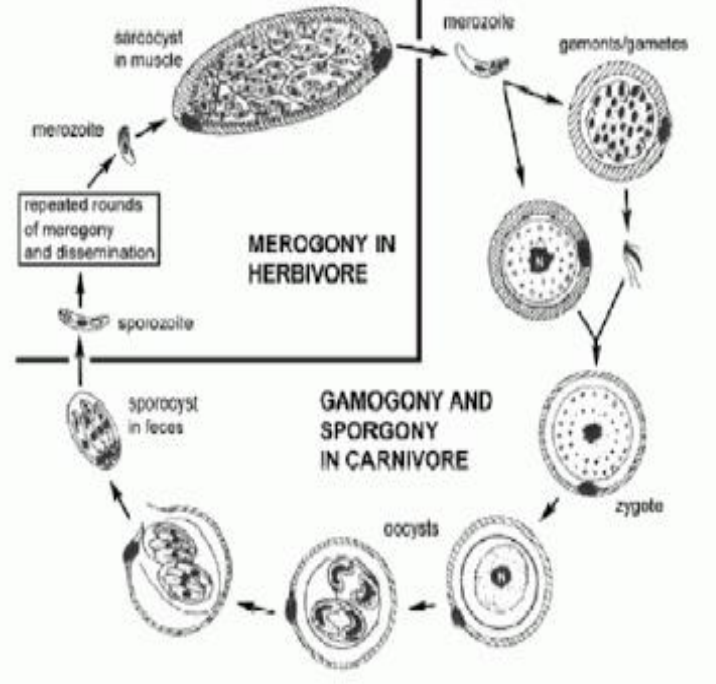
Genus: sarcocystis, of family sarcocystidae, of order Eimerida.
The life cycle of Sarcocystis species is heteroxenous, meaning it involves different hosts for its different life stages.
Definitive Host: such as carnivores including dogs and humans, harbors the sexual stages of the parasite, where gamogony (sexual reproduction) and sporogony (formation of oocysts) occur in the small intestine.
Intermediate Hosts: The intermediate hosts (which include herbivores, omnivores, and birds) play a crucial role in the life cycle as the parasite undergoes asexual reproduction, known as merogony.
In these hosts, the sporozoites that are ingested from oocysts excyst (are released) in the small intestine and invade the cells. They replicate asexually, generating meronts (by merogony) in endothelial of blood capillaries and after 3 rounds, subsequently forming cysts filled with bradyzoites, the slow-growing stage of the parasite → sarcocyst in muscle.
Transmission: When a carnivore consumes the infected tissues of the intermediate host, it can become infected with Sarcocystis again, thus continuing the life cycle.
this is the life cycle of?
life cycle of S. Bovicans
Definitive Host (Dog)
Ingestion: The dog ingests tissue cysts containing bradyzoites from infected intermediate hosts (such as cattle).
Gametogony: In the dog's intestines, the bradyzoites invade epithelial cells and develop into gametes.
Sporulation: Oocysts are formed, which then sporulate within the dog's intestine.
Excretion: Sporulated oocysts are excreted in the feces.
Intermediate Host (Cow)
Ingestion: The cow ingests sporulated oocysts from contaminated food or water.
Sporozoite Release: The oocysts release sporozoites in the cow's intestines, which then invade endothelial cells.
Schizogony/merogony: Sporozoites undergo multiple rounds of nuclear division to form schizonts/meronts in the cow's tissues.
Cyst Formation: Schizonts/meronts release merozoites, which will develop into cysts containing bradyzoites within the muscles and other tissues of the cow.
The cycle continues when the definitive host ingests these cysts, and the process begins anew.

This is the life cycle of?
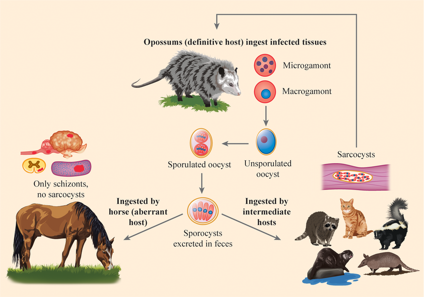
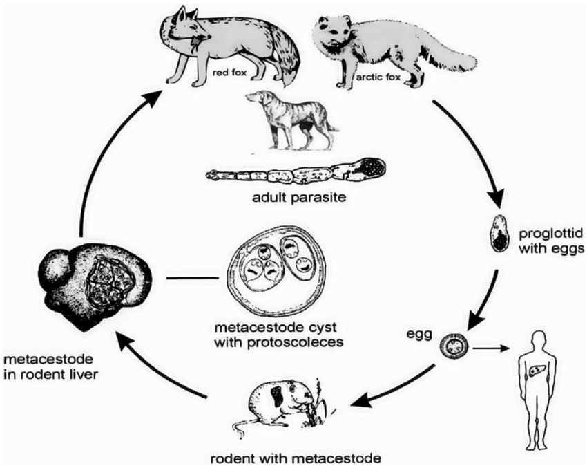
Life Cycle of Sarcocystis neurona.
FH: opposums (sexual reproduction - gamogony and sporulation)
IH: raccons, armadillos, skunks, cats - asexual reproduction and sarcocysts forms)
Aberrant host/dead-end host - horse (no transmission from these hosts).
Definitive Host (DH):
The opossum ingests tissue cysts containing bradyzoites from infected intermediate hosts.
nside the opossum, the parasite undergoes sexual reproduction, forming unsporulated oocysts.
Sporulation:
The unsporulated oocysts develop into sporulated oocysts within the opossum's intestines.
Excretion:
The sporulated oocysts are excreted in the feces of the opossum.
Intermediate Hosts (IH):
such as raccoons, armadillos, skunks, and felines ingest the sporulated oocysts from the environment.
Within the intermediate hosts, sporozoites are released → intestine → blood → merogony (in host cells) → merozoites produced → multiple rounds → migrate to muscle → forming bradyzoites → forms sarcocysts in the muscle tissues.
Aberrant Host (EQ - Horse):
Horses ingest sporocysts from the environment. In horses, only schizonts form, and no sarcocysts are produced.
Disease in Horses: The schizonts cause neurological disease in horses, known as Equine Protozoal Myeloencephalitis (EPM).
This is the life cycle of?
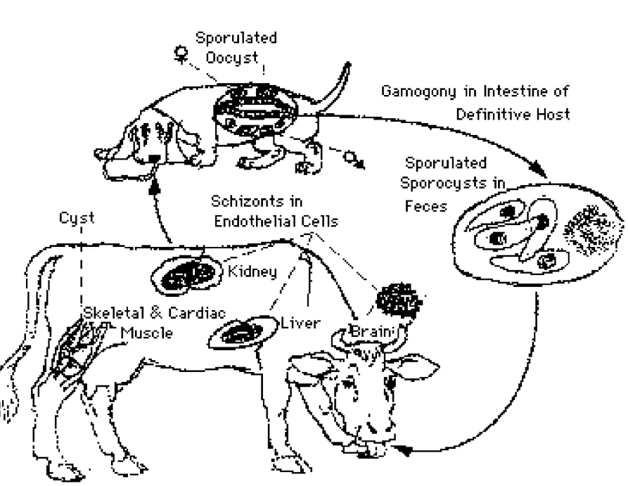
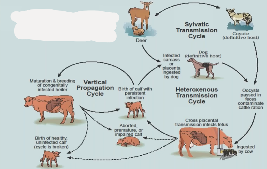
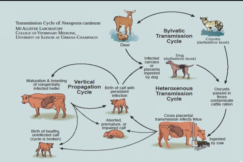
Lifecycle of Neospora caninum.
FH: carnivores, dogs, coyote, gray wolfs. (sexual reproduction, oocysts)
IH: mammals, cattle, sheep, goat etc. (asexual, tachyzoites, bradyzoites + tissue cysts)
Definitive Host (Dog):
Dogs ingest tissue cysts from infected intermediate hosts (e.g., cattle).
intestine: sexual reproduction of parasite → oocyst → excreted in the feces → contaminates.
Intermediate Hosts (Cattle):
Cattle ingest sporulated oocysts from contaminated feed, water, or soil.
Sporozoites are released and invade tissues, leading to the formation of tachyzoites.
Tachyzoites → damage → converts later into tissue cysts with bradyzoites, in nervous tissue + muscle.
Vertical Transmission in Cattle:
Infected cows can transmit the parasite to their fetus via the placenta.
This vertical transmission can result in aborted, premature, or weak calves.
Wildlife Hosts (Sylvatic Cycle):
Deer and other wild animals can also act as intermediate hosts, maintaining the parasite's presence in the environment.
This is the life cycle of?
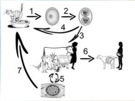
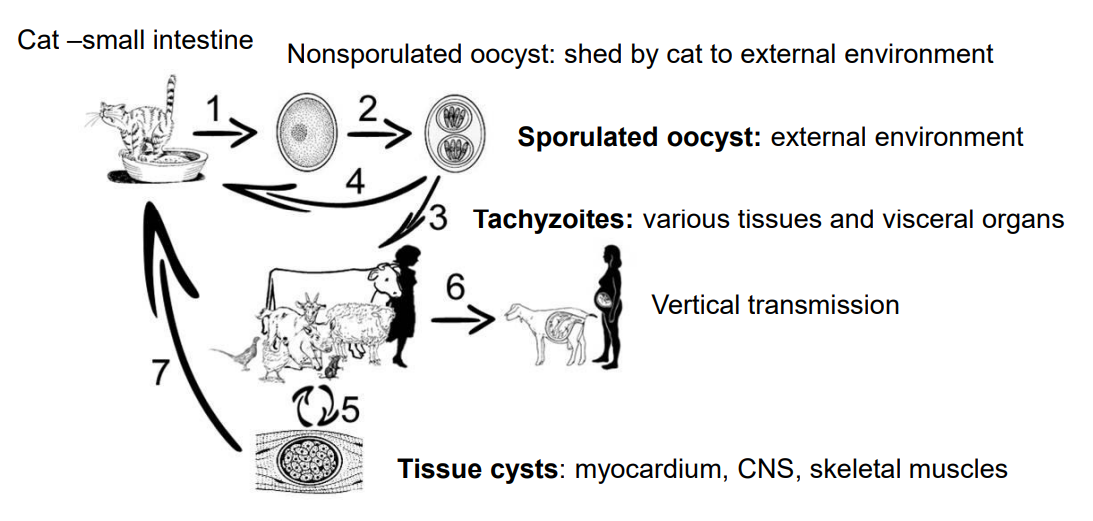
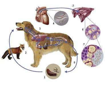
Toxoplasma gondii
Definitive Hosts (Cats and Wild Felids):
In the intestines of cats and wild felids → schizogony (asexual reproduction) and gamogony (sexual reproduction).
This results in the production of nonsporulated oocysts, which are shed in the feces into the environment.
Sporulation:
Once in the environment, these oocysts undergo a process called sporulation, becoming sporulated oocysts, which are infectious.
Intermediate Hosts (Mammals and Birds):
ingest sporulated oocysts, the oocysts release tachyzoites.
Tachyzoites rapidly invade various tissues, forming structures called pseudocysts.
Vertical (Transplacental) Transmission: (nr. 6 in picture)
Tachyzoites can also be transmitted from an infected mother to her fetus through the placenta, leading to congenital infection.
Tissue Cysts (Bradyzoites) (nr. 5 in the picture).
In the tissues of intermediate hosts, tachyzoites eventually transform into bradyzoites, which form tissue cysts.
These cysts can reside in muscles, the myocardium (heart muscle), and the central nervous system (CNS). → can be eaten by cat/wild felids → life cycle continues. (nr. 7 in picture).
This is the life cycle of?
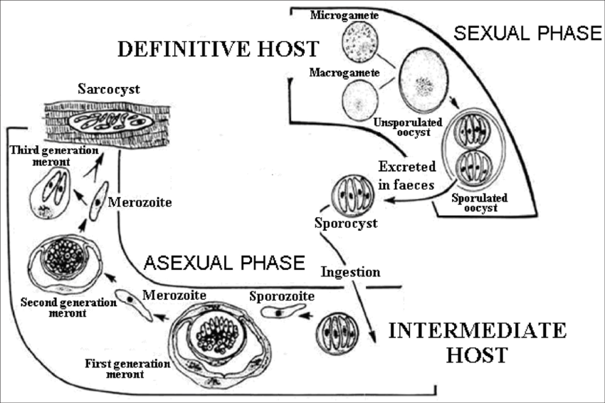
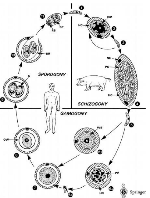
Sarcocystis spp.
FH: humans, mammals (gamogony - sexual, sporogony)
IH: ex. pigs (schizogony/merogony - asexual)
1. Schizogony: Intermediate Host (e.g., pig):
Endothelial Cells: The parasite undergoes rapid multiplication (2x) via endopolygony, where multiple rounds of nuclear division occur within a single host cell.
Leukocytes and Muscle Cells: Here, the parasite multiplies slowly via endodyogony, where two daughter cells form inside the parent cell.
Young Cysts (Metrocytes): These develop within muscle tissues during the slow multiplication phase.
Mature Cysts: Over time, these young cysts mature and contain bradyzoites, which are the infective forms for the definitive host.
2. Gamogony: Definitive Host (e.g., dog, cat, human):
Lamina Propria of the Small Intestine: When the definitive host ingests tissue cysts from the intermediate host, the bradyzoites are released and invade the intestinal cells.
The parasite differentiates into male and female gametes. These gametes fuse to form zygotes, completing the sexual reproduction phase.
3. Sporogony: Definitive Host (e.g., dog, cat, human):
Small Intestine: The zygotes develop into oocysts within the small intestine. This process is known as endogenous sporulation.
Excretion: oocyst → sporulated with sporozoites → excreted in the feces → infect new intermediate host.
This is the life cycle of?
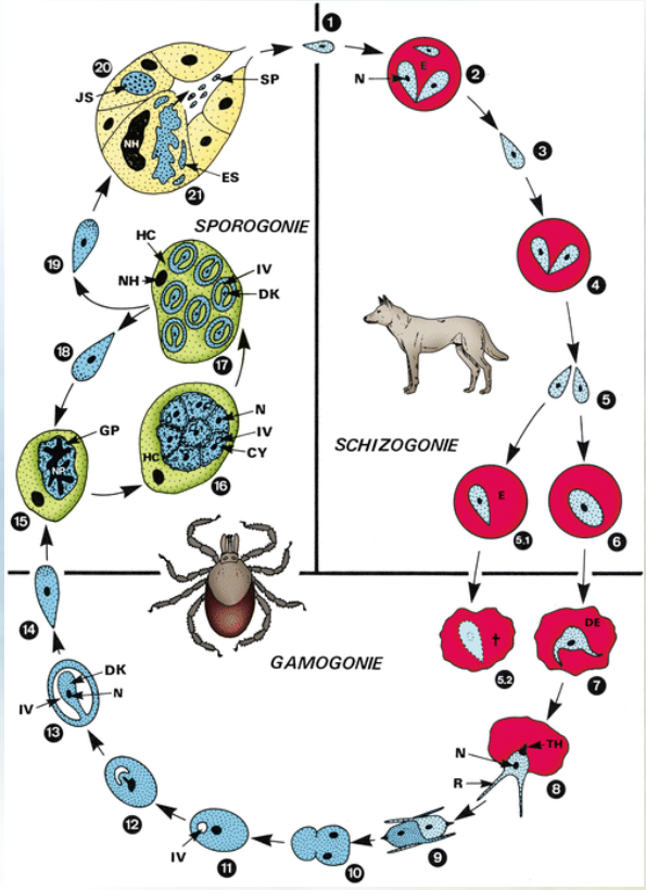
Theileria (genus) of the family Theileriidae.
FH: tick, all theileria species use ixodid (hard) ticks. (Gamogony-sexual, sporogony)
IH: mammals (merogony - asexual, binary fission, trophozoites/piroplasms)
Species: T. parva, annulata, orientalis, equi, ovis etc.
Common genera of ticks: Ixodes spp. Rhipicephalus spp.
Life Cycle of Theileria
Tick Feeding and Sporozoite Release: cycle starts when tick feeds on mammalian host ex. cattle. Tick releases sporozoites into the host bloodstream.
Sporozoite Attachment and Entry: sporozoites will attach to host lymphocytes, entering them through a zippering mechanism.
Transformation and Schizont/meront Formation: schizonts/meronts are formed inside the lymphoblast (mature vesion of lymphocyte), these mature later.
Proliferation of Schizont/meront-Infected Cells: the infected lymphoblasts proliferates.
Merozoite Release: Schizonts/meronts eventually differentiate into merozoites, which are then released when the host cell ruptures.
Piroplasms in Red Blood Cells: merozoites infect RBCs → forming piroplasms (intracellular parasites).
Tick Infection: when another tick feeds on the infected host → ingestion of the piroplasms
larvae and nymphs acquire the infection: will get infection through feeding or from infected mother (trans-stadial transmission).
gametes → zygote → kinete → sporogony → infected nymphal and adult ticks.
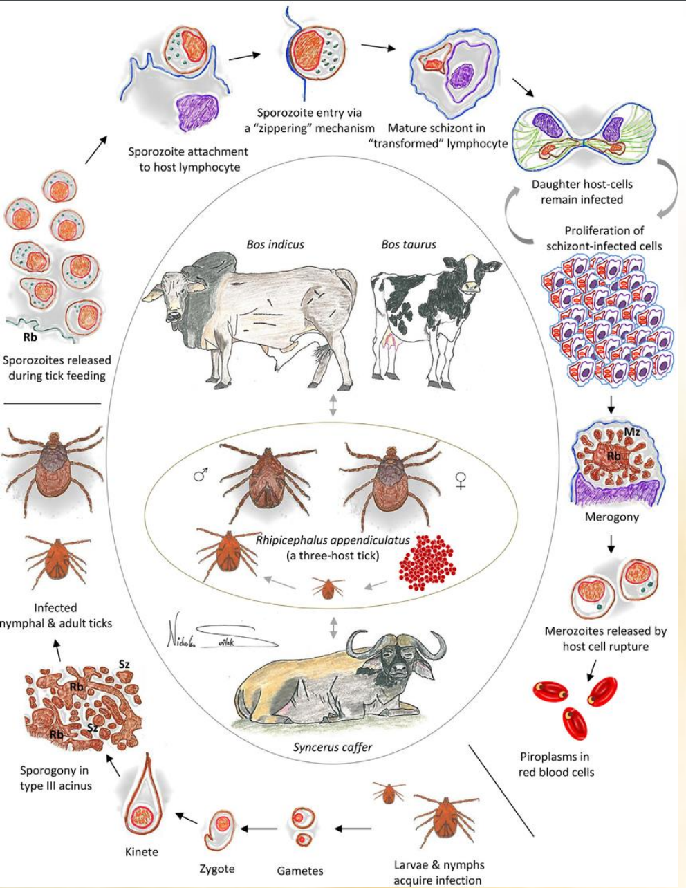
this is the life cycle of?
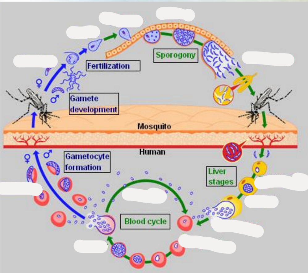
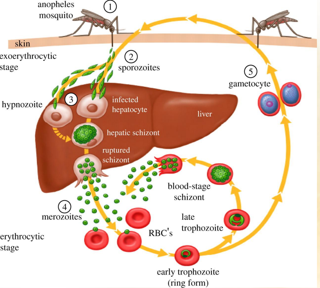
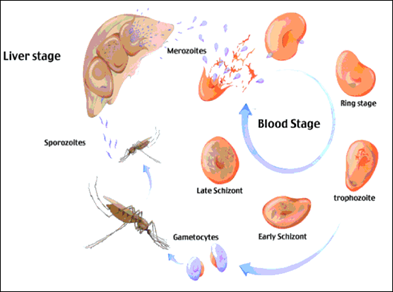
Cycle of Malaria parasite. (plasmodium malariae)
invasive forms: sporozoites and merozoites.
asexual stages: liver schizonts, blood schizonts.
sexual development: gametocytes and mosquito stages, where the gametocytes → gametes → zygote → ookinetes → oocysts.
Mosquito Bite: An infected mosquito injects sporozoites into a human.
Liver Stage: Sporozoites travel to the liver, becoming trophozoites, infect liver cells, and develop into schizonts, which release merozoites.
Blood Stage: Merozoites infect red blood cells, develop into trophozoites, mature into schizonts, and release more merozoites. Some merozoites develop into gametocytes.
Mosquito Uptake: Another mosquito bites the infected human, ingesting gametocytes.
Mosquito Development: Gametocytes develop into gametes, fertilize to form a zygote, which becomes an ookinete, then an oocyst. Oocysts produce sporozoites.
Completion: Sporozoites migrate to the mosquito's salivary glands, ready to infect another human, continuing the cycle.
this is the life cycle of?
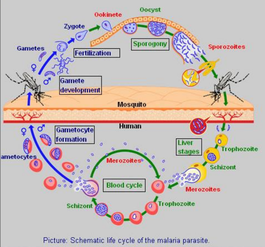
Plasmodium (genus), of family plasmodidae of order Haemosporida.
Mosquito Bite: An infected mosquito injects sporozoites into a human.
Liver Stage: Sporozoites travel to the liver, becoming trophozoites, infect liver cells, and develop into schizonts, which release merozoites.
Blood Stage: Merozoites infect red blood cells, develop into trophozoites, mature into schizonts, and release more merozoites. Some merozoites develop into gametocytes.
Mosquito Uptake: Another mosquito bites the infected human, ingesting gametocytes.
Mosquito Development: Gametocytes develop into gametes, fertilize to form a zygote, which becomes an ookinete, then an oocyst. Oocysts produce sporozoites.
Completion: Sporozoites migrate to the mosquito's salivary glands, ready to infect another human, continuing the cycle.
which parasite has this life cycle?
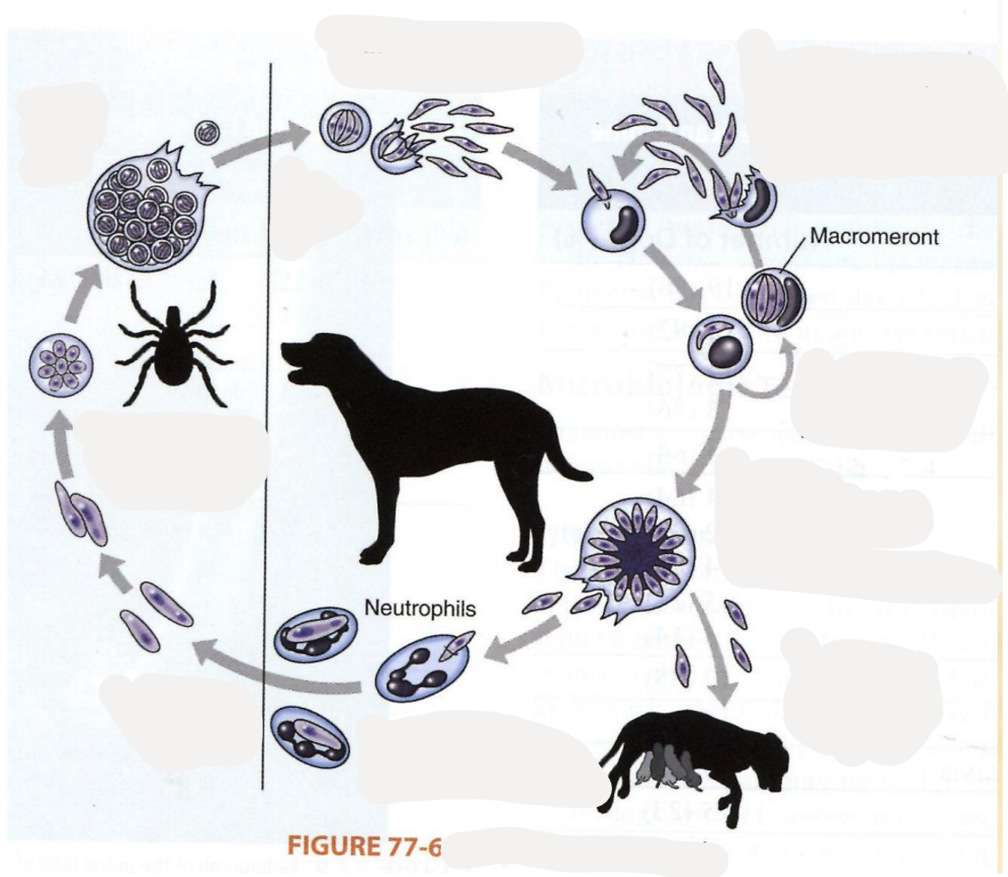
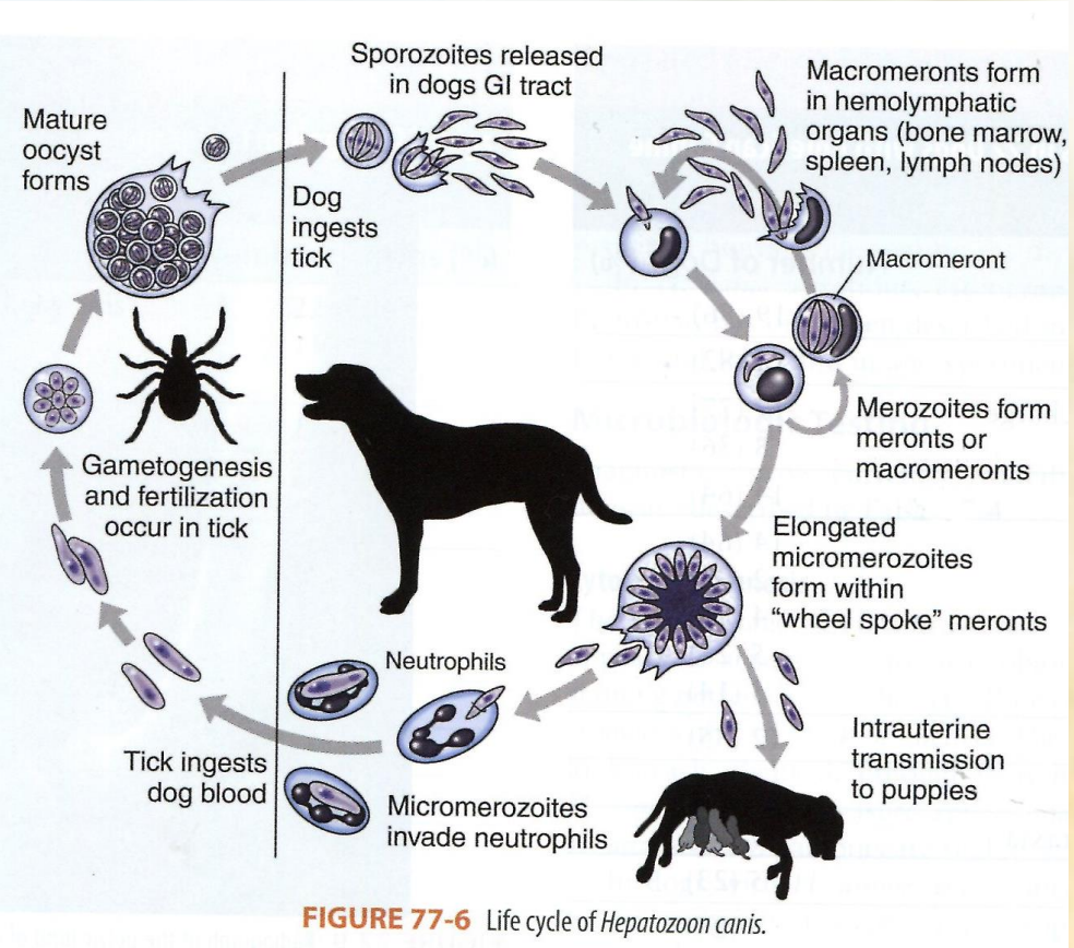
Hepatozoon canis (species of genus hepatozoon) of family hepatozoidae, order aleleida.
asexual reproduction in ca, fe (IH)
sexual reproduction: sporogony in tick (vector) DH.
Trans-stage transmission.
Dog ingests tick containing mature oocyst.
sporozoite release: oocyst release sporozoites in the dog`s GIT.
Macromeronts formation: sporozoites travel to the hemolymphatic organs like bone marrow, spleen, lymph nodes, forming macromeronts.
merozoite production from the macromeronts.
micromerozoites formation
neutrophil invasion: the micromerozoties invade the neutrophils in the dog`s blood.
tick feeding: a tick ingest the infected blood, taking up the micromerozoites.
gametogenesis + fertilization happen in the tick → mature oocyst formation.
cycle continuation - tick carries mature oocyst → infect another dog.
Describe life cycle of ——-?
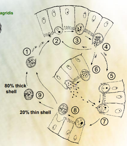
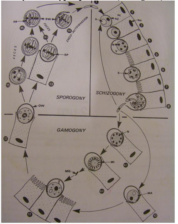
cryptosporidium
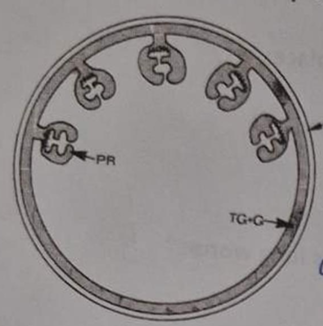
Schizogony:
Host ingest oocyst, with 4 sporozoites.
Sporozoites released in small intestine
Attach (not penetrate) to microvilli.
Multiply and form schizont 🡪 division 🡪 8 merozoites
One more round of schizogony. (total of 2 rounds of schizogony)
Gametogony:
Formation of multinucleated microgamonts and uninucleated macrogamonts
microgamont develop into 16 non-flagellated microgametes that fuse with the macrogamete to form 🡪 zygote
Sporogony:
the zygote can develop into 2 types of oocyst:
80% develop into thick walled oocyst. Pass to the environment with feces.
20% develop into thin- walled oocyst and release their sporozoites in the intestine causing autoinfection.
write/draw life cycle ——? (picture was shown, then describe species, life cycle)
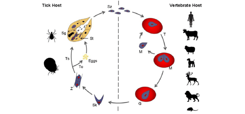
babesia spp. parasite causing babesiosis in mammals, involving a tick vector for transmission.
Sexual Cycle in Tick Host
tick eats gametocytes during blood meal on mammalian host.
gametocytes → male + female gametes in gut of tick.
zygote develops (gamogony) → meiosis of zygote, → kinete develops
2 options further:
Trans-ovarial transmission: in female tick, kinete will penetrate through ovaries and the eggs, thus when female lays eggs, larvae of tick hatch and are infected with babesia → continues in saliva.
trans-stage transmission: kinete penetrating several tissues, multiplicates, then invading salivary glands → sporogony happens → sporoblast is formed → sporozoites, ready to infect next vicitim. “larva taking blood from infected dog”.
Asexual Cycle in Mammalian Host
Sporozoites invade the red blood cells (RBCs) of host, when tick takes a blood meal.
Sporozoites develop into trophozoites.
Trophozoites divide by binary fission, producing merozoites.
Merozoites proliferate into new trophozoites and new merozoites.
Certain merozoites differentiate into male and female gametocytes within the RBCs.
Gametocytes are acquired by ticks during their feeding process.
Sporogony: tick`s salivary glands
Gamogony: Tick's gut. (intestine)
Schizogony/Merogony: Erythrocytes (red blood cells) of the mammalian host. (IH).
Life cycle of ——-? Describe steps.
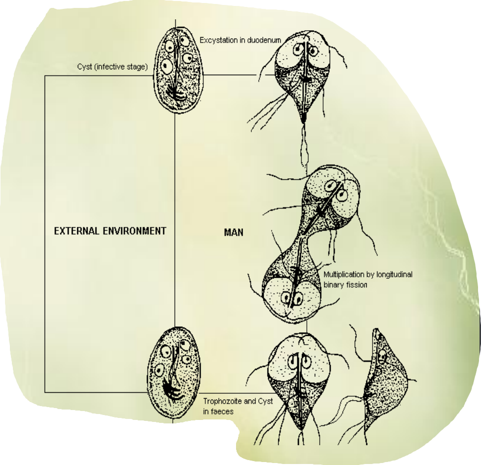
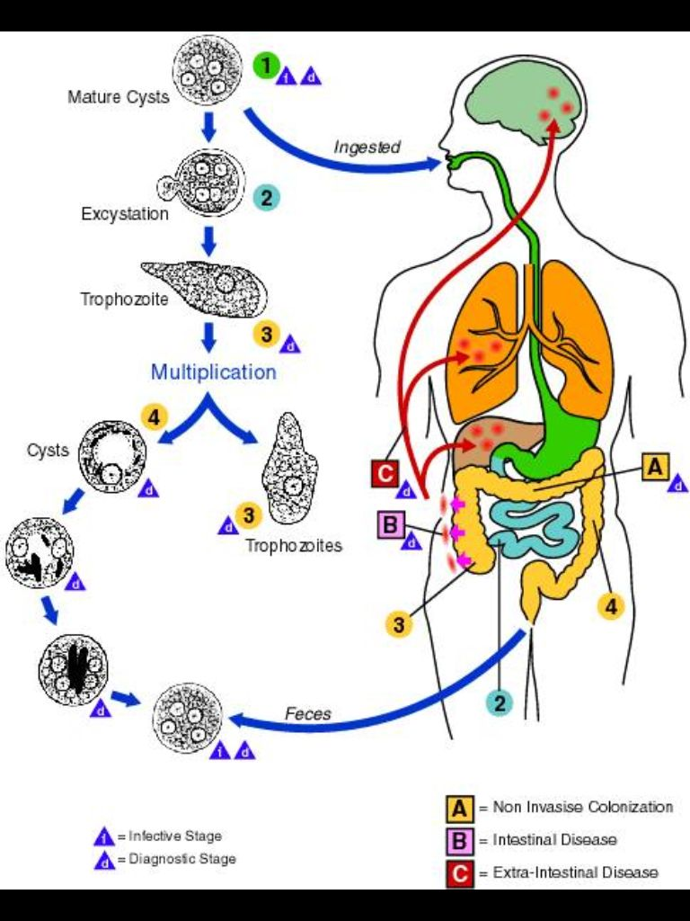
Entamoeba spp.
Ingestion of mature cysts (person eats from contaminated food/water)
excystation (once ingested, cyst travels to SI, where excystation happens, trophozoites are released (active + feeding stage).
Trophozoites → Large intestine → multiply by binary fission, some relain in lumen of intestine, while others invade intestinal mucosa → intestinal disease.
encystation (some trophozoites encyst, forming new cyst → feces → infect new host.
2 forms can develop:
forma minuta (chronic, latent, 8-20 micrometers, going to environment as cyst, 4 nuclei)
forma magna (10-60micrometer, invasive, no cysts, necrosis, abscess → 2nd infection)
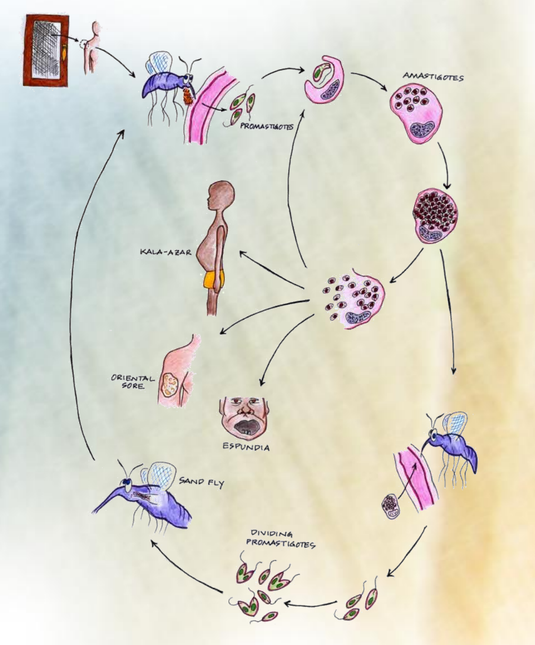
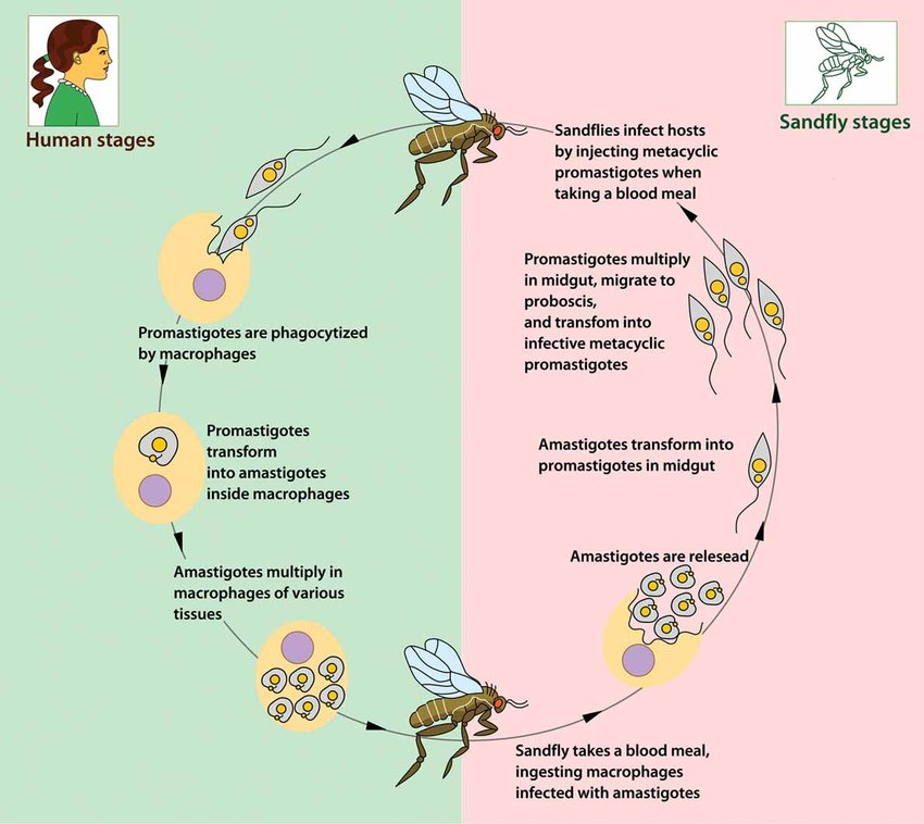
Describe life cycle of Leishmania.
Insect: vector (phlebotomus/Lutzomyia)
promastigote in insect - lepomonad form (injects promastigote stage into skin)
The promastigotes are then phagocytized by macrophages into blood
promastigotes → amastigotes inside the macrophages
amastigotes multiply by binary fission
sandfly takes a blood meal, ingesting macrophages infected with amastigotes
amastigotes → released → transform into promastigotes in midgut → multiply and transform into infective metacylic promastigotes
sandflies infect hosts with these.
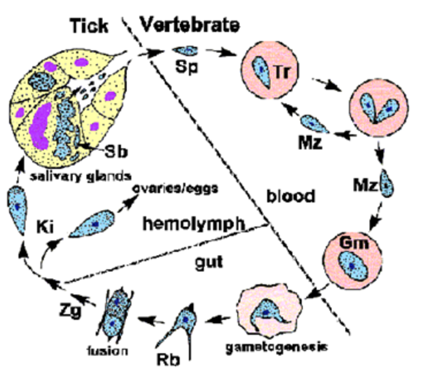
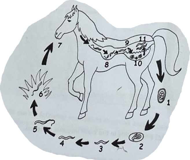
What is the life cycle of babesia canis?
Transmission: A tick carrying sporozoites in its saliva bites a dog, injecting the sporozoites into the bloodstream.
Entry into RBC: Sporozoites enter red blood cells (RBCs) and undergo schizogony (asexual reproduction). (schizogony in the vertebrae)
Trophozoites: Inside the RBCs, sporozoites level up into trophozoites, which multiply by binary fission. → RBC rupture → releasing merozoites into the blood stream → gametocytes
Gamogony in Tick: When a tick feeds on an infected dog, it ingests gametocytes
gametes→ zygote formed → kinete
Sporogony: Zygotes form and undergo sporogony in the tick's salivary glands, producing sporozoites.
can be trans-stage (by salivary) or trans-ovarial (by eggs/ovaries)
Transmission to New Host
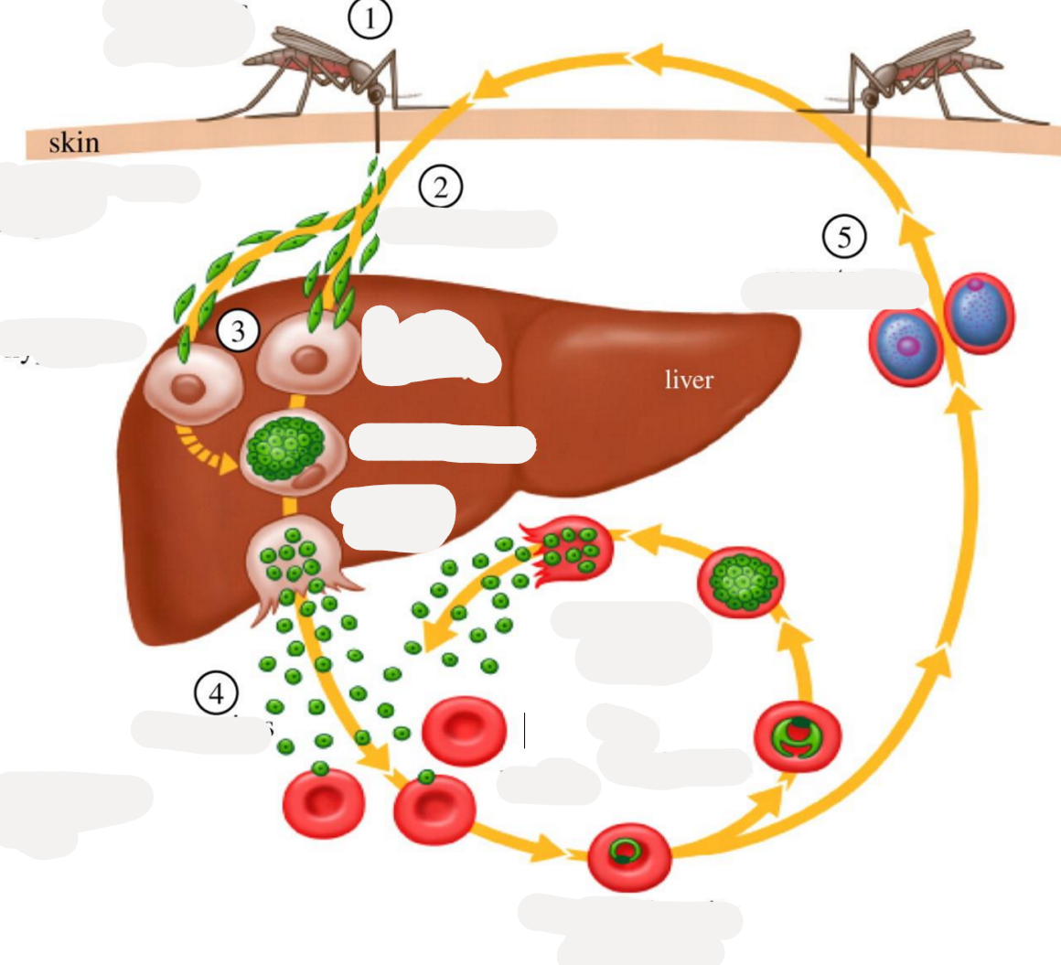
the life cycle of plasmodium.
Sporozoites are injected into the host through the bite of an infected mosquito.
They enter and infect liver cells, developing into schizonts.
Schizonts release merozoites into the bloodstream.
Erythrocytic phase:
Merozoites invade red blood cells (RBCs) and develop into trophozoites.
Trophozoites undergo asexual reproduction (schizogony) resulting in more merozoites which rupture RBCs to enter new ones.
Gamogony (in mosquito):
Some merozoites develop into microgamonts (male) and macrogamonts (female).
If a mosquito takes a blood meal, it ingests both types of gamonts.
Microgametes fertilize macrogametes to form a zygote (ookinete).
Sporogony (in mosquito):
The zygote develops into an oocyst in the mosquito's stomach, which eventually ruptures to release thousands of sporozoites.
These sporozoites migrate to the mosquito's salivary glands.
Transmission:
When the mosquito bites another vertebrate host, sporozoites are injected into the bloodstream, continuing the cycle.
Life cycle include: one sexual and three asexual
vector of human plasmodium: anopheles (also vector of malaria)
Gametogony P. vivax happens in: in the intestinal cells of mosquitoes
neosporium caninum - what are its lifecycle and pathogenesis?
Lifecycle of Neospora caninum.
Indirect, FH - carnivores (gamogony), IH: cattle (merogony, tissue cysts). Sporogony in environment.
Definitive Host (Dog):
Dogs ingest tissue cysts from infected intermediate hosts (e.g., cattle).
intestine: sexual reproduction of parasite → oocyst → excreted in the feces → contaminates.
Sporulation - environment.
Intermediate Hosts (Cattle):
Cattle ingest sporulated oocysts from contaminated feed, water, or soil.
Sporozoites are released and invade tissues, leading to the formation of tachyzoites.
Tachyzoites → damage → converts later into tissue cysts with bradyzoites, in nervous tissue + muscle.
Vertical Transmission in Cattle:
Infected cows can transmit the parasite to their fetus via the placenta.
This vertical transmission can result in aborted, premature, or weak calves.
Wildlife Hosts (Sylvatic Cycle):
Deer and other wild animals can also act as intermediate hosts, maintaining the parasite's presence in the environment.

Life cycle of?
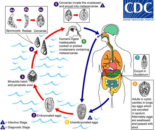
Fasciola Hepatica - Trematode (Fasciolidae)
Indirect, location: bile duct, liver
IH: water snail (lymnaeidae)
FH: Ru, eq, man
Process:
adults release unembryonated eggs → biliary ducts of FH → feces → water → embryonation
eggs release miracidia → infects freshwater snail (IH)
miracidia → sporocysts → rediae → cercariae
cercariae → into water → finds water plants to encyst to form metacercaria
Metacercaria (infective stage) → FH
After FH eats it → excyst in duodenum → intestine → liver parenchyma, develops into adults within bile ducts.
Life cycle of?
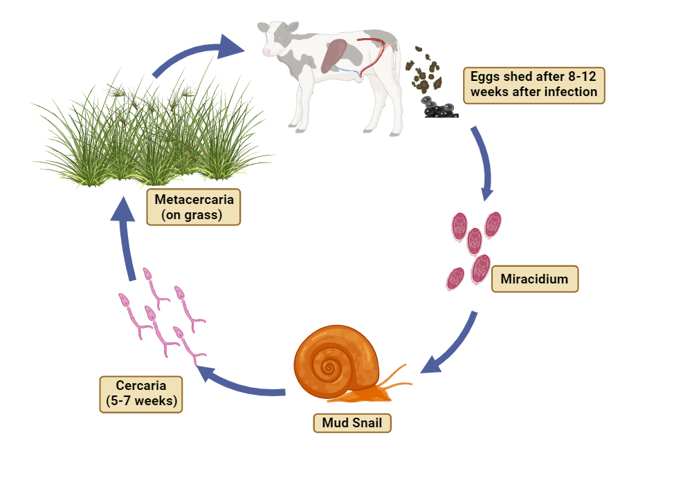
Paramphistomum cervi (trematode, Paramphistomidae)
Indirect lC.
FH: mammals, birds, reptiles (commonly in RU).
IH: mud snail (Planorbidae),
Location: juvenile - SI, adults in rumen.
Infective stage: Metacercariae.
Process:
Eggs with feces, eggs develop an embryo (miracidium) → hatches from egg and seeks snail
inside snail, transforms into sporocyst → redia → cercaria → leaves and become metacercariae on vegetation.
FH is infected by ingestion. Young flukes gets through intestine → rumen, matures.
Life cycle of?
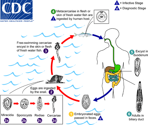
Opisthorchis tenuicollis (felineus)/Clonorchis sinensis (similar) - Opistorchiidae, trematode
FH: fish-eating mammals.
IH: 1st: water snail (Bithynia), 2nd: fish (bream, barbel).
Location: liver, bile ducts.
Infective: Metacercariae with fish tissue.
Process:
eggs shed with feces → snails ingest eggs
in snail: cercariae develops → fish infected by the free swimming cercariae ecysting in skin/flesh
metacercariae are formed within fish in muscle
FH ingest fish, migrates to bile ducts, matures.
life cycle of?
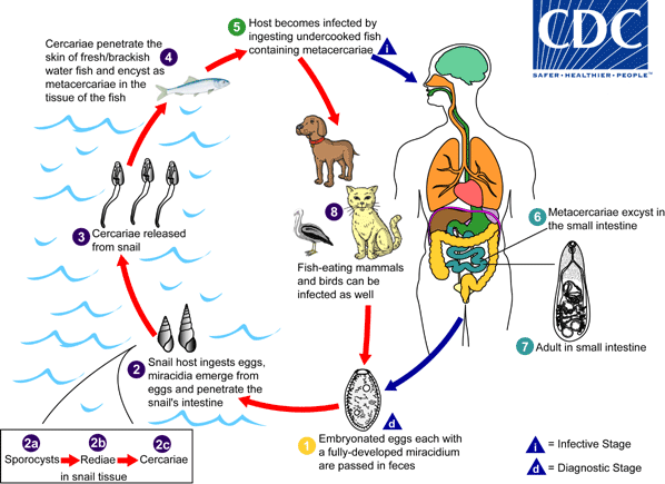
Metagonimus yokogawai (opistorchiidae, trematode)
FH: fish-eating mammals, humans.
Location: Small intestine.
IH: 1st water snails (semiliscospira) and 2nd: sweet fish (asia, balkans).
Process:
embryonated eggs each with miracidium is passed down with feces
snail ingest eggs → develops into sporocyst → redia → cercariae
cercariae is released from snail → infects fish
in fish: encyst to metacercariae
FH infected by undercooked fish with metacercariae → excyst in SI.
H. Heterophyes - similar, but it releases toxins in FH → severe catarrhal inflammation (abd. pain, diarrhea)
life cycle of?
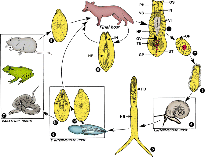
Alaria spp. (alata/canis/mercianae) - Diplostomidae
FH: dogs, cats, red foxes, humans. Found in Small intestine.
IH: 1st: Water snails (planorbis spp.), 2nd: frog, toads.
Paratenic host: frogs, snakes, mice, birds.
infective: Mesocercariae.
Process:
Adults live in SI → eggs passed in feces
hatch in water, releasing miracidia
1st IH (snails) will eat the miracidia, and it will develop into cercariae (Furcocercariae) → released
2nd IH (tadpole) will eat furcocercariae → tadpole develops into frog → mesocercariae (special larval stage)
FH becomes infected after eating frog/snake. Mesocercariae will migrate through lungs, trachea → adults in SI.
life cycle of?
Paragonimus westermani (paragonimidae, Trematode)
Lung fluke. Indirect cycle.
FH: dogs/cats/humans.
IH: 1st: water snails (melania), 2nd: crabs, crayfish.
Location: Lungs.
infective: Metacercariae.
Process:
eggs released and hatch in water, releasing miracidia
infect snail → develops into cercariae
infects crabs/crayfish → metacercariae develops
FH infected by ingestion of raw or undercooked 2nd IH.
life cycle of?
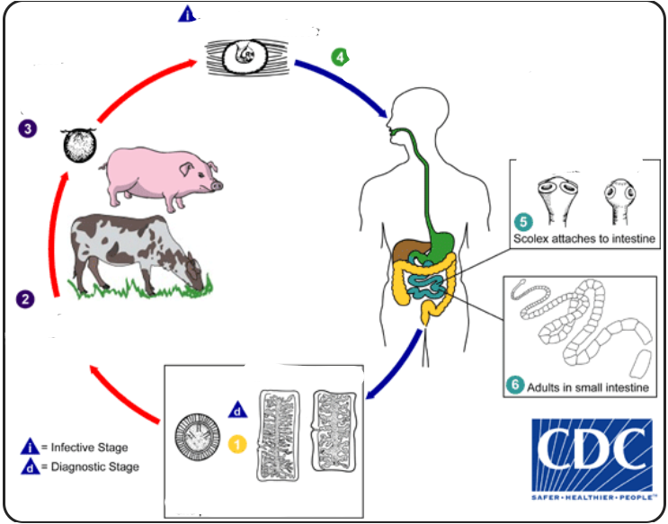
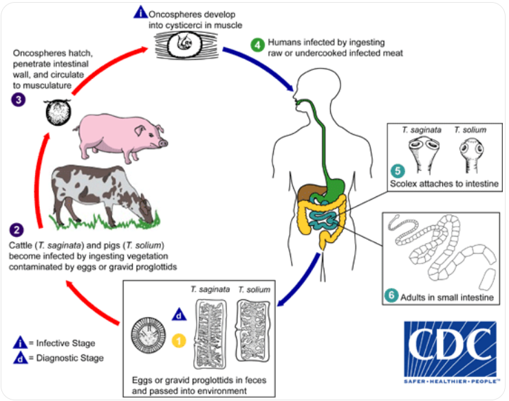
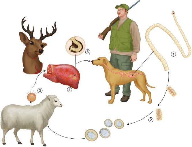
Taenia solium (cyclophyllidae, cestode)
FH: human, IH: pig,
Location: muscle in IH, SI in man.
Infective stage: cysticercus.
Process: eggs/gravid proglottids are shed with feces → pigs ingest eggs → oncospheres hatch in their intestine → cysticercus develop in their muscle → humans get infected by eating undercooked pork. In the human intestine, cysticercus develops into adults.
Taenia saginata
FH: human, IH: cattle. Location: muscle in IH, SI in human. Infective: cysticercus bovis. Process: same principle as t. solium
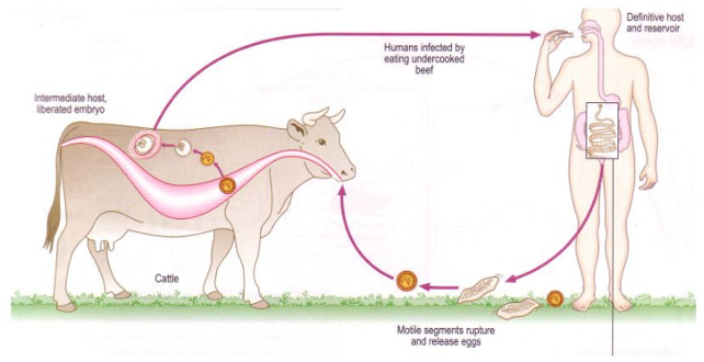
Life cycle of? what is the infective stage called?
Taenia saginata + cysticercus bovis (cyclophyllidea, cestode)
Taenia - Indirect LC, located in SI
IH: Cattle (T. saginata)
FH: humans
Process:
Eggs → feces from FH
Enters IH (cattle) → oncospheres released from egg → muscles
In muscle, develops into cysticercus bovis
FH eats infected uncooked/raw meat
parasite → SI → matures → released gravid proglottids with eggs by feces
life cycle of?
Taenia hydatigena (Cyclophyllidea, cestode)
FH: dog/fox, IH: livestock, Location: abdominal cavity, liver.
Infective stage: cysticercus tenuicollis.
Process: Adults reside in SI of FH, gravid proglottids are shed with eggs in feces → ingested by IH → oncospheres hatch in the intestine of IH → cysticercus tenuicollis develop in the tissues (bladderworm with single invaginated scolex) → FH ingest the infected organs of IH → Tapeworm matures.
life cycle of?
Taenia pisiformis (cyclo, cestode)
FH: dogs/fox,
IH: Lagomorphs (rabbits)
location: serosa, abdominal cavity.
Infective stage: Cysticercus pisiformis.
Process: adults in SI of FH → releases gravid proglottids in feces → lagomorphs ingest the eggs → oncospheres hatch → serosa, abdominal cavity → cysticercus pisiformis develops → FH ingest the infected tissues → SI → matures.
life cycle of?
Taenia ovis (cyclo, cestode)
FH: dog/fox, IH: sheep, Location: small muscles. infective stage: Cysticercus ovis.
Process: adults in SI of FH → releases gravid proglottids in feces → sheep ingest eggs → oncosphere hatch in intestine → small muscles → cysticercus ovis develops → FH ingest infected tissues → tapeworm matures in SI.
describe infective stage and process of taenia serialis.
Cyclophyllidae, cestode.
FH: dog/fox, IH: lagomorphs (rabbits)
location: tissue
infective stage: coenurus serialis.
Process: adults in SI of FH → gravid proglottids are shed with feces → Lagomorphs ingest the eggs → oncospheres hatch in intestine → tissues → coenurus serialis develops → FH becomes infected → scolices evaginate + attach to SI, Matures.
describe process and name infective stage of taenia taeniaeformis.
Cyclophyllidae, Cestode.
FH: CATS, IH: rodents/rabbits. Location: liver.
Infective stage: Strobilocercus/cysticercus fasciolaris.
Process: Adults in SI of FH → releases gravid proglottids in feces → IH eats eggs → oncospheres hatch → liver → cysticercus fasciolaris develops → FH becomes infected, attaches to SI and matures.
life cycle of?
Echinococcus multilocularis, cyclophyllidae, cestode
FH: carnivores - fox, dog, wild canids, racoon, cat.
IH: rodents/humans.
Location: liver, lung but also brain, bone marrow, muscle, lymph nodes and heart.
Infective: alveococcus.
Process: Adults in SI of FH → releases eggs in feces → IH ingest eggs → oncosphere hatch in intestine → migrates → alveococcus (alveolar cyst) develops → FH becomes infected when ingesting the organs → SI and matures.
Life cycle of? What is the name of metacestode stage?
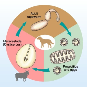
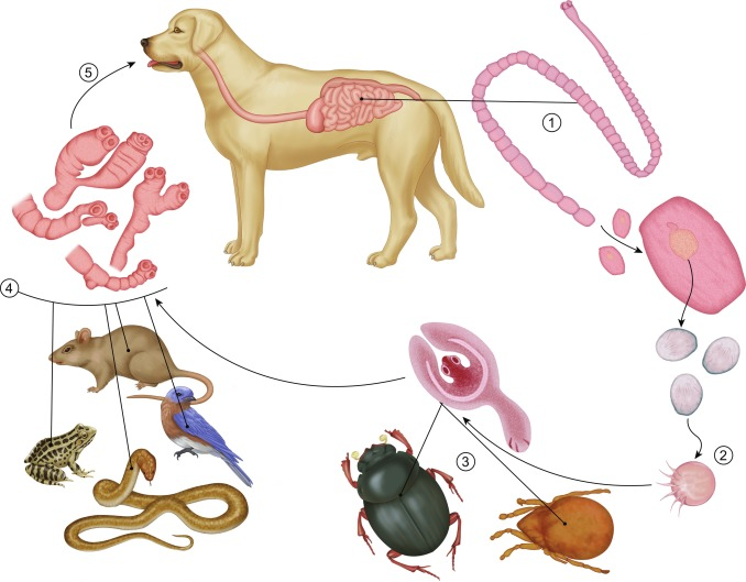
Dipylidium caninum (dipylidiidae, Cestode)
Indirect LC
FH: dog, cat, wild canids, fox, humans
Location: SI
IH: fleas, louse
Metacestode stage: Cysticercoid (infective stage)
Process:
Adult in FH → SI → gravid proglottids → sheds with eggs by feces
fleas ingest the eggs → egg develop into cysticercoid (infective larvae)
Flea larvae mature into adult → cysticercoid become infective
FH ingest flea → larva is released from flea → adult in intestine

life cycle of?
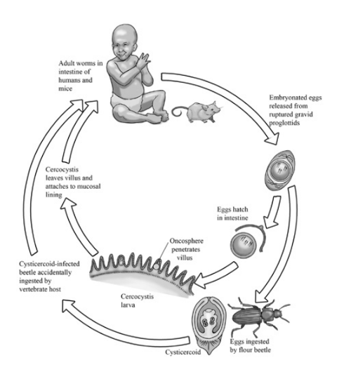
Hymenolepsis nana (of family Hymenolepididae) - cestode, cyclophyllidae
FH: Rodents, humans
IH: can complete cycle without IH, but can also have beetles, flea, insects as IH
Location: SI
Process:
Direct cycle:
eggs ingested by humans
eggs release oncospheres → intestinal villi → develops into cysticercoids
cysticercoids → mature into adult → in SI → makes gravid proglottids → releasing eggs in feces
Indirect cycle (if IH is involved):
IH: eggs can be eated by arthropods like beetles or fleas → cysticercoids develops
humans get infected by eating these infected insects
life cycle of?
Strongyloididae (nematode)
Strongyloides westeri - horse
Strongyloides ransomi - pig
Strongyloides papillosus - ru
Strongyloides stercoralis - human
Strongyloides spp.
FH: depends on species - S. westeri (horse - foals), stercoralis (man, dog), papillosus (ru), ransomi (pig).
Location: SI of FH.
Males and females reproduce sexually in the environment.
Eggs - hatch in intestine of host.
2 forms of larvae - rhabditiform (L1, L2) and filariform (L3).
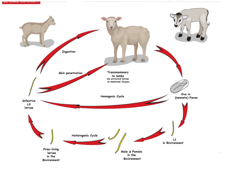
Infection through skin and oral. Direct LC.
Process: Eggs released from FH → releases L1 → develops into L3 → autoinfection or passed into feces where it develops into L3 in the environment or free living adults - produce more.
life cycle of?
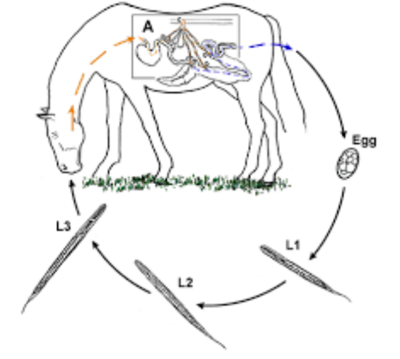
Strongylus spp. (strongylus vulgaris) - nematode
Direct LC, located in large intestine (Colon, cecum)
FH: horse
Process:
Eggs in feces, hatches in environment
Larva → L2 → L3
host ingest L3 larvae when grazing
Larvae → intestine → mucosa and molt into L4
L4 → small arteries → along endothelium to cranial mesenteric artery + branches
After few months → L5 and returns to intestinal wall
matures → eggs release with feces
Life cycle of?
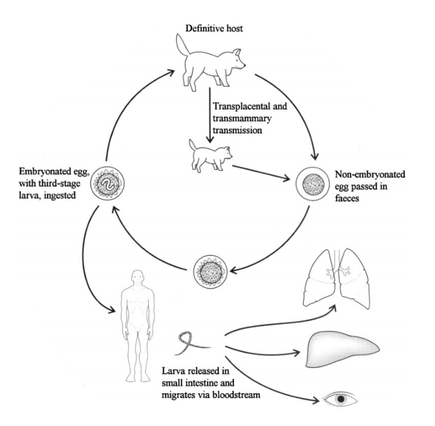
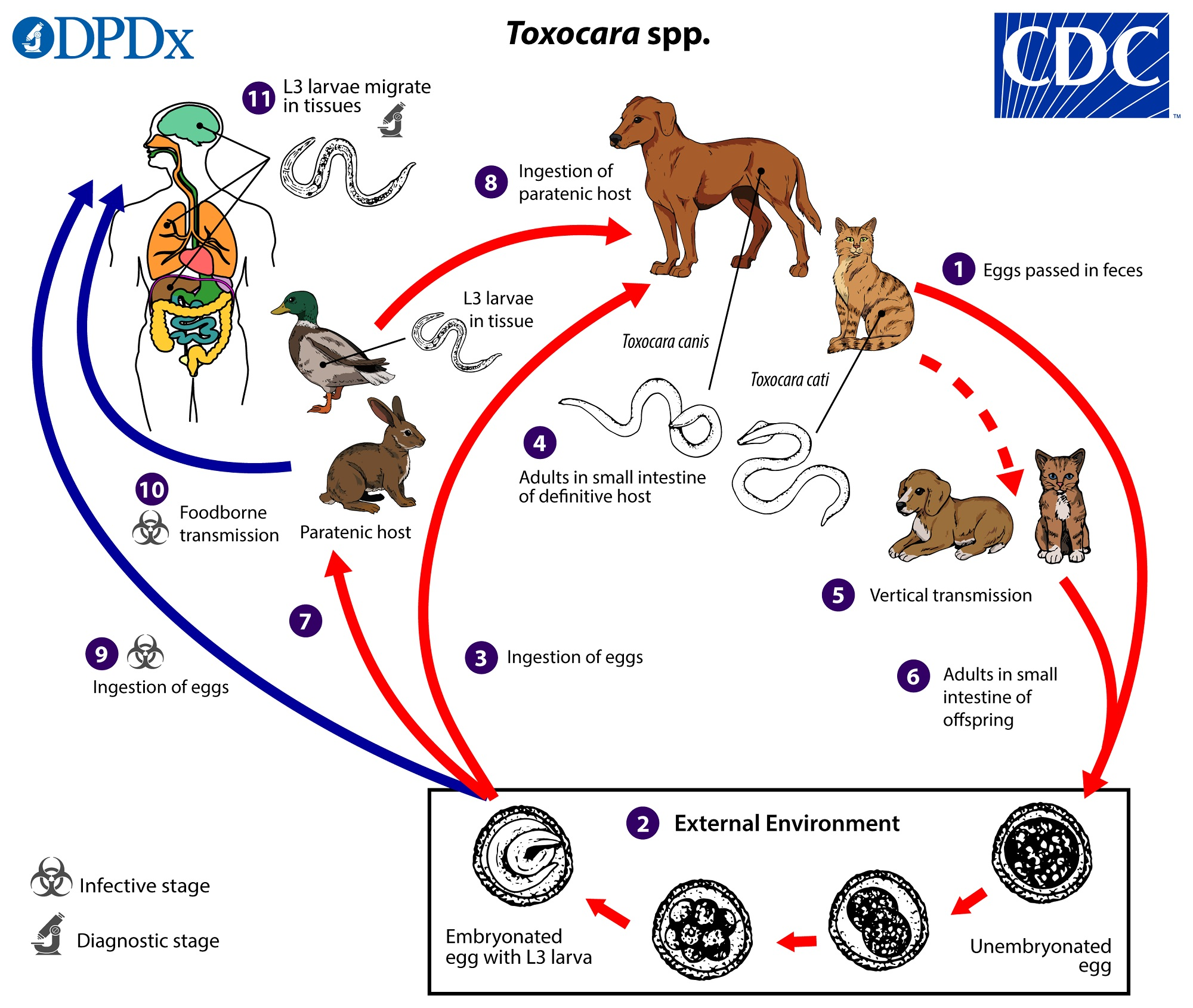
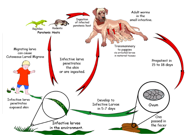
Toxocara canis (canis as there is dog in pic, Nematode)
direct LC, located in SI, diagnostic stage - unembryonated eggs in feces, infective - embryonated egg with L3
entero-hepato-pulmonal migration
FH: car (canis - dog, cat - cati, leonina - both), accidentally man
paratenic host: various small mammals, can be ingested by dogs (no development of parasite but remains infective)
Process:
eggs shed in feces of FH
eggs embryonate in environment with L3 larvae
Eggs ingested by host → can be FH (dog) or paratenic host (mouse, rabbit etc.)
In FH - larvae mature into adults in SI
vertical transmission: adults can be transmitted from mother to offspring
L3 larvae can get to other tissues → liver, lungs and brain
Life cycle of?
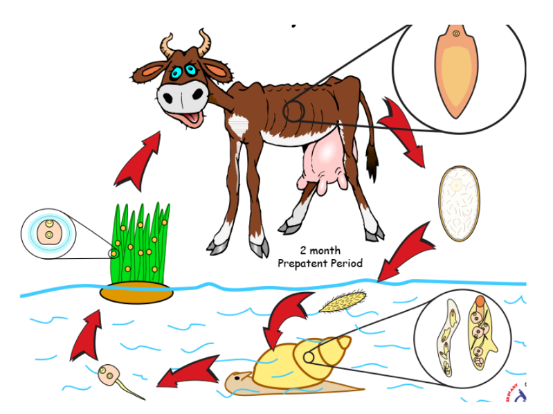
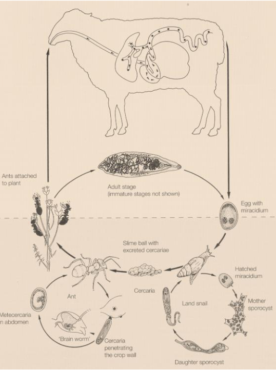
Dicrocoelium dendriticum (dicrocoelidae) - trematode
Indirect LC, located in Bile ducts, gall bladder
IH: 1st: land/terrestrial snail, 2nd: Ant
FH: reptiles, birds, mammals
infective: metacercariae
Process:
Eggs with miracidia in feces of FH (ru) → eggs eaten by 1st IH (snail)
miracidia → sporocyst → cercariae → respiratory chamber → shed from snail
ant eat slime ball with cercariae → intestine → metacercariae
FH eats ant, metacercariae excyst in SI. Worms migrate to bile duct and mature.
Life cycle of? What is the metacestode?
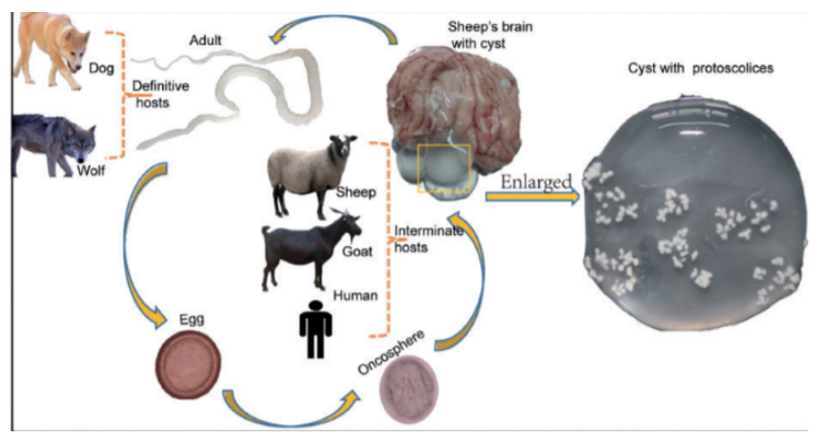
Taenia multiceps + coenurus cerebralis (Cyclophyllidae, cestode)
FH: Dog, fox, wild canids
IH/larvocyst: Sheep, cattle, goat, pig, horse, deer, camel (coenurus cerebralis)
Location: Brain, spinal cord
Process:
eggs shed in feces of infected FH
eggs are eaten by IH → oncospheres released in intestine → circulate in blood, until they find suitable organs → brain in this case (spinal cord, eyes also).
develops into coenurus cerebralis with protoscolices
FH becomes infected by ingesting tissue of an infected IH with the coenurus.
Protoscolices → attach to SI wall → adult cestodes in FH
Humans get infected after accidental ingestion of eggs in food/water.
coenurus: large fluid-filled bladder with invaginated scolices attached to wall.
Life cycle of?
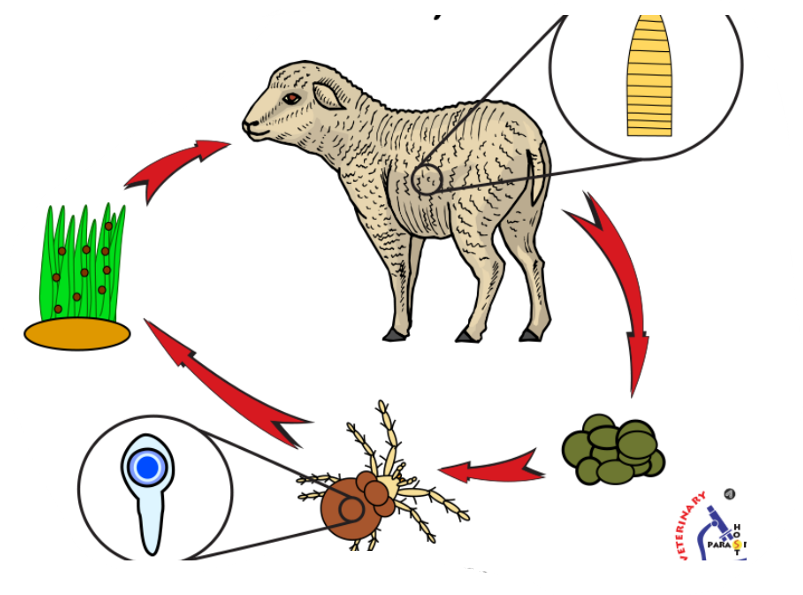
Moniezia spp. - M. expansa/M. benedeni (Cyclophyllidea, cestode)
in this case, it would be M. expansa as this is for small/young ru.
While benedeni is typ. found in medium-large ru
indirect LC, located in SI
IH: oribatid mites (Oribatidae)
FH: Ru
Process:
Eggs with oncosphere are released from FH → ingested by IH where they hatch into cysticercoid in the body cavity
FH ingest infected mite and the cysticercoid travel to SI where they mature into adults
Life cycle of?
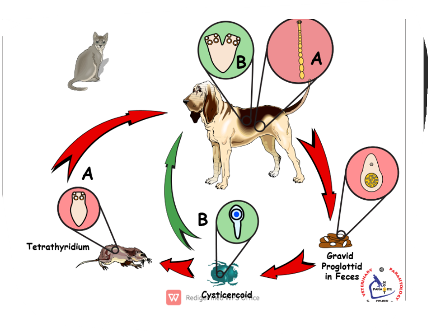
Mesocestoides Lineatus (cyclophyllidea, cestode)
Indirect LC, 2 IH, Located in SI
FH: dogs, cats, some wild canids
1st IH: arthropods, such as ants, beetles, oribatid mites etc. - cysticercoid develops here.
2nd IH: small vertebrates like reptiles, amphibians, birds and small mammals - Tetrahyridium.
Process:
1st IH ingests eggs with oncosphere/proglottid → develops into cysticercoid
2nd IH ingest infected arthropod with cysticercoid → tetrahyridium
FH ingest the IH → parasite travels to SI → matures → release gravid proglottids in the feces with many eggs.
life cycle of?
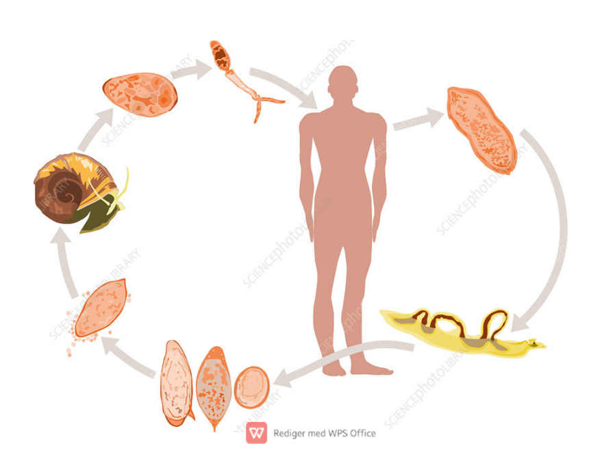
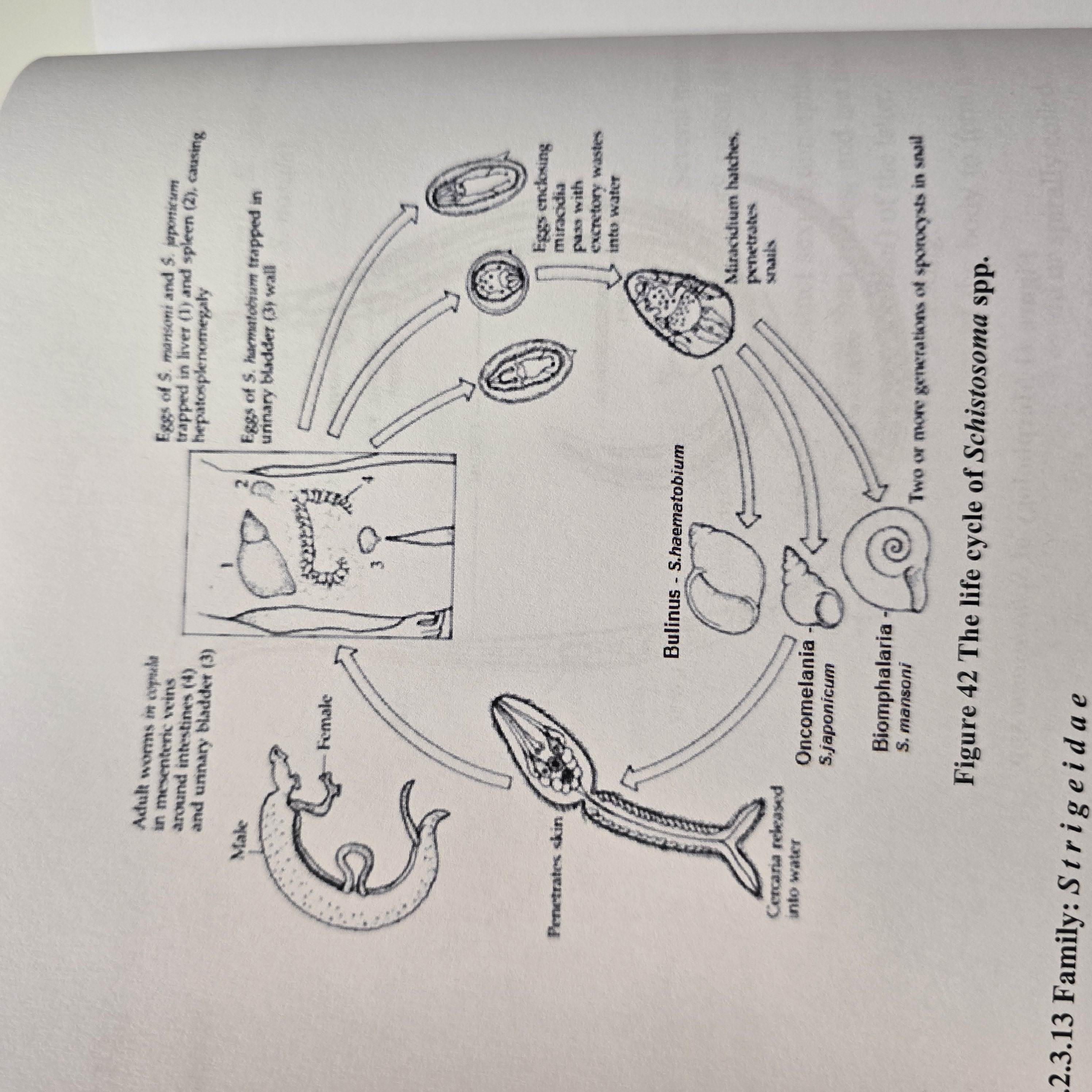
Schistosoma spp. (S. haematobium, Mansoni, japonicum) - trematode, fam. schistosomatidae.
Indirect LC, located in Blood vessels
IH: water snail
FH: man, other mammals (cattle, dog, cat, rodents, pigs, horses, goats)
process:
eggs hatch into miracidium → IH → pathogony → sporocysts → cercaria with forked tail (furcocercaria) → exits
FH gets infected by furcocercaria → looses their tail and become schistosomulae → circulation → matures.
Worms migrate to mesenteric or vesicular veins depending on species.
S. mansoni: mesenteric veins of LI
S. haematobium: veins of bladder
S. japonicum: mesenteric veins of SI
Embryonated eggs with miracidium released by urine/feces.
Life cycle belongs to? what is the infective stage?
Trichuris vulpis (trichuridae, trichuris species, nematode)
Direct LC, located in large intestine
FH: Ru, car, su, man (but here it is T. vulpis so FH is dog).
also have:
suis - pig
ovis - small ru
globulosa - ru
infective: egg with L1 larvae and diagnostic: eggs
Process:
Unembryonated eggs are shed by host`s feces
L1 larvae develop inside the egg in the environment
Host ingest embryonated eggs
eggs hatch and L1 are released in the SI → large intestine (All 4 moults - happens)
The larvae matures into adults within LI → release eggs.
Life cycle of?
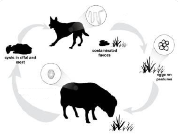
Echinococcus granulosus (also have E. multilocularis but not in this picture case though) - Cyclophyllidae, cestode
indirect LC, located in liver, lung (both), SI of FH
multilocularis: heart, brain, lymph nodes, SI of FH (primarily liver, but widely also)
IH: granulosus: RU (sheep, cattle, other livestock)
multilocularis: rodents
FH: dogs, wolves, fox, dingoes
multilocularis: fox, dog, wild canids, racoons, cats
Process:
IH ingest embryonated eggs → hatch in SI and release oncospheres
oncospheres → through blood or via lymph to liver, lungs or other tissues → develops into hydatid cyst
cyst grows and releases brood capsules with scolices
multilocularis → develops into a multiocular or alveolar cyst here (metacestode stage)
FH ingest infected tissues with cysts
Scolices attach to intestinal mucosa in SI → matures and eggs are released with feces
Life cycle of?
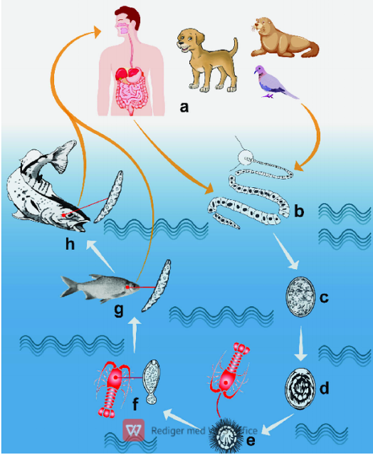
Diphyllobothrium latum (pseudophyllidea, cestode)
Indirect LC, located in SI
IH: 1st: water arthropod, 2nd: fish
FH: fish-eating mammals
infective stage: plerocercoid for FH, procercoid for fish
Process:
eggs embryonate in water → coracidium hatches from eggs taken up by 1st IH
inside 1st IH, coracidium → procercoid larvae
fish ingest infected arthropod and the procercoid → plerocercoid larvae in the muscle
Small infected fish can be then eaten by a bigger carnivorous fish
FH eats raw/undercooked fish → plerocercoid develops into immature worm and then into mature adult in the SI
Unembryonated eggs release from proglottids with feces
life cycle of?
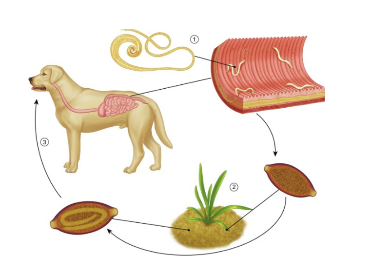
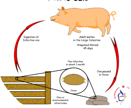
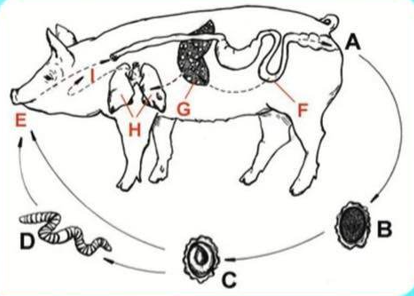
Ascaris Suum (Ascarididae, nematode)
Direct LC, located in SI
Hepato-pulmonary passage of larvae after hatching
Host: pig
Process:
Unembryonated eggs in feces
moults to L3 larvae (infective stage)
ingestion of infective egg by host
hatched larvae → invades intestinal mucosa → hepato-pulmonary migration (to lungs)
further maturation in lungs → swallowing of larvae → SI, develops into adult worms
life cycle of?
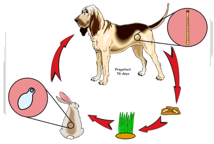
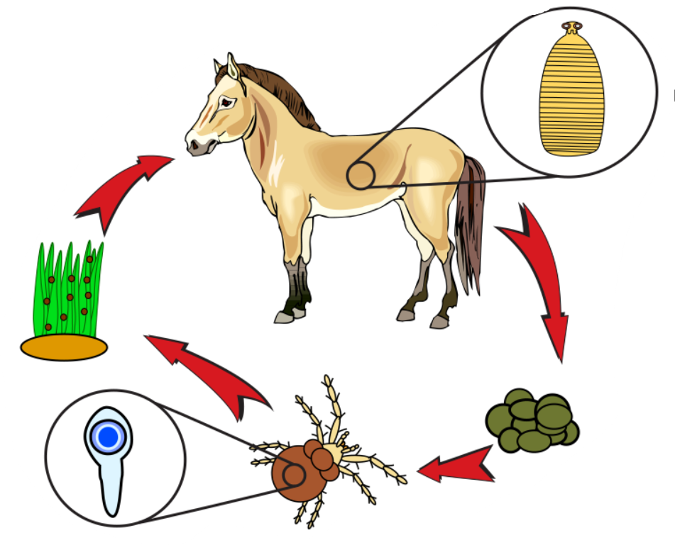
Anoplocephala spp. (Perfoliata + magna) - also paranoplocephala mamillana. (Cestode, Anoplocephalidae).
Difference is that perfoliata is in SI and LI of horse, cysticercoid in oribatid mites. While magna is in SI of horses, scolex is small, no lappets.
indirect LC
IH: oribatid mites
FH: horse
infective stage: cysticercoid
Process:
grazing horse ingest mite infected with cysticercoid
Cysticercoid develops to adult in intestine of horse
gravid proglottids and embryonated eggs pass in horse feces
eggs eated by oribatid mites → cysticercoid develops here
mites crawl on vegetation → eaten by horse
life cycle of?
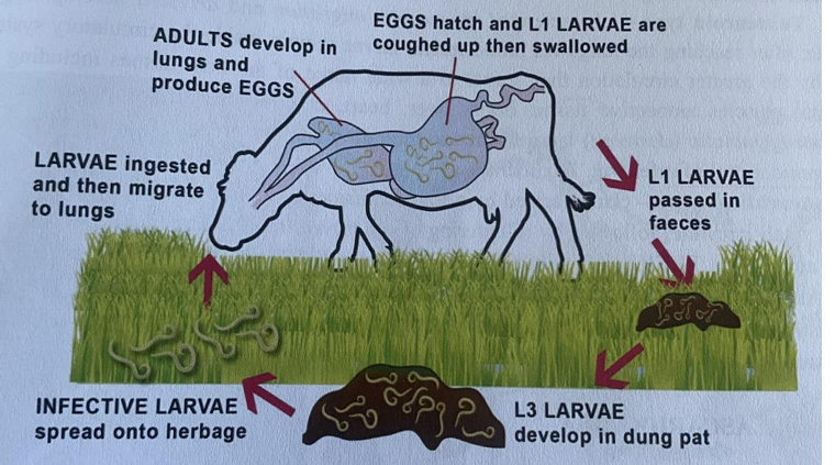
Dictyocaulus viviparus (dictyocaulidae, nematode)
FH:
viviparus (cattle)
Filaria (small ru)
arnfieldi (horse, donkeys)
Location: Trachea and bronchi
Direct LC, infective stage: L3
Process:
L1 larvae are passed in feces.
L1 → L2 → L3 ➔ FH ingest free L3 larvae
Penetrate intestinal walls and enter lymphatic vessels.
Reaches mesenteric lymph gland.
L3 → L4 ➔ migrates to the lungs.
Migrate through the parenchyma to the airways.
L4 → Adult ➔ Adult lays eggs in the bronchi
Eggs coughed up and swallowed. ➔ L1 hatch in the intestine
LC of?
Ancylostoma caninum/Tubaeforme (Ancylostomatidae, Nematode)
Direct LC
FH: caninum - car/man, tubaeforme - cat
Location: small intestine
Infective stage: L3 eggs
Infection is by: ingestion, per skin, transmammary + transplacental.
Process: adults in SI of FH → eggs passed in feces → eggs develop → L1 into soil → L3 develops → infection of FH → if by skin, then it will enter blood, lungs then coughed up and swallowed → SI.
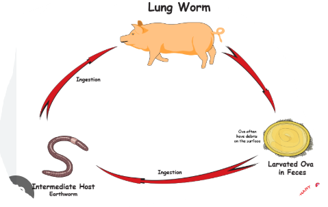
Describe LC of metastrongylus species (pudendotectus, apri, salmi, confusus)
Metastrongylidae - nematodes, lung worms.
FH: pigs (Pudendotectus, apri, salmi, confusus)
Location: respiratory system of pig (lungs)
Indirect LC, IH: earthworms
Infective stage: L3
Process:
Adults in bronchi of pigs → release eggs with L1 → coughed up → swallowed → feces → soil
The infected feces are ingested by IH → L3 develops here
Pigs will eat earthworms → migrate to lungs via the hepatic-portal system → develops into L5.
Explain LC of angiostrongylus (Vasorum).
Angiostrongylidae, nematode.
Indirect LC.
Host: dogs/fox
Location: pulmonary arteries and right side of heart.
IH: snails/slugs
Process:
adults in pulmonary arteries and heart → eggs excreted → IH ingest L1 → inside, they develop into L3 (infective)
FH eats IH → intestinal wall → lymph nodes → liver → heart → pulmonary arteries and mature.
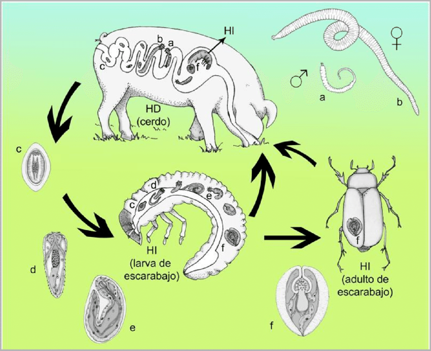
Explain LC of macracanthorhynchus hirudinaceus.
Of Acanthocephala.
Indirect LC
FH: pigs
Location: SI
IH: beetles, larvae
Infective stage: cystacanth larvae
Process:
adults in SI of pigs → releases Acanthor larvae eggs in feces
eggs are ingested by IH → develops into acanthella larvae → cystacanth larvae (infective)
FH ingest beetles (IH) → matures.
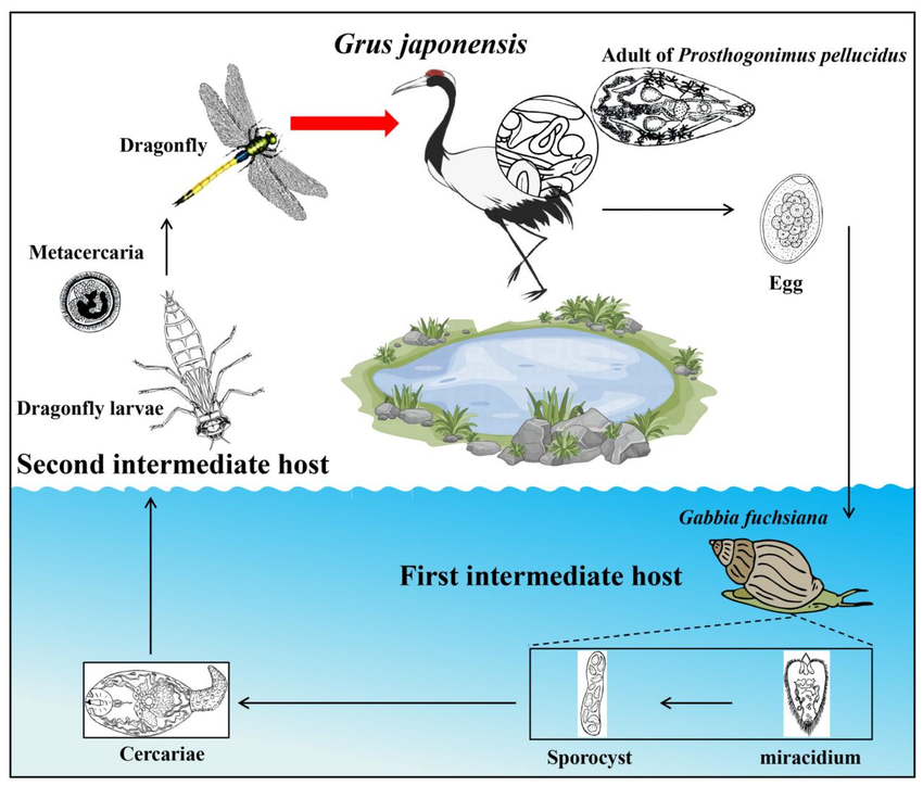
Explain LC of Prosthogonimus Macrorchis.
fam. Prosthogonimidae, within order Plagiorchiidae. Trematode.
Indirect LC
FH: birds
IH: 1st: snails, secondary: dragonflies.
Location: oviduct and bursa.
infective stage: Metacercariae (trematode typical).
Process:
Adults in bursa + oviduct in bird → eggs released with feces
snails ingest the eggs (miracidium → redia → cercariae)
2nd IH eats snails (encyst as metacercariae)
FH eats dragonflies → excyst in bird intestine → bursa or oviduct - matures.
LC of this?
cyathostomum genus
“small strongyles”, cycliocyclus
Nematode, from family Strongylidae.
Direct LC, located in Large intestine of horses
Process:
Adult cyathostomins live in LI → eggs pass in feces (unembryonated)
larva develops within each egg, then hatches
released larva develops → moults twice to infective L3 stage.
Infection of horse is by ingestion of these larvae → mucosal migration → moults to L4 in LI
Larvae migrates to lumen → moults to L5/adult and release eggs in feces
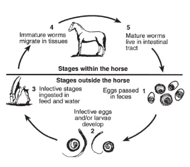
Infective stage and migration of parascaris Equorum?
Migration of strongylus vulgaris, equinus and edentatus?
Tidligere: P. equorum, strongylus spp. qs:
Infective stage of parascaris, migration: L3, larva within egg. Migration: after horse eats egg with L3 → hatches in SI → goes through intestinal wall → migrate via veins to the liver (where it molts to L4) → 2 weeks → migrates to heart and lungs → travels to bronchi → trachea, coughed up and then swallowed → mature to L5 and then adults in SI.
Migration of strongylus vulgaris, equinus, edentatus
S. vulgaris: L3 larvae penetrates the intestinal mucosa → L4 in the submucosa → arteries and migreate to cranial mesenteric artery → molt to L5 → migrate to intestinal wall, forming nodules in cecum and colon → adults.
S. equinus: L3 → wall of SI, cecum, colon → encapsulating in nodules → L4 leaves and goes to liver → migrates back to LI → molting to L5 during this return.
S. edentatus: L3 excyst in the SI → gut wall → molts to L4 → migrate to root of mesenteric artery → liver, lungs → returns to cecum wall and right ventral colon.