L8- AN2102 Abdominal Viscera
1/35
There's no tags or description
Looks like no tags are added yet.
Name | Mastery | Learn | Test | Matching | Spaced | Call with Kai |
|---|
No analytics yet
Send a link to your students to track their progress
36 Terms
What is the liver and where is it located?
The largest gland in the body (~1.5 kg), located in the right hypochondriac and epigastric regions, extending slightly into the left hypochondriac area.
What shape is the liver and what does its superior surface rest against?
Wedge-shaped; superior surface conforms to the diaphragm.
What are the main functions of the liver?
Produces bile, performs metabolic and detoxification processes, stores glycogen, vitamins, and iron.
What is the falciform ligament and what does it contain?
A double layer of peritoneum connecting the liver to the anterior abdominal wall and diaphragm
Contains the round ligament (ligamentum teres hepatis)—the remnant of the umbilical vein.
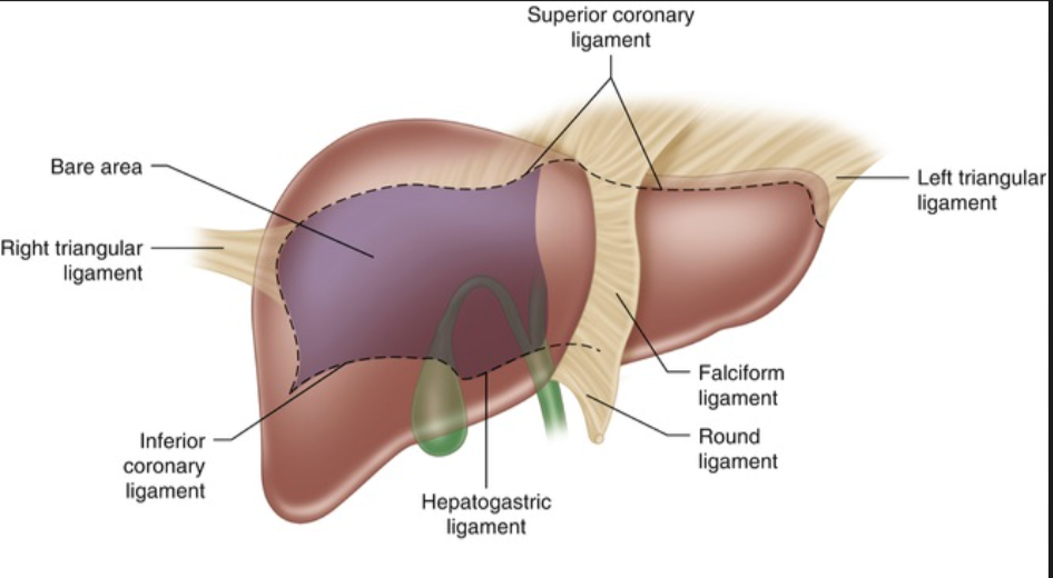
What are the coronary ligaments?
Peritoneal reflections anchoring the liver to the diaphragm; they border the bare area of the liver.
What forms the right and left triangular ligaments?
The lateral ends of the coronary ligaments; attach the liver to the diaphragm.
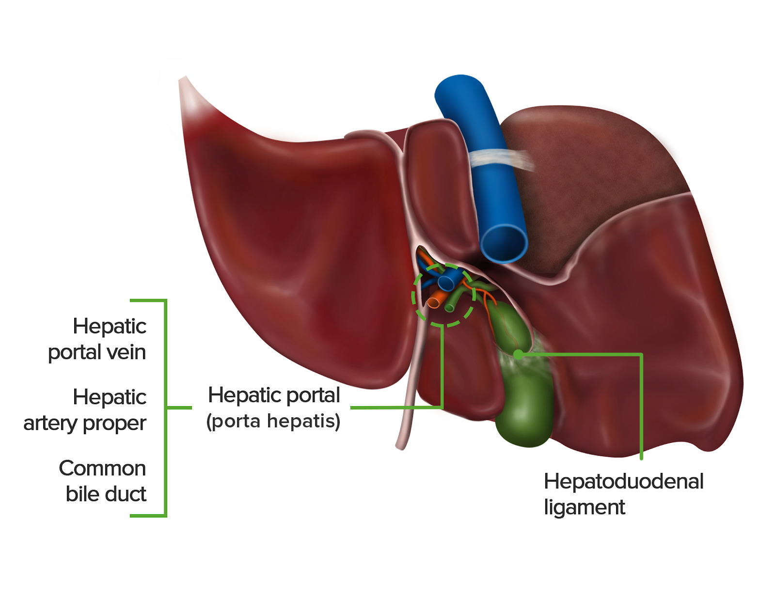
What is the lesser omentum and what does it connect?
A double layer of peritoneum connecting the liver to the stomach and first part of the duodenum.

What are the two parts of the lesser omentum?
Hepatogastric ligament and hepatoduodenal ligament.

Which ligament contains the Portal Triad?
The hepatoduodenal ligament.

What structures make up the Portal Triad?
Proper hepatic artery, portal vein, and common bile duct (A–V–D: artery, vein, duct from med ).
What divides the diaphragmatic surface into right and left anatomical lobes?

What marks the inferior border of the liver clinically?
It lies just below the right costal margin in the midclavicular line.

What structures form the “H-shaped” arrangement on the visceral surface?
Gallbladder fossa, IVC groove, fissure for round ligament, and fissure for ligamentum venosum.

What is the porta hepatis? (hepatic portal)
The hilum of the liver where the portal triad enters/exits.
Which lobes lie on the visceral surface?
Right lobe, left lobe, quadrate lobe, caudate lobe.
What is the bare area of the liver?
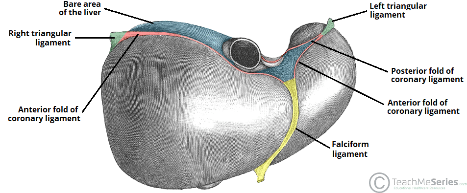
A posterior region without peritoneal covering; in direct contact with the diaphragm.
What is significant about functional lobes?
Each has its own hepatic artery, portal vein branch, and bile duct.
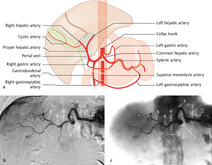
What artery supplies the liver?
Proper hepatic artery → divides into right and left hepatic arteries.
What is the function of the portal vein?
Brings nutrient-rich, oxygen-poor blood from the GI tract to the liver.
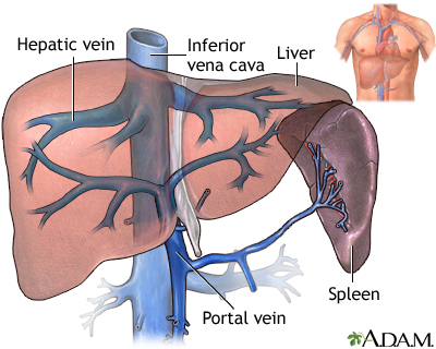
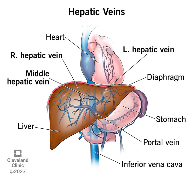
What veins drain the liver into the IVC?
The hepatic veins (usually 3 main ones).
Where is bile produced?
In hepatocytes.
What are bile canaliculi?
Tiny channels between hepatocytes that collect bile.
What do canaliculi drain into?
Intrahepatic bile ducts located in portal triads.
What ducts leave the liver at the hilum?
The right and left hepatic ducts.
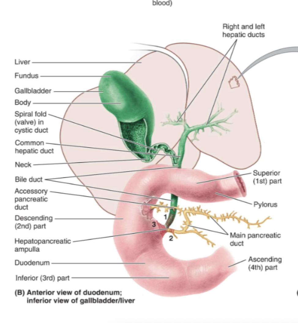
What duct is formed when the right and left hepatic ducts join?
The common hepatic duct.
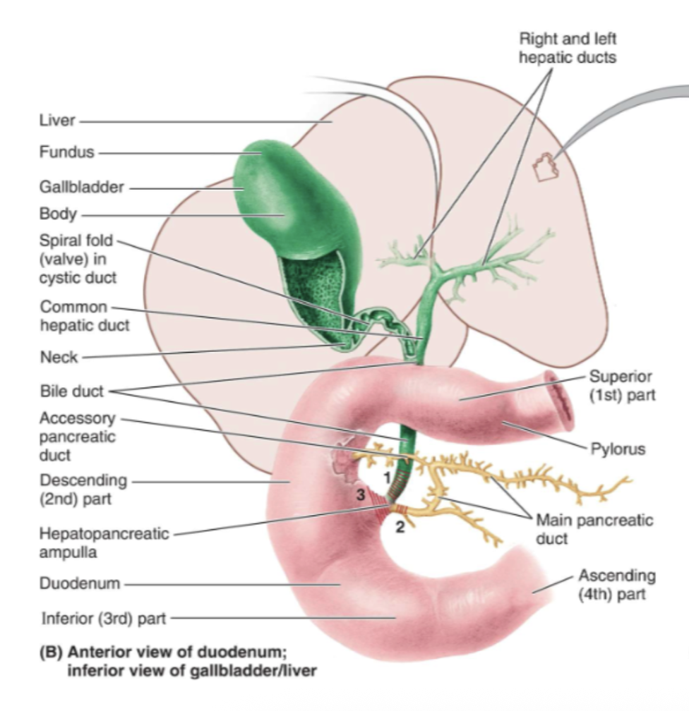
What happens when the cystic duct meets the common hepatic duct?
They form the bile duct (formerly “common bile duct”).
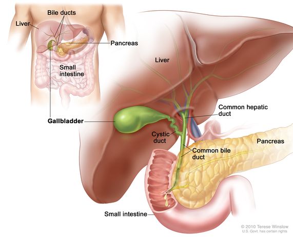
Where do the bile duct and pancreatic duct join?
At the hepatopancreatic ampulla (ampulla of Vater).
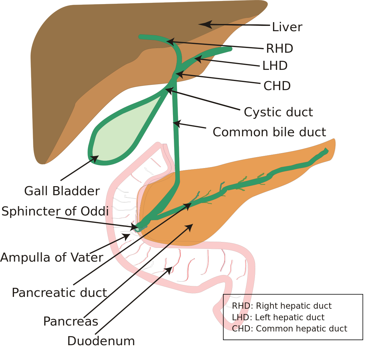
Where does bile enter the duodenum?
Through the major duodenal papilla.
What are the parts of the gallbladder?
Fundus, body, and neck.
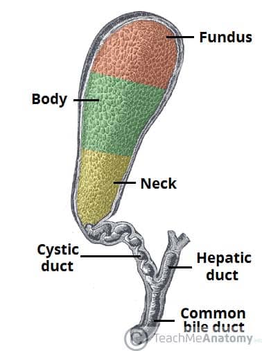
What is the function of the gallbladder?
Stores and concentrates bile.
What is the spiral fold in the cystic duct?
A mucosal fold that may hinder passage of small gallstones.

What is jaundice and what causes it?
Yellowing of skin/sclera due to ↑ bilirubin from hemolysis, liver dysfunction, or bile duct obstruction.
What is hepatomegaly?
Hepatomegaly is an enlarged liver that becomes palpable below the right costal margin.
Common causes include:
Congestive heart failure: Liver becomes congested with venous blood.
Infections (hepatitis): Inflammation increases liver size.
Fatty liver disease: Alcoholic or non-alcoholic.
Tumors or metastases: Increase liver volume.
What is cirrhosis?
Cirrhosis is chronic scarring (fibrosis) with nodular regeneration of liver tissue.
It leads to:
Loss of liver function (metabolic failure).
Distorted architecture → blocks portal blood flow.
Portal hypertension, causing major complications like ascites and varices.
What problems develop from portal hypertension?
Portal hypertension (↑ pressure in portal venous system) causes blood to bypass the liver through collateral pathways, resulting in:
Esophageal varices → risk of massive bleeding.
Rectal varices → hemorrhoid-like bleeding.
Caput medusae → dilated abdominal wall veins.
Ascites → fluid accumulation in the peritoneal cavity.
What conditions can lead to hepatic cancer?
Hepatic cancer (hepatocellular carcinoma) typically develops after chronic liver injury, especially:
Chronic Hepatitis B or C infections → long-standing inflammation.
Cirrhosis of any cause → ongoing cell turnover and DNA damage.
These conditions increase mutation risk and promote uncontrolled growth.