Genetics and Cell Division: Chromosomes, Meiosis, and Genetic Variation
1/84
There's no tags or description
Looks like no tags are added yet.
Name | Mastery | Learn | Test | Matching | Spaced | Call with Kai |
|---|
No analytics yet
Send a link to your students to track their progress
85 Terms
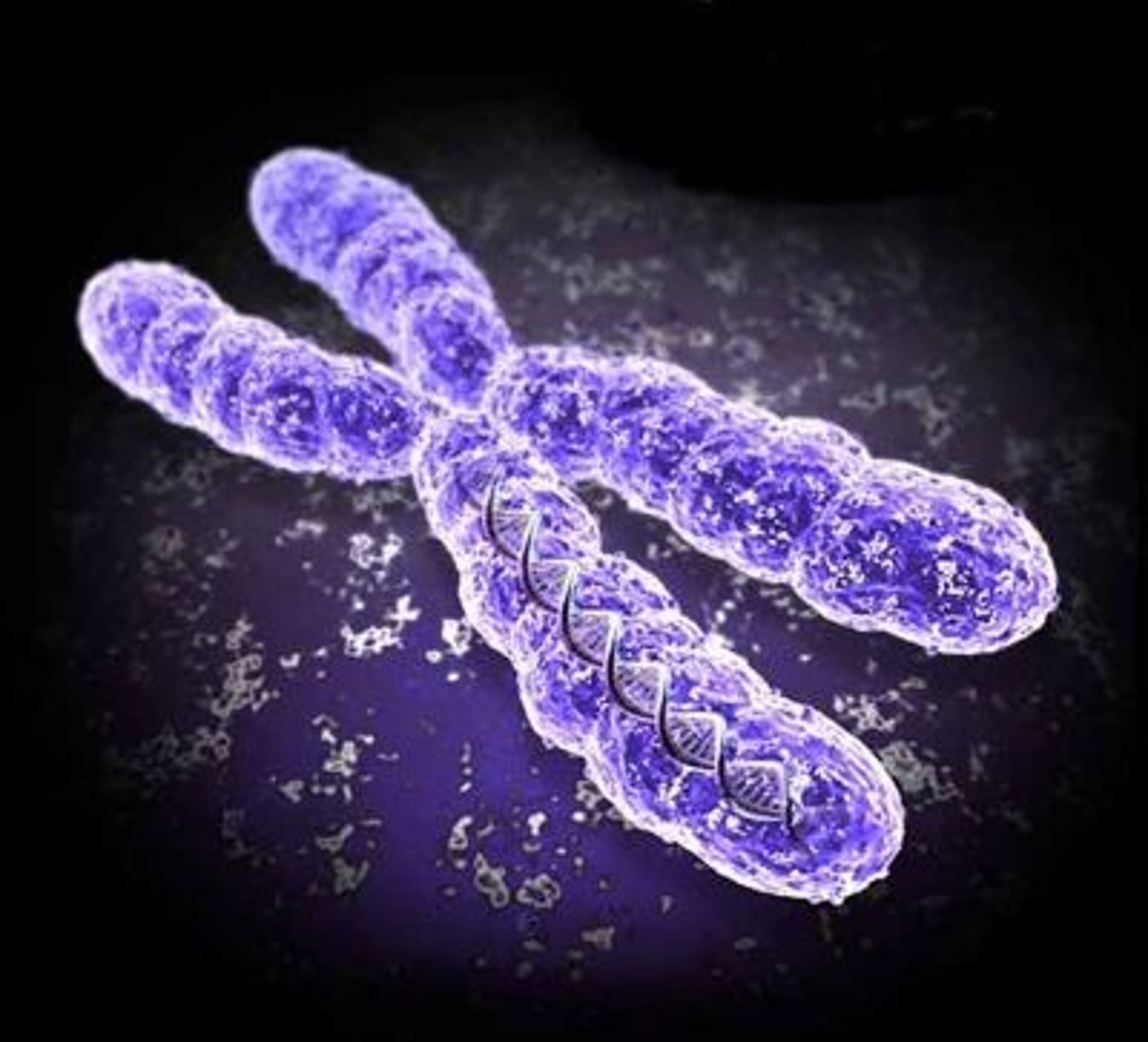
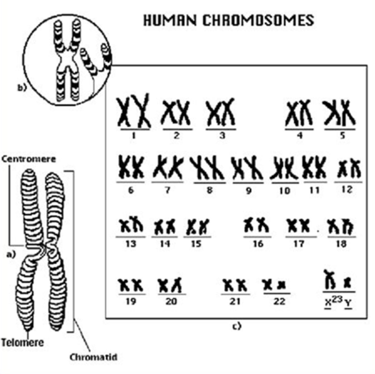
chromosome
Any of the usually linear bodies in the cell nucleus that contain the genetic material. Made up of DNA and proteins
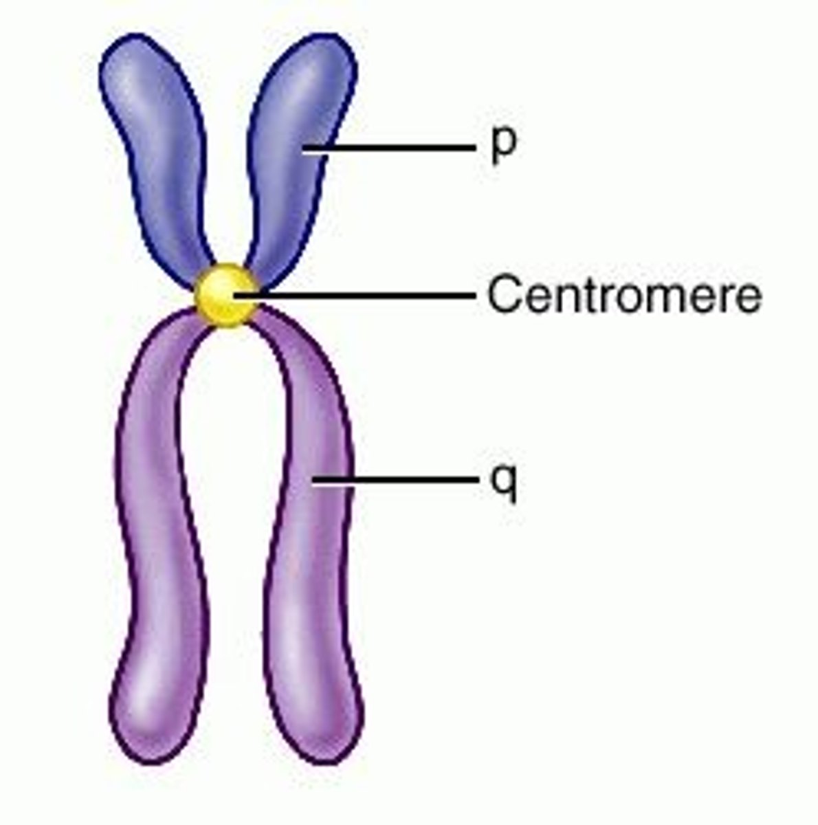
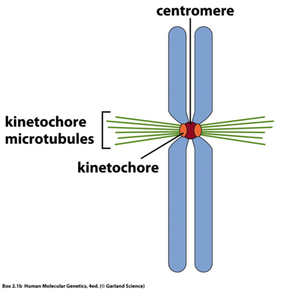
centromere
Area where the chromatids of a chromosome are attached
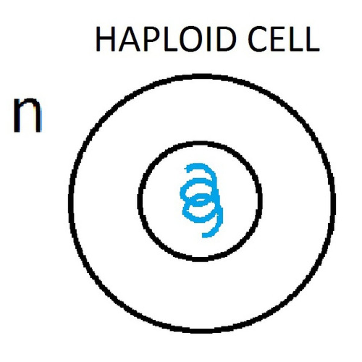
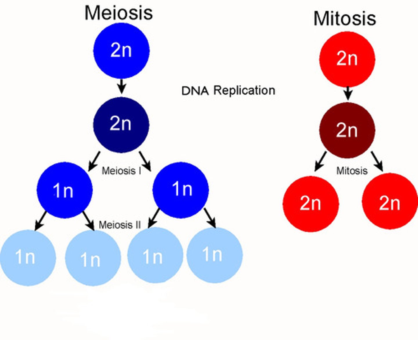
haploid
An organism or cell having only one complete set of chromosomes.
diploid
2 sets of chromosomes
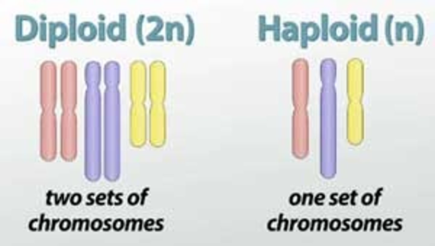
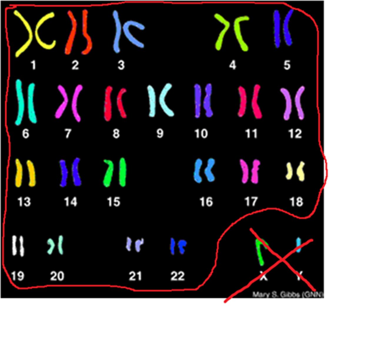
diploid number
46 chromosomes (2n)
haploid number
23 chromosomes (n)
ploidy
number of sets of chromosomes in a cell
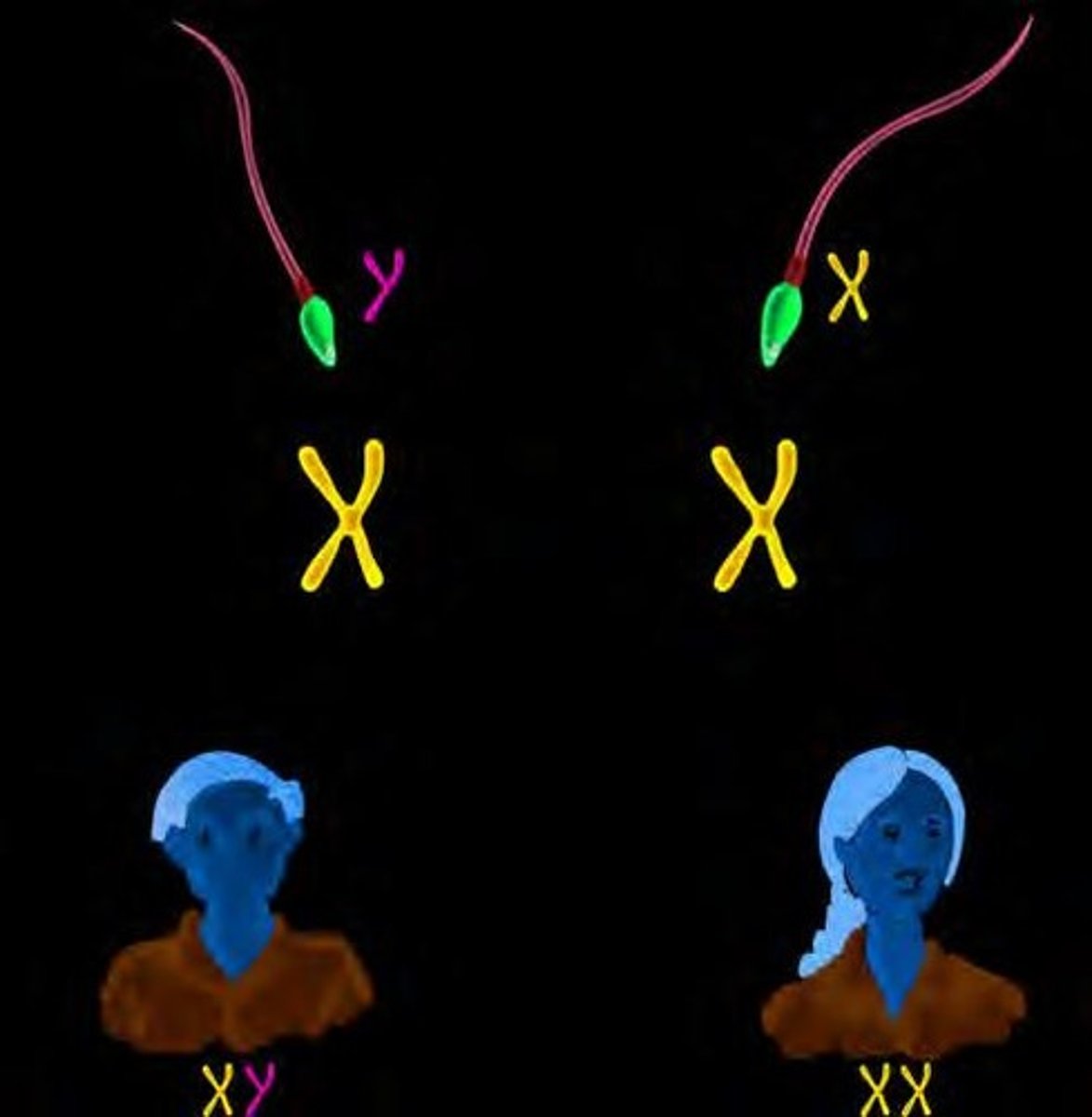
sex chromosomes
Chromosomes that determine the sex of an individual (X & Y)
autosomes
Any chromosome that is not a sex chromosome
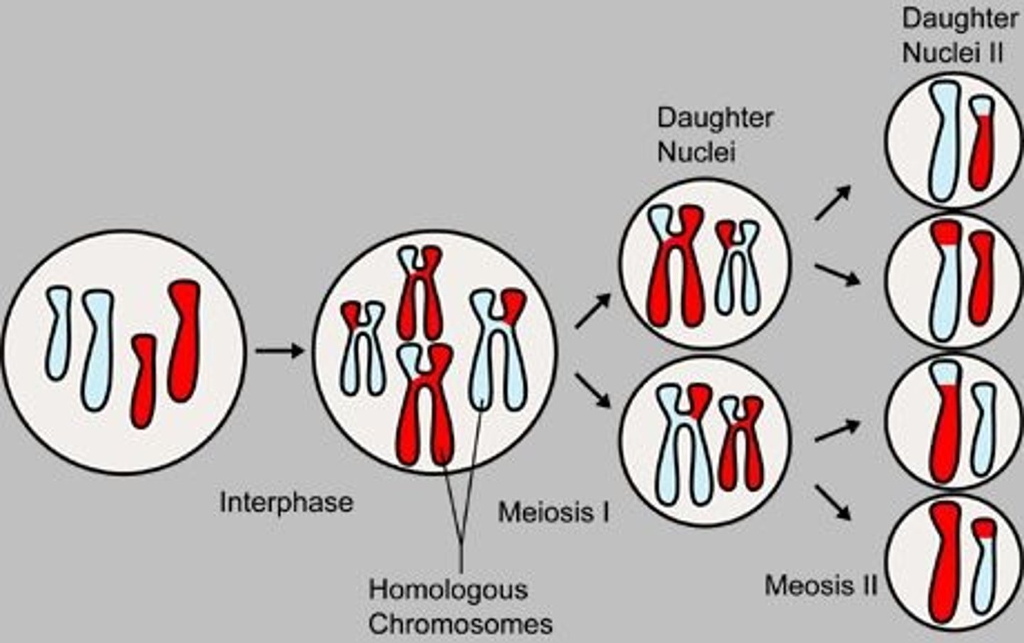
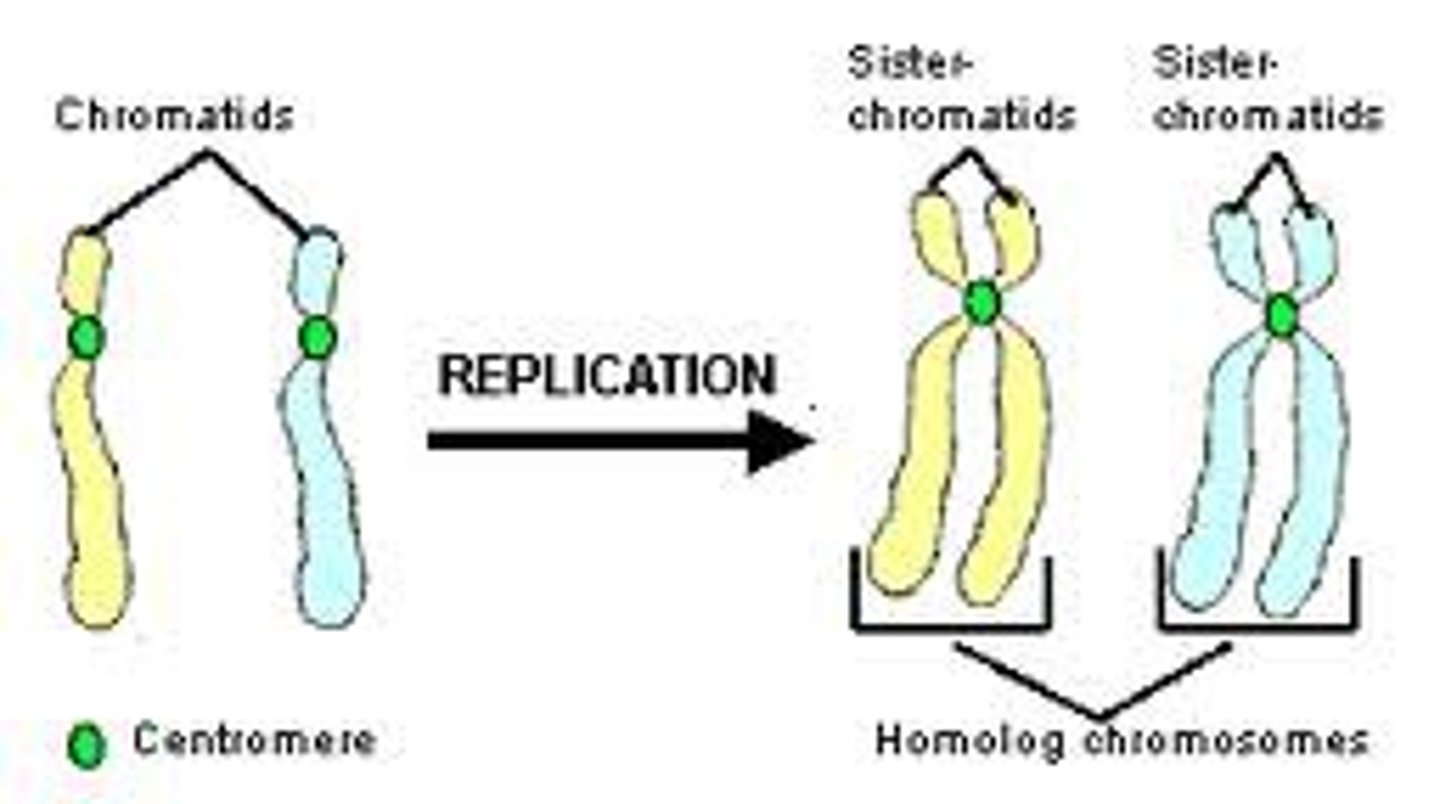
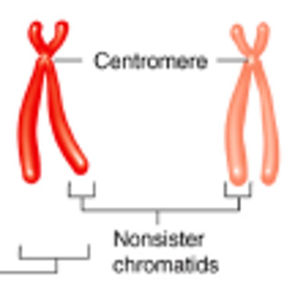
homologous chromosomes
Pair of chromosomes that are the same size, same appearance and same genes.
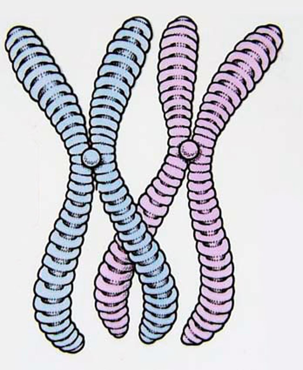
sister chromatids
Identical copies of a chromosome; full sets of these are created during the S subphase of interphase.
non-sister chromatids
chromatids belonging to homologous chromosomes
fertilization
Process in sexual reproduction in which male and female reproductive cells join to form a new cell (egg meets sperm cell)
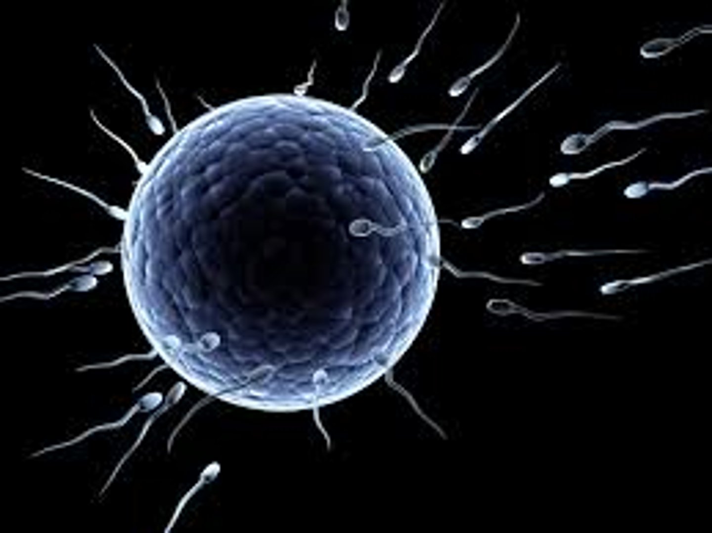
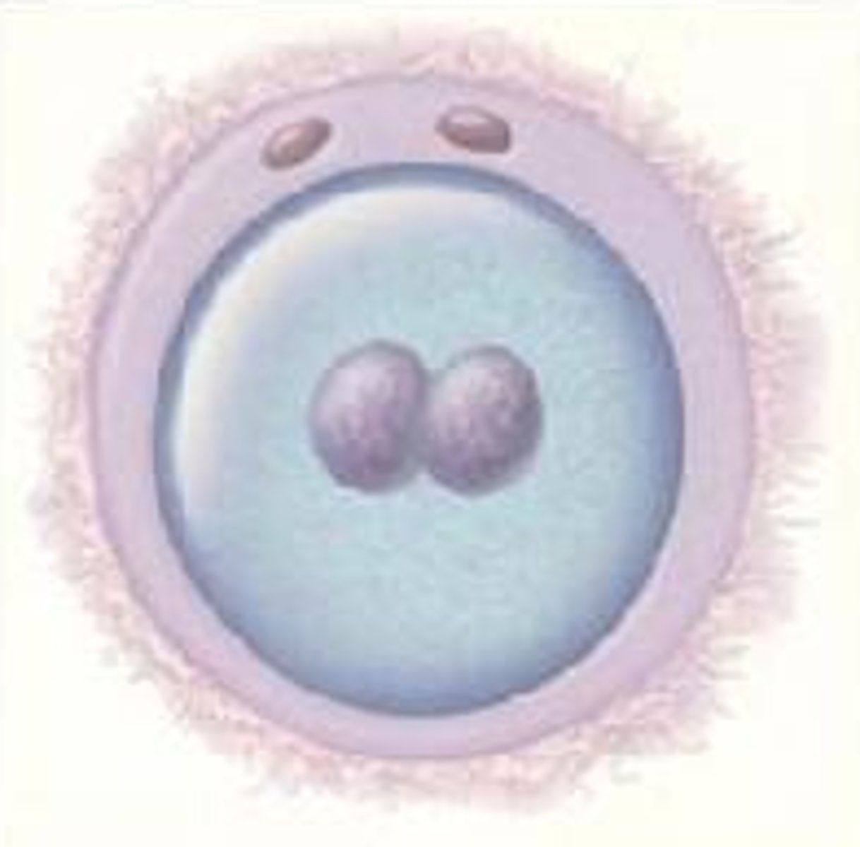
zygote
fertilized egg
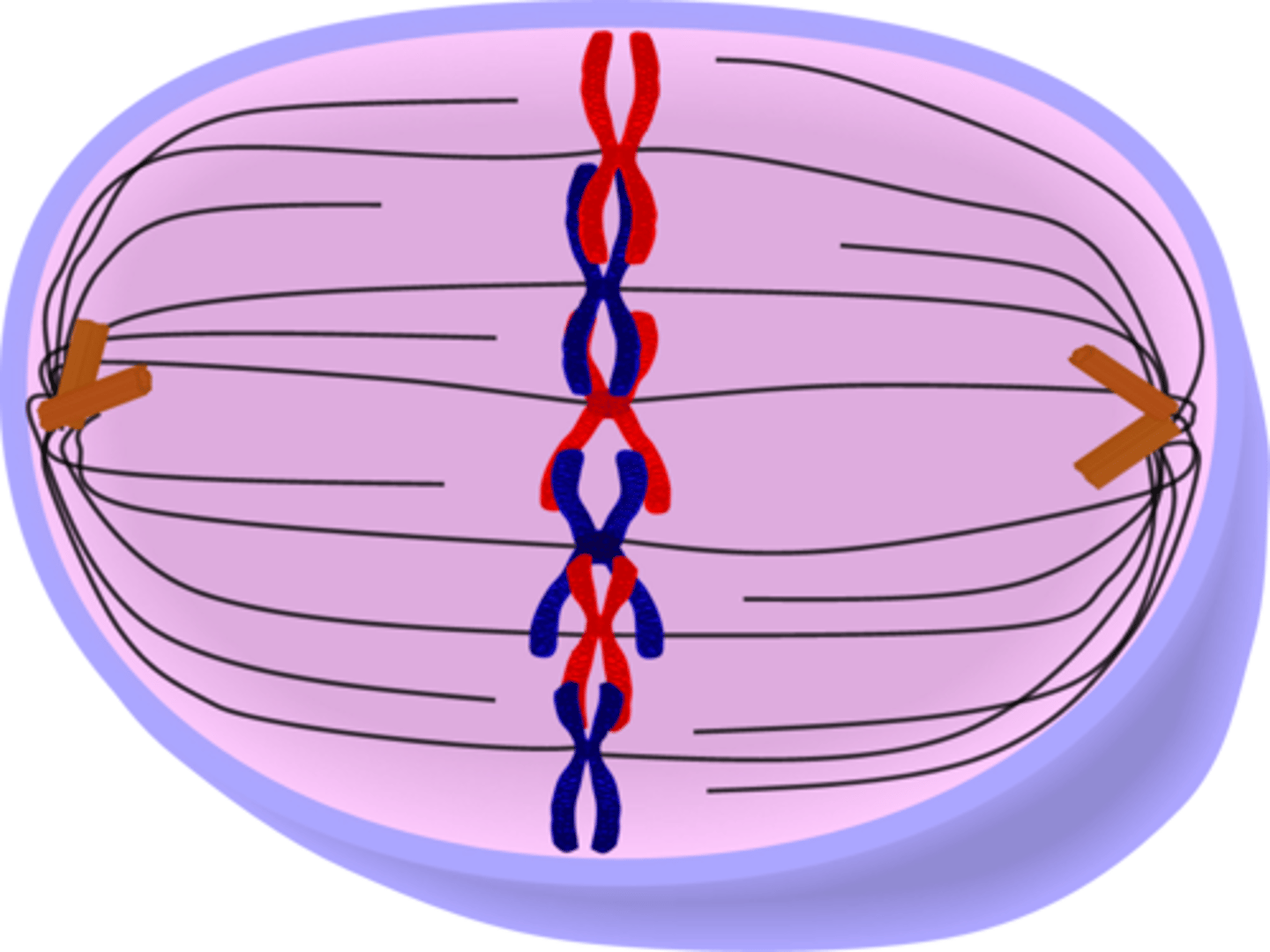
synapsis
the pairing of homologous chromosomes during meiosis
chiasma
site of crossing over
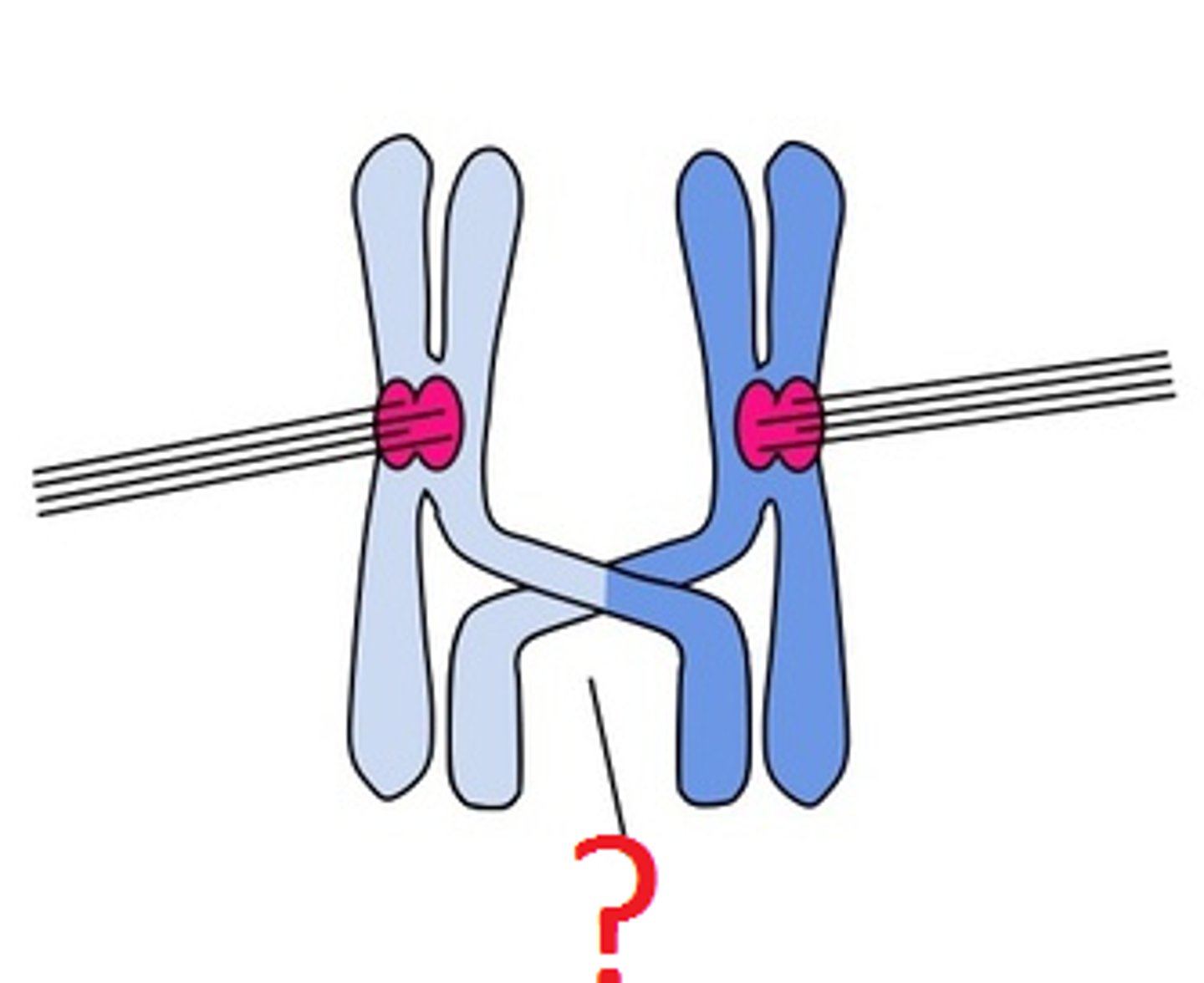
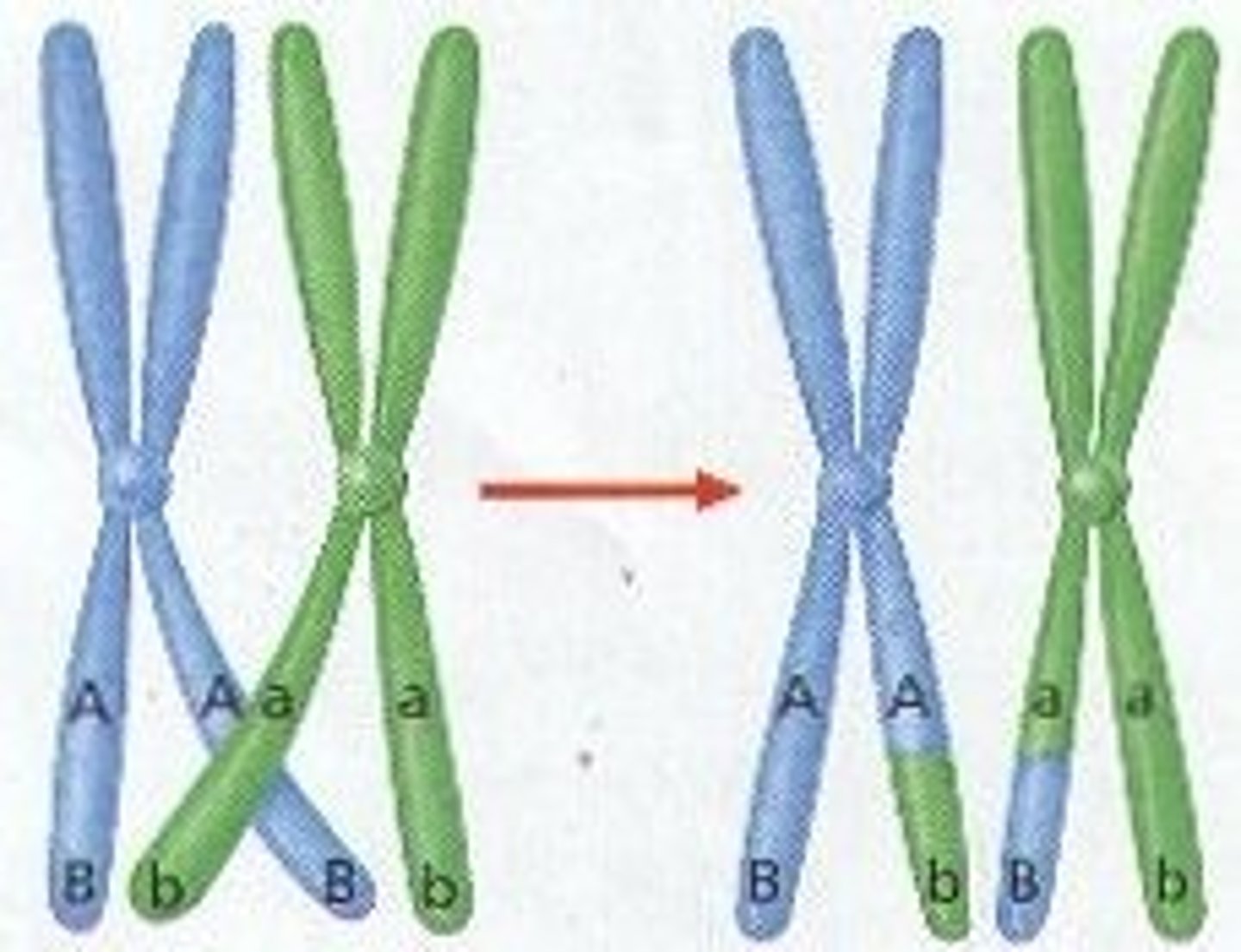
crossing over
the exchange of genes between homologous chromosomes, resulting in a mixture of parental characteristics in offspring.
spindle apparatus
the whole structure, including the spindle fibers, centrioles, and aster fibers
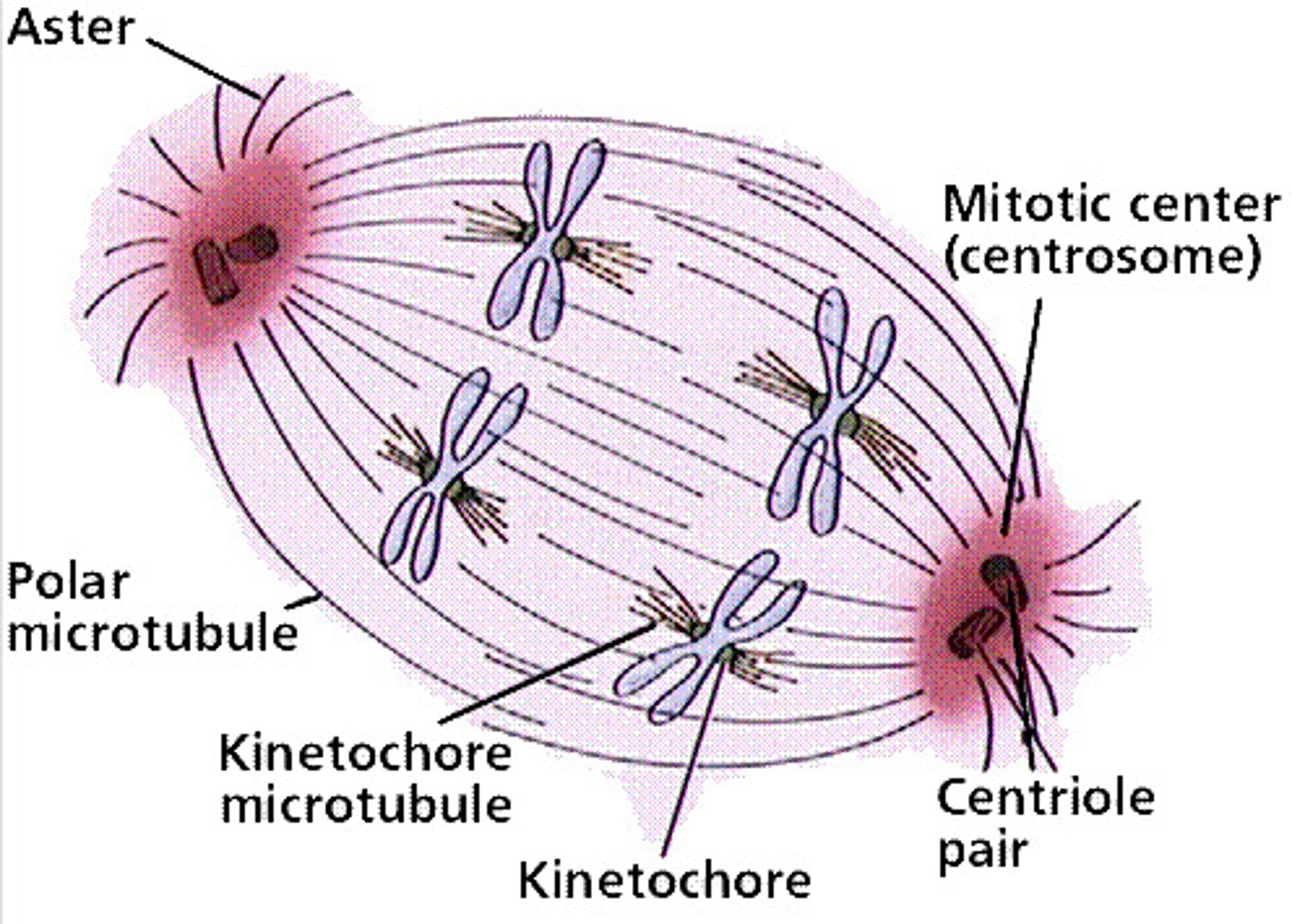
kinetochore
A specialized region on the centromere that links each sister chromatid to the mitotic spindle.
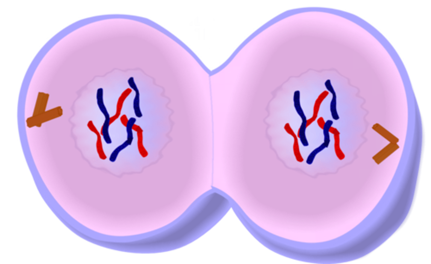
cytokinesis
division of the cytoplasm
describe each phase of interphase
G1- Growth
S- DNA synthesis
G2- Growth and preparation for mitosis
M- Mitosis (cell divison): formation of 2 daughter cells
describe the roles of mitosis and meiosis
Meiosis is to make genetic variety and create sex chromosomes. Mitosis produces body and identical cells by cell division, it's great for the repair of damage.
explain how genetic variation arises from crossing over
exchanging segments of DNA by paired homologous chromosomes during prophase 1 of meiosis
independent assortment
One of Mendel's principles that states that genes for different traits can segregate independently during the formation of gametes
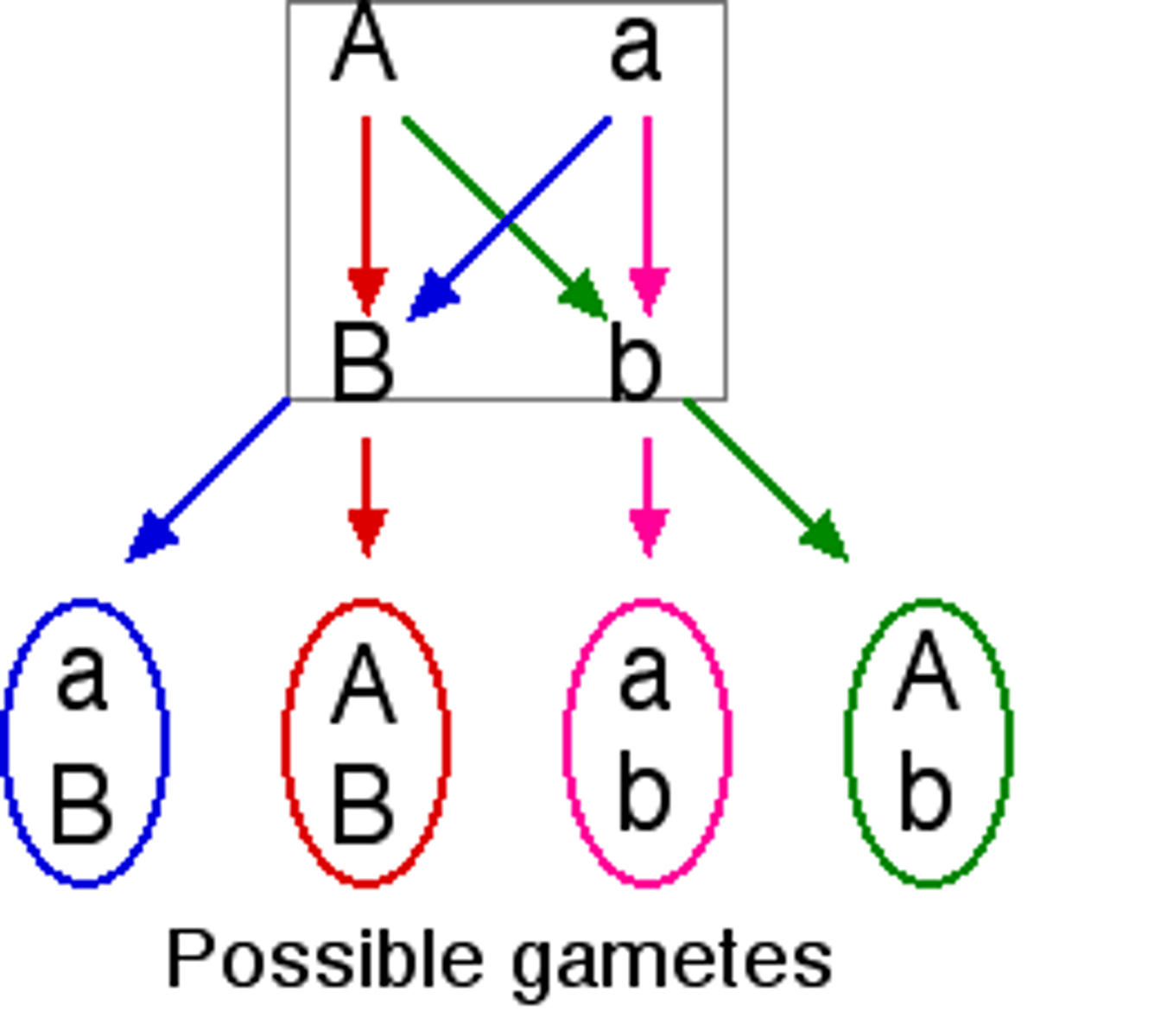
random fertilization
source of genetic variation caused by the unlimited number of possible sperm & egg combinations
prophase/prometaphase (mitosis)
- chromosomes condense and become visible
-spindle fibers emerge from the centrosomes
-nuclear envelope breaks down and then nucelolous disappears
- chromosomes continue to condense
-kinetochores appear at the centromeres
- mitotic spindle microtubules attach to kinetochores
- centrosomes move toward opposite poles
interphase (mitosis)
G1, S, and G2 phase of cell cycle
DNA is replicated in the S phase. its Used for growth, DNA replication, and cell functions.
metaphase (mitosis)
- mitotic spindle is fully developed, centrosomes are opposite poles of the cell
- chromosomes are lined up in the middle
- each sister chromatid is attached to a spindle fiber originating from opposite poles
anaphase (mitosis)
- cohesin proteins binding the sister chromatids together, break down
- sister chromatids (now are chromosomes) are pulled toward opposite poles
- spindle fibers are lengethened and elongate the cell
telophase (mitosis)
- chromosomes arrive at opposite poles and begin to decondense
- nuclear envelope material surronds each set of chromosomes
- mitotic spindle breaks down
nondisjunction
Error in meiosis in which homologous chromosomes fail to separate.
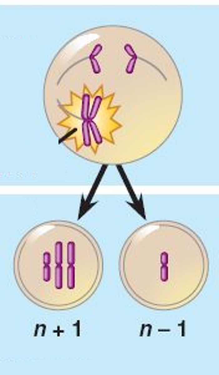
anueploidy
Abnormal number of chromosomes.
monosomy
missing a chromosome (2n-1)
trisomy
gained a chromosome; 3 copies
haploinsufficiency
The appearance of a mutant phenotype in an individual cell or organism that is heterozygous for a normally recessive trait
trisomy 18
edward's syndrome; second-most common autosomal trisomy; seen with clenched fists
trisomy 13
Patau syndrome - 50% die within 1 month; 95% die by age 3
trisomy 21
Down syndrome caused by an extra chromosome 21.
gene dosage
The number of times a given gene is present in the cell nucleus.
where can nondisjunction occur
- during anaphase of meiosis 1 and 2
- during anaphase of mitosis
meiosis vs mitosis
- meiosis has 2 cell divisions, mitosis only one
- in meiosis homologous chromosomes pair up on cell's equator, in mitosis homologous chromosomes never pair up
- in anaphase 1 of meiosis sister chromatids are still paired, in anaphase in mitosis, sister chromatids are separated
- meiosis results in a haploid cell, mitosis results in a diploid
- meiosis has two cell divisions, mitosis only one
why is anueploidy in animals usually lethal
causes massive gene dosage imbalances; it causes genetic disorders and even death
why does nondisjunction in meiosis occur more frequent in women than men
since women are born with all their eggs and remain "stuck" in prophase 1 for decades, it can lead to misalignment during the final division.
example of human autosomal aneuploidy
Down syndrome, Edwards syndrome, and Patau syndrome
example of sex-chromosome aneuploidy
Turner syndrome and Klinefelter syndrome
gene
A segment of DNA on a chromosome that codes for a specific trait
locus
Location of a gene on a chromosome
allele
Different forms of a gene
trait
specific characteristic of an individual
dominant allele
An allele whose trait always shows up in the organism when the allele is present.
recessive allele
An allele that is masked when a dominant allele is present
homozygous
Having two identical alleles for a particular gene
heterzygous
having two different alleles for a trait
genotype
genetic makeup of an organism
phenotype
physical characteristics of an organism
testcross
cross between an organism with an unknown genotype and an organism with a recessive phenotype
incomplete dominance
A pattern of inheritance in which two alleles, inherited from the parents, are neither dominant nor recessive. The resulting offspring have a phenotype that is a blending of the parental traits.
complete dominance
a relationship in which one allele is completely dominant over another
multiple alleles
three or more forms of a gene that code for a single trait
recessive lethal allele
an allele that negatively affects the survival of a homozygote
dominant lethal allele
the presence of just one copy of the allele results in the death of the individual
autosomal dominant
inheritance pattern of a dominant allele on an autosome; Hungtion's disease
autosomal recessive
two copies of an abnormal gene must be present in order for the disease or trait to develop
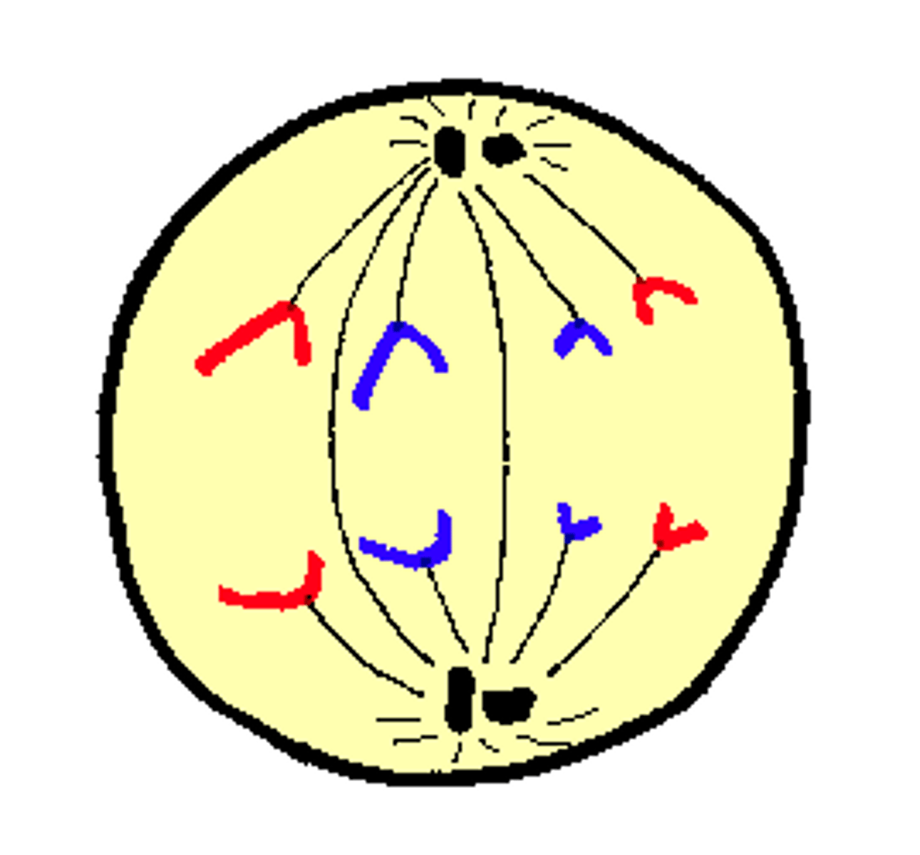
prophase 1
Chromosomes become visible; the nuclear envelope breaks down; and homologous chromosomes undergo crossing-over. they begin to transfer their DNA to each other.
metaphase 1
Paired homologous chromosomes line up across the center of the cell
anaphase 1
Homologous chromosomes are being pulled away by the spindle fibers
telophase 1
2 daughter cells are formed, each daughter cell contains only one chromosome of the homologous pair.
prophase 2
A new spindle forms around the chromosomes, but no homologous chromosome pairs or crossing over
metaphase 2
Centromeres of chromosomes line up randomly at the equator of each cell.
anaphase 2
sister chromatids separate and move to opposite poles
telophase 2
A nuclear membrane forms around the chromosomes in each of the 4 new cells then cytokinesis splits the cytoplasm
wild type allele
Generally, but not always a dominant allele. It's the most prevalent allele in the population.
loss of function mutation
causes a complete or partial loss of function
gain of function mutation
produces a new trait or causes a trait to appear in inappropriate tissues or at inappropriate times in development.
null mutation
results in complete loss of function
neutral mutation
a mutation that has no effect on survival or reproduction
masking genes can
alter the appearance of the expected phenotype from a genotype
Aa x Aa phenotypic ratio
3:1
Aa x aa phenotypic ratio
1:1
Aa x Aa (recessive lethal) phenotypic ratio
2:1 (lethal so it doesn't survive)
AaBb x AaBb (no gene interaction) phenotypic ratio
9:3:3:1
AaBb x AaBb (recessive epistasis) phenotypic ratio
9:3:4
AaBb x AaBb (dominant epistasis) phenotypic ratio
12:3:1
AaBb x AaBb (complementary gene interaction) phenotypic ratio
9:7
AA x aa phenotypic ratio
1:0