3.4-Mass transport in animals 1
1/47
There's no tags or description
Looks like no tags are added yet.
Name | Mastery | Learn | Test | Matching | Spaced | Call with Kai |
|---|
No analytics yet
Send a link to your students to track their progress
48 Terms
what do arteries do?
carry blood at high pressure, travelling fast, and pulsing- that is, the pressure fluctuates with the heartbeat
what is the structure of arteries?
relatively small lumen
walls are:
very thick
have an inner coat of a single layer of endothelium
have lots of elastic tissues and muscle fibre
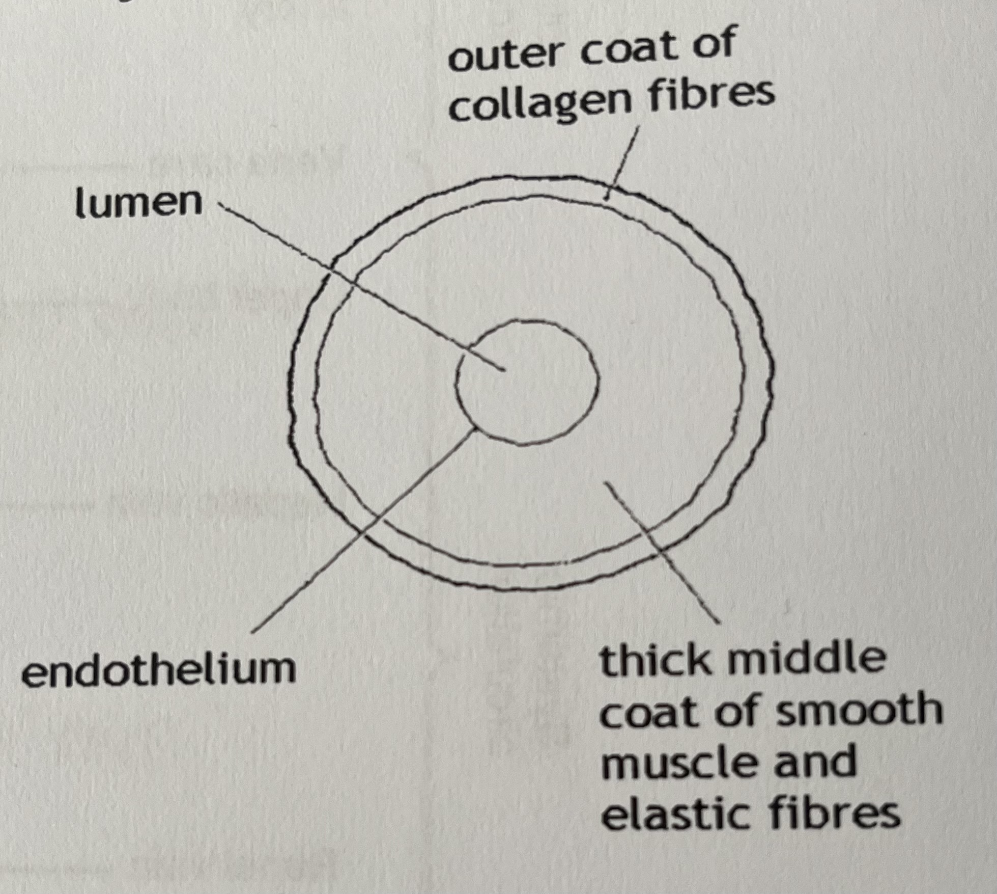
Why is the endothelium smooth?
To reduce friction between blood and blood vessel wall
when do the elastic fibres stretch?
when high pressure blood is pumped out by the heart
makes the artery wider and reduces the pressure a little
when do elastic fibres recoil?
when the pressure falls during diastole
elastic recoil keeps the blood under pressure and pushes it along
reduces fluctuations in pressure and helps smooth out blood flow as the blood moves along
Why do arteries close to the heart have more elastic fibres in their walls than arteries further away from the heart?
Greater blood pressure closer to the heart- elastic fibres allow artery walls to stretch to accommodate surges in pressure
Elastic recoil of arteries helps to smooth out blood flow- keeps blood under some pressure and moving during diastole
what are arterioles and what is their function?
Smaller vessels which arteries branch into when they reach the tissues to which they are transporting blood and they control blood flow to the capillaries of different organs
what is the structure of arteriole walls?
have lots of muscle tissue
Are thinner than arteries
Have an inner coat of a single layer of endothelium
what is vasoconstriction?
When the muscles contract, narrowing the lumen diameter
This reduces blood flow to the capillaries they serve
what is vasodilation?
When the muscle relaxes, to widen the lumen diameter
This increases the blood flow to the capillaries they serve
What happens during exercise?
Arterioles supplying blood to the muscles in the legs would be dilated ( wide), whilst those carrying blood to the gut would be constricted ( narrower )
What is the advantage of arterioles in the skin dilating when we are too hot?
Allows blood to flow to surface of the skin for heat to be lost by radiation
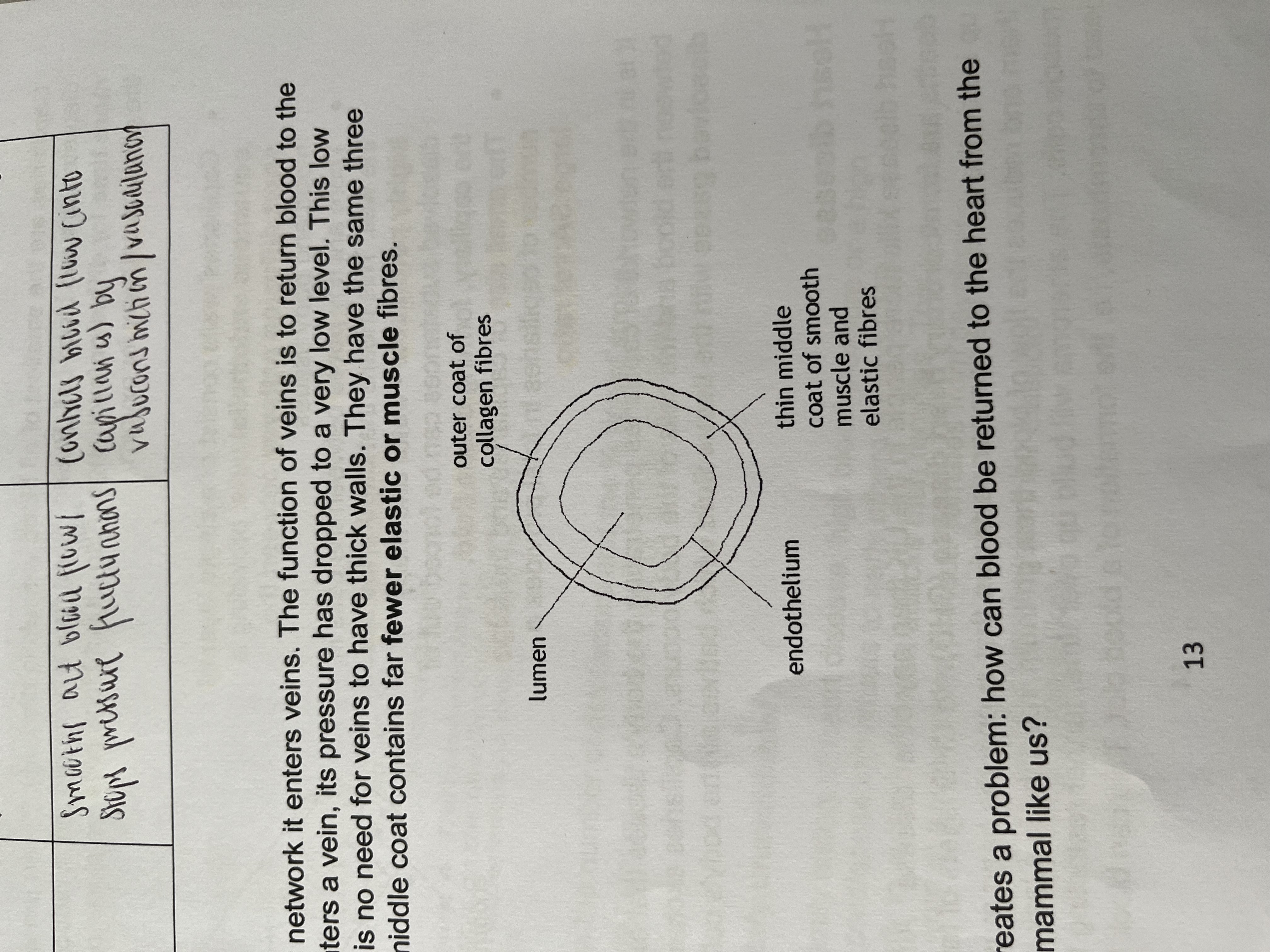
what is the function of veins?
return blood to the heart
why don’t veins have thick walls?
by the time blood enters a vein, pressure has dropped to a very low level
what is the structure of the walls of veins?
very thin
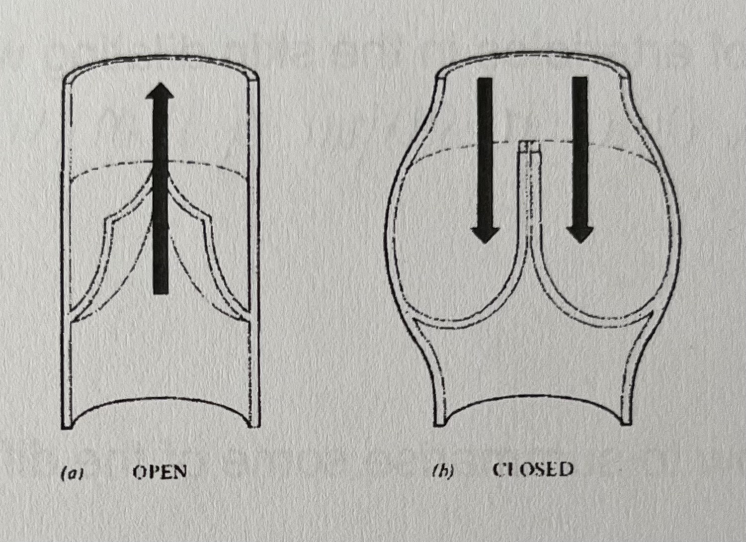
Have valves
why do veins contain semi-lunar valves?
to keep blood flowing in the right direction
these valves only open to allow blood to flow towards the heart and close to stop it falling back down again
what are adaptations of capillaries?
Same diameter as red blood cell- blood flow reduced- allows more time for diffusion/ exchange
All red blood cells close to capillary wall- reduces diffusion pathway for oxygen
walls- consist of single layer of squamous endothelial cells, providing a short diffusion pathway between the body and blood tissues
tiny pores between the cells in the wall- making them highly permeable- water and many dissolved substances can be forced out of the capillary, forming tissue fluid
Small size and large number- provides large sa:v ratio
What is the importance of capillary beds?
It is in the networks of capillaries penetrating the body’s tissues that exchange of materials between the blood and the cells of the body occurs
Capillaries exchange nutrients and dissolved gases with the tissue fluid
How is tissue fluid formed?
High hydrostatic pressure at the arterial end (caused by ventricular contraction)
Causes water and small soluble substances to be forced out through the capillary wall
Plasma proteins are too large and remain in the capillary
There is a decrease in water potential
Water at the venous end re enters by osmosis
Remaining fluid is absorbed by the lymphatic capillary + transported around the body by the lymphatic system
What causes a drop in blood pressure in the capillary?
Loss of fluid from the capillary to the tissue fluid
Resistance as blood flows thru narrow lumen
What causes the water potential to decrease in the capillary?
Large plasma proteins remain in capillary + loss of fluid
what is the heart made of?
cardiac muscle
what is pulmonary circulation?
pulmonary artery carries deoxy blood from the heart to lungs, then pulmonary vein carries oxy blood back to heart
what is systematic circulation?
aorta carries oxy blood from the heart to the body, then vena cava carries deoxy blood to the heart
what is the pathway of blood from the body, back to the body?
vena cava carries deoxy blood from the body, to the right atrium, right ventricle, pulmonary vein to the lungs where it becomes oxygenated
pulmonary artery, left atrium, left ventricle, aorta to the rest of the body, back to the vena cava
what is the advantage for the human heart to have two separate pumps rather than one?
RHS generates pressure to send blood thru the lungs where pressure drops
returned to the LHS of the heart, generates more pressure so blood can travel around the body- fast enough to generate high HR
what do the coronary arteries do?
delivery oxy blood to the cardiac muscle that makes up the walls of the heart
why does the cardiac muscle need a rich supply of blood?
needs good supply of O2 and glucose for high rates of aerobic respiration to produce lots of ATP for muscle contraction
what does the septum do?
separate the oxy blood on the left and the deoxy blood on the right- makes gas exchange in lungs and body tissues more efficient
why are the left ventricle walls thicker than the right ventricle walls?
left ventricles need to contract with more force and pump blood all round the body, whereas the right ventricle only pumps blood to the lungs
What is the function of the AV and SL valves?
Prevent backflow of blood when they close
AV- allow blood to be pumped from atrium to ventricles
SL- allow blood to be pumped from ventricles to artery
what is a cardiac cycle?
one complete sequence of contraction and relaxation
what is atrial systole, ventricular systole and diastole?
atrial systole- atria contract
ventricular systole- ventricles contract
diastole- relaxation of heart muscle
where does blood flow from?
region of high pressure to low pressure
what happens during atrial systole?
atrial contraction
atrial pressure increases (higher than ventricular pressure)
AV valves open
blood pumped from atria to ventricles
blood stays in ventricles
what happens during ventricular systole?
ventricular contraction
ventricular pressure increases (higher than atria)
AV valves close (prevent blood going back to atria)
SL valves open- ventricular pressure higher than arteries
blood pumped from ventricles to arteries
what happens during diastole?
relaxation of heart muscle i.e atria and ventricles
SL valves close- pressure higher in arteries than ventricles- prevent blood going back to ventricles
meanwhile, blood is being pumped from the veins to the atria to start the process again
why is the cardiac muscle myogenic?
contracts on its own without needing nervous stimulation
how is a heartbeat initiated and co-ordinated?
SAN- acts as a pacemaker and sends a wave of impulse across the atria, causing them to contract
non-conducting tissue prevents immediate contraction of ventricles
AVN delays impulse whilst blood leaves atria
AVN sends wave of impulse down Bundle of His/Purkyne fibres
causes ventricles to contract from base up
where is elastic tissue found?
in the arteries
what is a pulse?
the stretch and subsequent recoil of the arteries travels as a wave known as the pulse
Pulse rate also known as heart rate
Measured in bpm
what is stroke volume and what is it determined by?
volume of blood pumped out from one ventricle during each contraction
the fuller the ventricles, the greater the force of contraction
what is cardiac output?
total volume pumped out from one ventricle per minute
how to calculate cardiac output (dm3 min-1)
stroke volume (cm3) multiplied by heart rate (bpm)
if a trained athlete’s heart gets bigger over time, what happens to the stroke volume and heart rate?
resting stroke volume will be bigger, resting heart rate will drop
more capacity to increase cardiac output during exercise
what does an increase in cardiac output allow?
more oxygen and glucose to be delivered to working muscles
what does the renal vein do?
carry deoxy blood from kidneys to heart (vena cava)
what does the renal artery do?
carry oxy blood from heart (aorta) to kidneys