MTS Mycology
1/19
There's no tags or description
Looks like no tags are added yet.
Name | Mastery | Learn | Test | Matching | Spaced | Call with Kai |
|---|
No analytics yet
Send a link to your students to track their progress
20 Terms
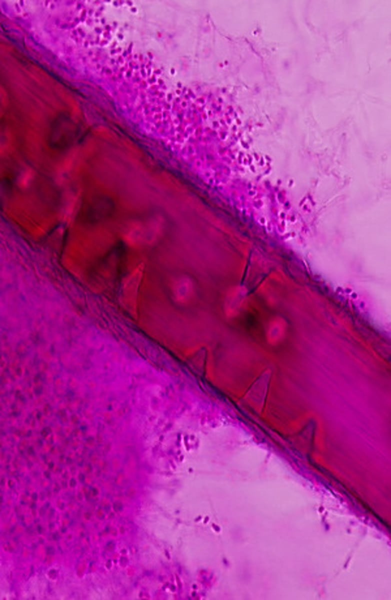
This result of a hair perforation test is typical for which of the following fungi?
Note: select an answer in the list below. Click the Submit button to continue.
Epidermophyton floccosum
Malassezia furfur
Trichosporon beigelii
Trichophyton rubrum
Trichophyton mentagrophytes
Trichophyton mentagrophytes produces hyphal structures that invade hair shafts forming cone-shaped perforations. This is a useful test to differentiate dermatophytes.
This India ink preparation suggest which of the following organisms?
Blastomyces dermatitidis
Candida albicans
Cryptococcus neoformans
Histoplasma capsulatum
Paracoccidioides brasiliensis
Cryptococcus neoformans produces a polysaccharide capsule which is visible in an India ink preparation. India ink preparations can be performed directly on cerebrospinal fluid.
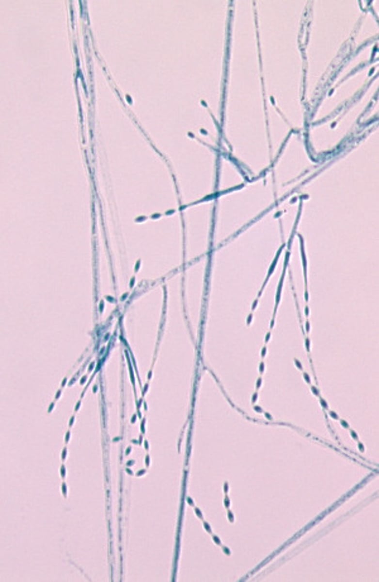
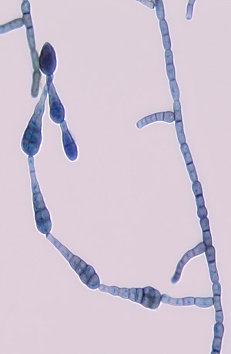
This microscopic image suggests which of the following fungi?
Coccidioides immitis
Histoplasma capsulatum
Phialophora verrucosa
Sporothrix schenckii
Wangiella dermatitidis
In the environment or when cultured on laboratory media, Coccidioides immitis grows as a mold. In this form, it produces hyphae which differentiate into arthroconidia alternating with empty disjunctor cells.
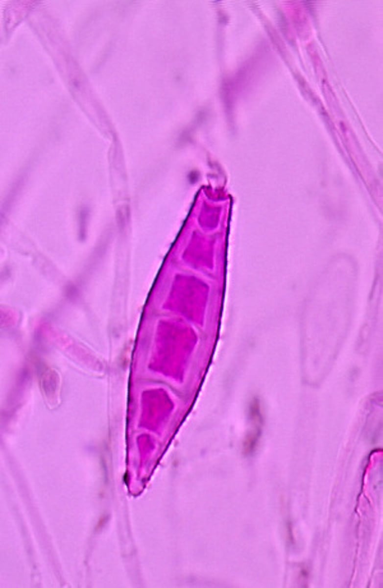
This microscopic image suggests which of the following dermatophytes?
Epidermophyton floccosum
Microsporum canis
Trichophyton mentagrophytes
Trichophyton rubrum
Microsporum canis produces rough, spindle-shaped, thick-walled macroconidia.
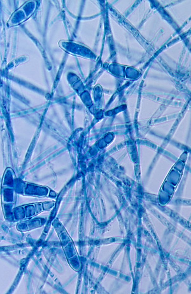
This microscopic image suggests which of the following?
Epidermophyton floccosum
Microsporum canis
Trichophyton mentagrophytes
Trichophyton rubrum
Trichophyton tonsurans
Epidermophyton floccosum produces clusters of smooth, club-shaped macroconidia. The cell walls of Epidermophyton are thicker than those of the genus Trichophyton.
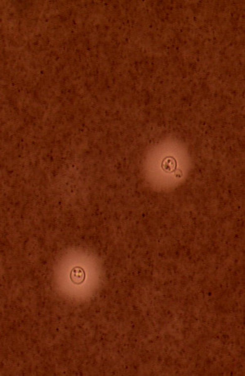
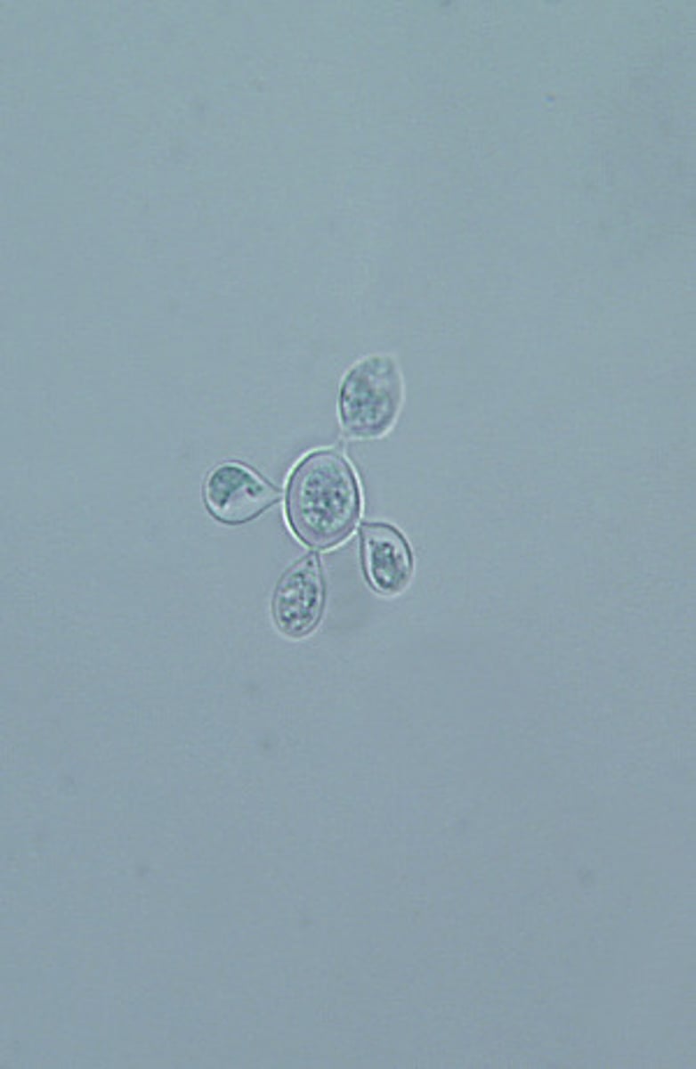
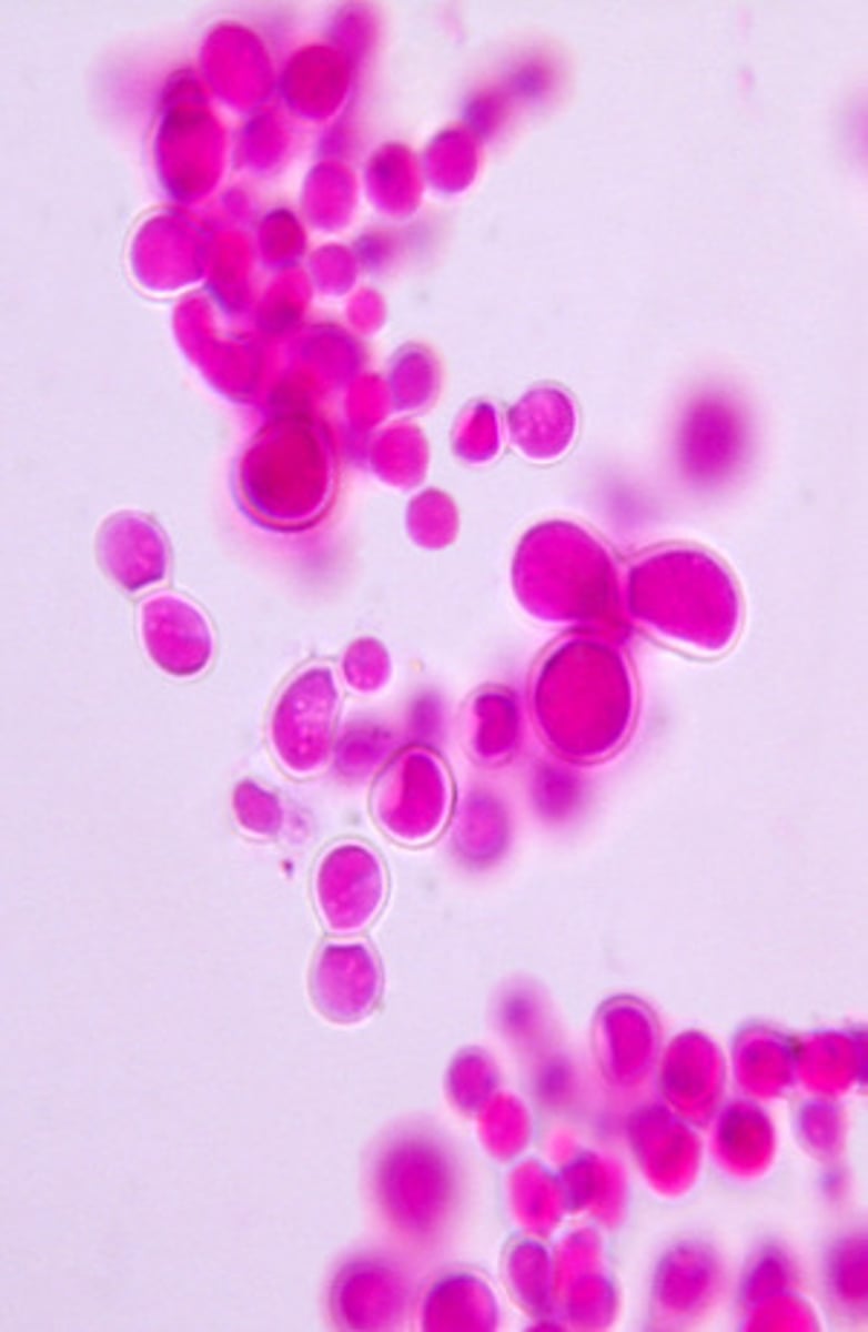
This yeast developed at 37ºC. Select the most likely identification.
Blastomyces dermatitidis
Cryptococcus neoformans
Coccidioides immitis
Histoplasma capsulatum
Paracoccidioides brasiliensis
Paracoccidioides brasiliensis is a dimorphic mold that produces yeast with multiple buds at 37ºC.
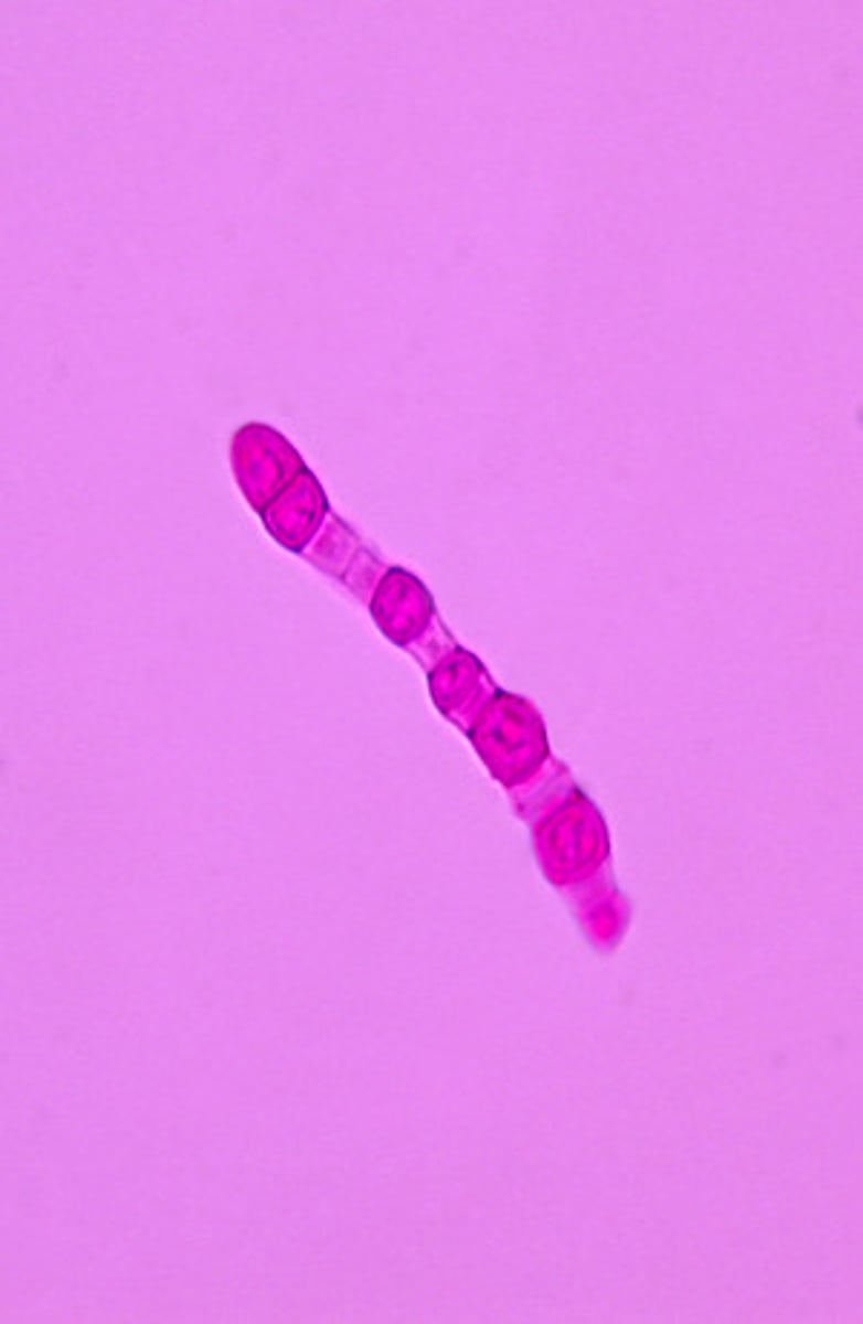
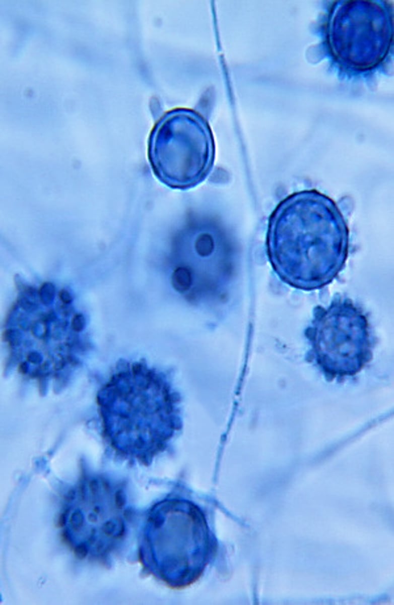
These macroconidia developed at room temperature. Select the most likely identification.
Blastomyces dermatitidis
Cryptococcus neoformans
Coccidioides immitis
Histoplasma capsulatum
Paracoccidioides brasiliensis
Histoplasma capsulatum produces warty (tuberculate) spherical macroconidia at room temperature.
This yeast developed at 37ºC. Select the most likely identification.
Blastomyces dermatitidis
Cryptococcus neoformans
Coccidioides immitis
Histoplasma capsulatum
Paracoccidiodes brasilensis
At 37ºC, Blastomyces dermatitidis produces broad-based budding yeast cells with thick walls.
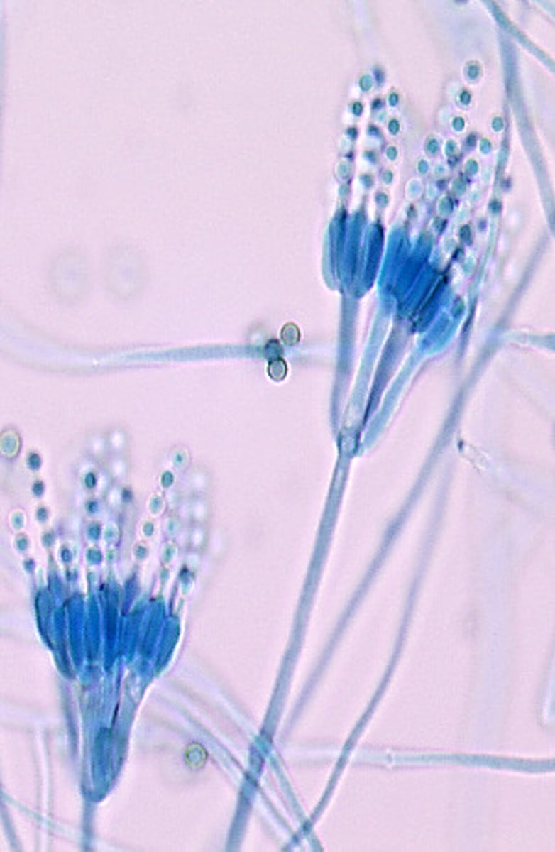
This microscopic image suggests which of the following molds?
Aspergillus flavus
Aspergillus fumigatus
Paecilomyces spp.
Penicillium spp.
Scopulariopsis spp.
Penicillium spp. produce clusters of thick phialides bearing chains of round conidia.
This microscopic image suggests which of the following fungi?
Aspergillus flavus
Aspergillus fumigatus
Paecilomyces spp.
Penicillium spp.
Scopulariopsis spp.
Paecilomyces spp. produce widely-spaced, elongated, slender phialides bearing chains of oval conidia.
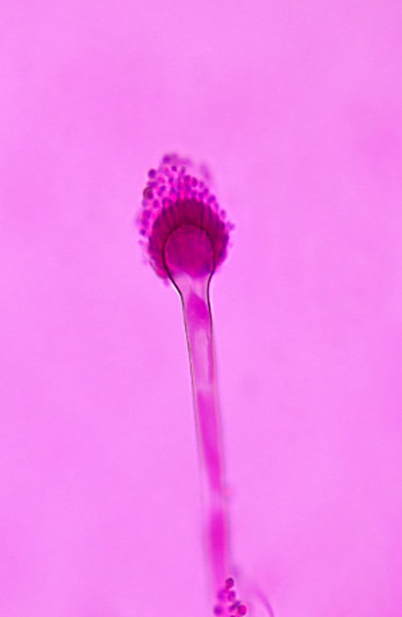
This microscopic image suggests which of the following molds?
Aspergillus flavus
Aspergillus fumigatus
Paecilomyces spp.
Penicillium spp.
Scopulariopsis spp.
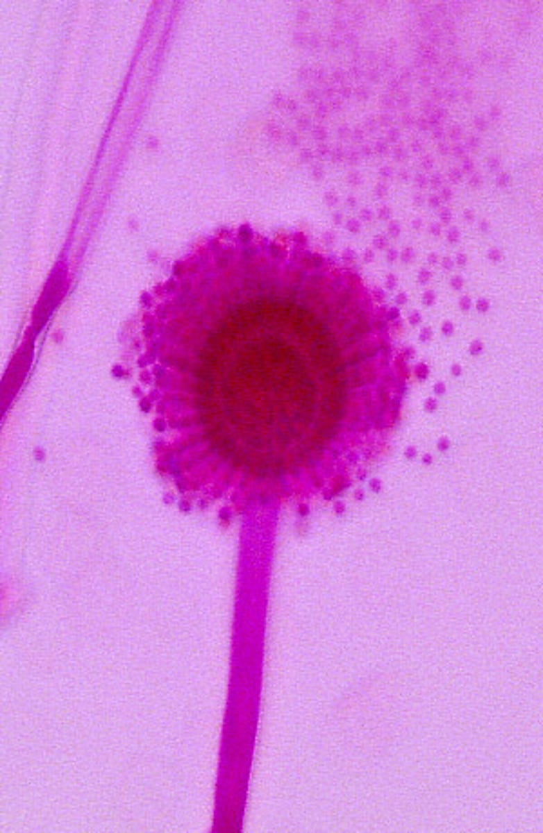
Aspergillus fumigatus produces uniserate phialides bearing chains of round conidia. The phialides are located only on the upper portion of the vesicle.
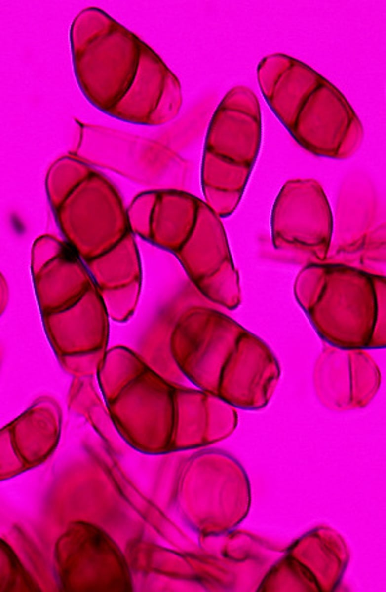
This microscopic image suggests which of the following molds?
Alternaria
Curvularia
Epicoccum
Pithomyces
Ulocladium
Alternaria spp. are dematiaceous molds that produce chains of racket-shaped muriform poroconidia.
This microscopic image suggests which of the following molds?
Alternaria
Curvularia
Epicoccum
Pithomyces
Ulocladium
Curvularia spp. are dematiaceous molds that produce curved poroconidia with a swollen central cell.
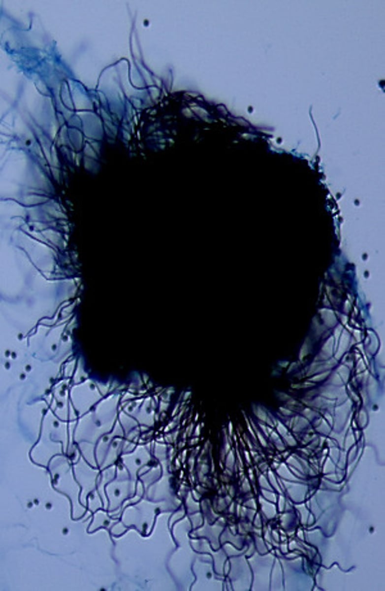
This microscopic image suggests which of the following molds?
Chaetomium spp.
Mucor spp.
Pseudallescheria boydii
Phoma spp.
Rhizopus spp.
Chaetomium spp. produce large fruiting bodies called perithecia. The perithecia are covered with thick hair-like projections called setae and contain oval ascospores.
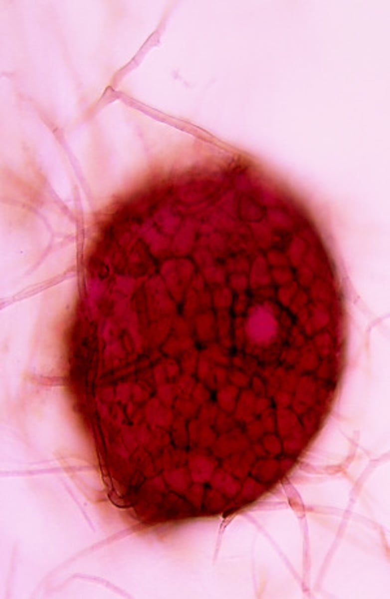
This microscopic image suggests which of the following molds?
Chaetomium spp.
Mucor spp.
Pseudallescheria boydii
Phoma spp.
Rhizopus spp.
Phoma spp. produce spherical pycnidia containing conidia which are released through an opening called an ostiole.
This microscopic image suggests which of the following molds?
Chaetomium spp.
Mucor spp.
Pseudallescheria boydii
Phoma spp.
Rhizopus spp.
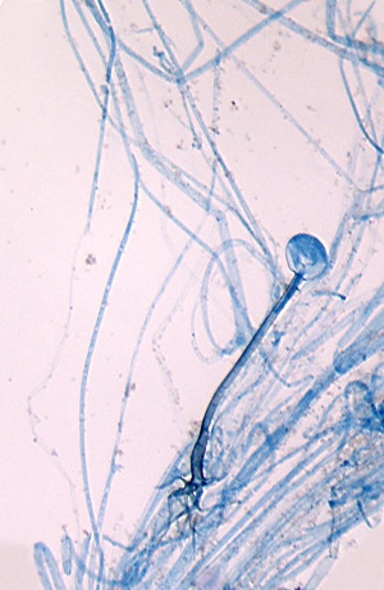
Rhizopus spp. produce sporanigia borne on long sporanigiophores. Rhizoids are produced directly opposite the sporangiophore.
This microscopic image suggests which of the following molds?
Aspergillus flavus
Aspergillus fumigatus
Paecilomyces spp.
Penicillium spp.
Scopulariopsis spp.
Aspergillus flavus produces rows of uni- or biserate phialides bearing chains of round conidia. The phialides are produced on the entire surface of the vesicle.
This microscopic image suggests which of the following organisms?
Blastocystis dermatitidis
Candida albicans
Cryptococcus neoformans
Pneumocystis carinii
Malassezia furfur
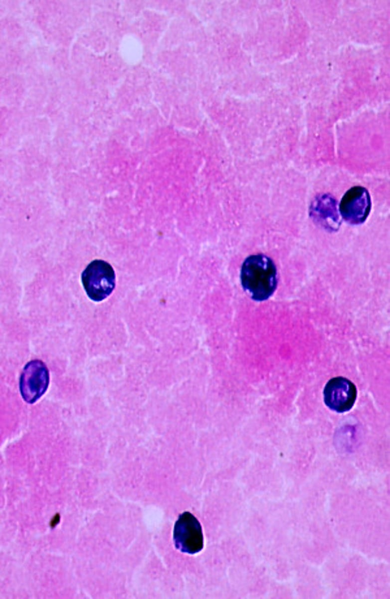
Pneumocystis carinii produces round to oval cysts that stain blue with Gram-Weigert.
This microscopic image suggests which of the following molds?
Exophiala jeanselmei
Fonsecaea spp.
Phialophora richardsiae
Phialophora verrucosa
Wangiella dermatitidis
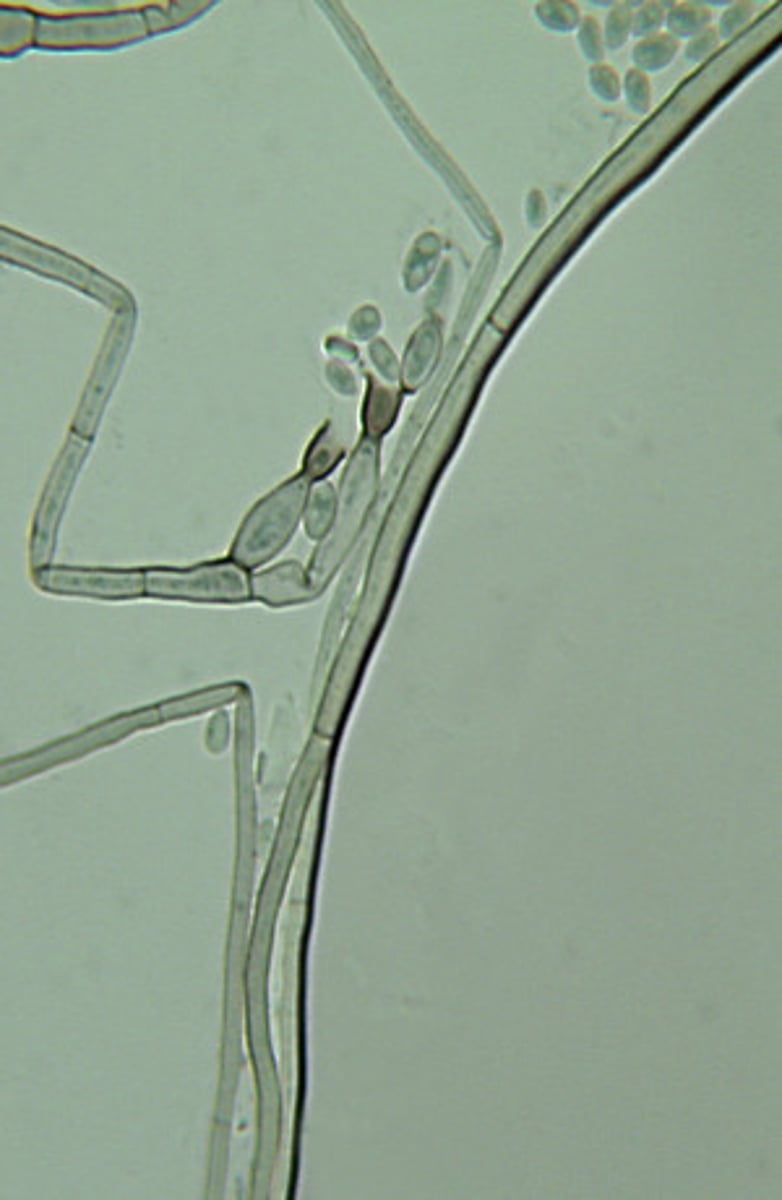
Phialophora verrucosa produces vase-like phialides with cup-shaped collarettes that form oval conidia.
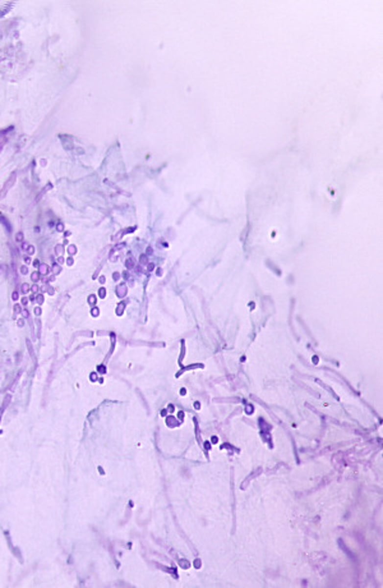
This microscopic image suggests which of the following organisms?
Candida albicans
Malassezia furfur
Microsporum canis
Trichophyton rubrum
Trichosporon beigelii
Malassezia furfur is most often diagnosed from direct examination. Infected skin scrapings show fungal elements with a characteristic "spaghetti and meatball" appearance.