BIOS: Anatomy of Speech
1/147
There's no tags or description
Looks like no tags are added yet.
Name | Mastery | Learn | Test | Matching | Spaced | Call with Kai |
|---|
No analytics yet
Send a link to your students to track their progress
148 Terms
what are the 3 planes of reference?
saggital, horizontal / transverse, frontal / conronal
what is the sagittal plane
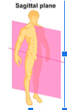
any vertical plane dividing the body into left and right
what is the midsaggital
a saggital plan that goes through the middle
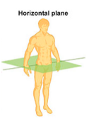
what is the horiztontal plane? aka transverse
cuts across the body horizontally
creates a superior and inferior
what is the frontal plane? aka coronal?
vertically dividies into:
front (anterior / ventral)
back (posterior / ventral)
explain:
median:
lateral:
proximal
distal
dorsal
ventral
anterior
posterior
rostral
caudal
Median = Symmetric in the middle
Lateral = not on median
Proximal = closer to the origin
Distal = further from the origin
Dorsal = towards the spine (diff for quadrupeds) / towards back
Ventral = towards the belly / towards front
Anterior = towards the front
Posterior = towards the back
Rostral = towards the front of the head.
Caudal = towards the tail / rear / coccyx
what is the dorsal part of the tongue?
the top part - as in its resting position, the tongue touches the roof of the mouth
what are the types of tissues
epithelial
connective
muscle
nervous
what is the def / features / examples of epithelial tissues?
covers body surfaces + body cavities
features: widely vary BUT no blood vessels, firmlt attatched to connective tissue and other side is exposed to: external environment/ inside of organ or vessel = lumen
can also be strongly attatched to each other = barrier
e.g skin outer layer, trachea, sweat glands
explain the definition of connection tissues/ function and examples
makes up the majority of the body - protects body organs / binds organs together
bones, fat, ligaments, tendons
what are key features of connective tissue?
intercellular matrix = protein fibres, ground substances, fluid = non-cellular + spongy + absorbs water
Resident cells that live/function within the tissue = repair, maintain + support
Fibres
Collagen = high tensile strength → doesn’t stretch much (most abundant fibre e.g ligaments, tendons)
Elastin = elastic, stretches without breaking (e.g arteries, ears)
Reticular = thin collagen fibres, form delicate structural networks → supports cells + soft tissue (e.g immune tissues, liver)
Cells
Fibrocyte = cells that help maintain fibres
Chondrocyte = cells that maintain cartilage
Osteocyte = cells that maintain cartilage
Ground Substance
Composed of gelatinous proteoglycan (protein + sugar) = can hold a lot of water = spongy = resists compressive forces
what are the 2 types of connective tissues?
loose connective tissue = lots of matrix, less fibre = fat
dense connective tissue = lots of fibre, less matrix = cartilage, bone
function / types of muscle tissue:
function: Responsible for movement (contraction)
types:
Skeletal Muscle = type of muscle tissue under voluntary control
Usually attached bone to bone
Muscles of facial expression
Origin is mainly up / proximal
Insertion = mainly down / distal
Cardiac Muscle = for the heart + also involuntary control
Smooth Muscle= under involuntary control
nervous tissue function
Initiates and transmits nerve impulses through neurons and nerve cells
3 types of joints based on structural classificaiton
fibrous joints
cartilaginous joints
synovial joints
def / features / examples of fibrous joints
Def | Joints connected by by dense connective tissue > made up by tightly packed collagen |
Features | Mostly immobile or very slightly movable
|
Examples | Cranial suture, radioulnar |
def / features / examples of cartilaginous joints
Features | Joints where bones are connected entirely by cartilage |
Function | Slightly movable (usually - otherwise immovable) |
Example | pubic symphysis, joints between vertebrae 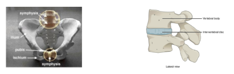 |
def / function of synovial joints
Def | Freely movable joints where bones are separated by a synovial cavity enclosed by a joint capsule |
Function |
|
key features of synovial joints (6)
Articular capsule / joint capsule = strong connective tissue reinforced by ligaments
Blends with periosteum = dense connective tissue that covers bone
Strong + flexible = prevents dislocation
Synovial Membrane:
Lines the articular capsule
Thin, shiny and vascular
Secretes synovial fluid
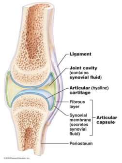
Synovial Fluid = decreases friction between the bones
Plasma-like w/out clotting factors
Low viscosity = well lubricated
Joint Cavity/synovial cavity = space between articulating bones > filled with synovial fluid
Articular Cartilage
hyaline cartilage that covers the two bones
Glassy, smooth = decreases friction
Contains collagen (high tensile strength) + elastin = resists pressure
Some joints also have:
Articular disc of cartilage = allows for a better fit between bones by dividing the joint into 2 compartments
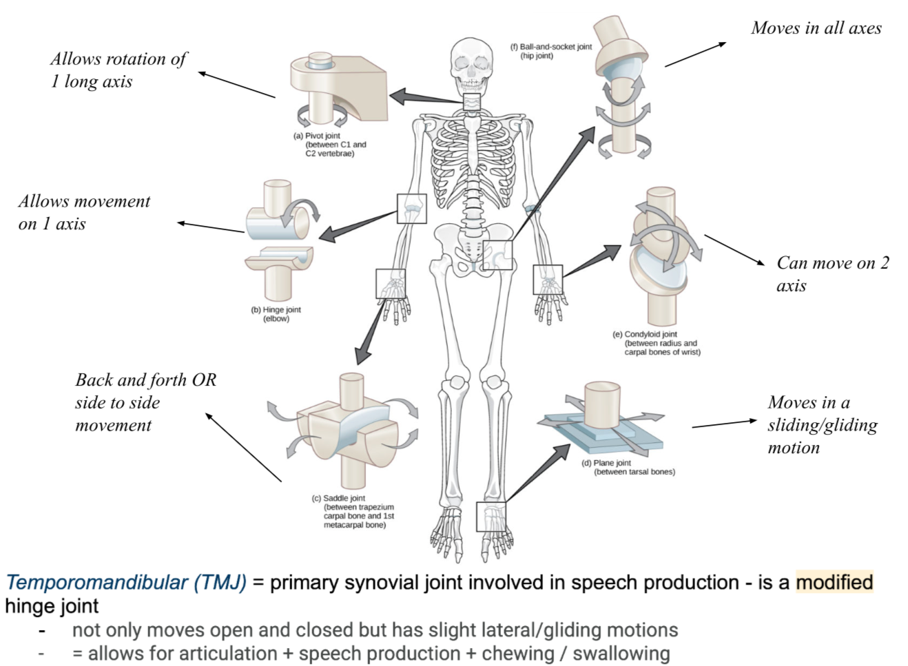
what are the 6 types of synovial joints and an example?
what is a ligament?
Ligament: a stabilising function that joins bone to bone
Proprioception: sense of we know where our body is
the muscles, ligaments will give signals to the sensory organs to help understand where the body is even if the eyes are closed
tell me about mechanical constraints:
Aligned with line of pull → will constraint at the end of range
Prevents unwanted movement + limits wanted movement
When moving the ligament on the opposite side of the direction you move will become tighter hence limiting movement, while the ligament on the side of direction will become loose.
what is flexion and extension?
Flexion | the angle of the joint is decreased = the bones move towards each other (flexing the biceps) | 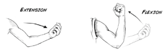 |
Extension | the angles at the joint increases = the bones of the joint move further away from each other |
what is abduction and adduction?
Abduction | movement of a limb away from the midline of the body When you abduct someone you take it away from its rightful place | 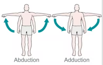 |
Adduction | movement of the limb towards the midline of the body |
what is rotation?
Rotation | Turning a joint around its horizontal axis
| 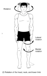 |
what is circumduction?
Circumduction | circular movement of a body part that makes a cone-shaped airspace e.g arm circles | 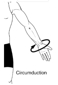 |
what is mandibular elevation and depression?
Mandibular Elevation | Closing the mouth = LIFTING the mandible | 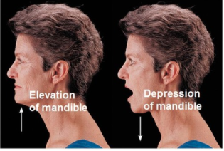 |
Mandibular Depression | Opening the mouth = LOWERING the mandible |
what is protraction and retraction?
Protraction | (of the jaw): the mandible moves anteriorly = pushes the chin forward | 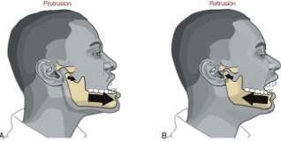 |
Retraction | The mandible moves posteriorly = pulls the jaw backward |
what are the 2 types of muscle contraction?
isometric
isotonic
what is isometric contraction?
Isometric = contracting with the same length/ no change
E.g sustaining vocal fold tension while holding a pitch
what is isotonic contraction and its two types?
Isotonic = contracting with a changed length
Concentric = shortens + bulges
Insertion moves CLOSER to origin
Closing lips for plosives (/p/ or /b/ sounds)
Eccentric = lengthens + overcomes forces
Controlling jaw opening slowly
what is articulation in terms of anatomy?
articulate refers to the point where two or more bones, or bone and cartilage, come together to form a joint.
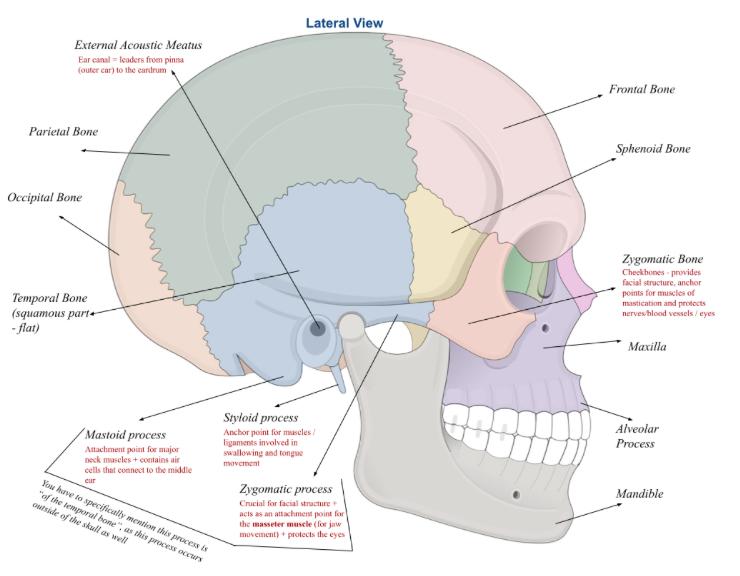
lateral view diagram:

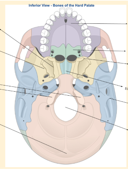
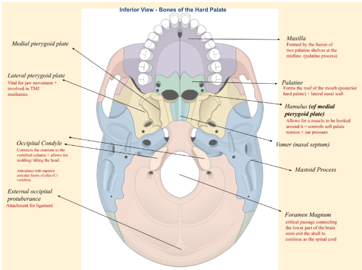
inferior view of hard palate:
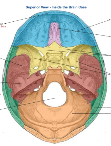
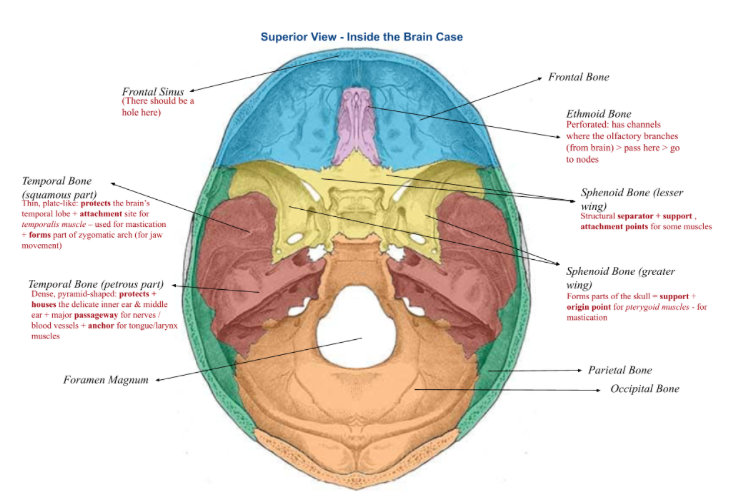
superior view of brain case
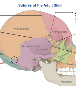
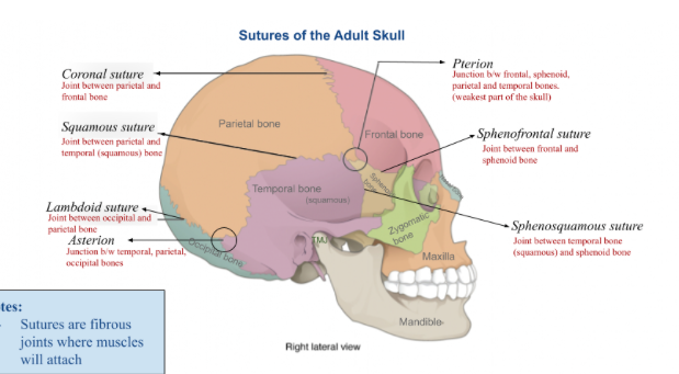
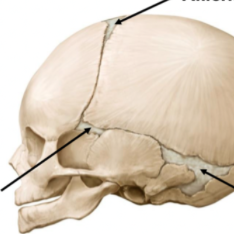
sutures of adult skull
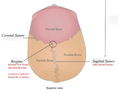
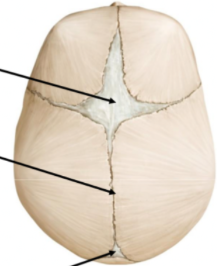
superior view of sutures

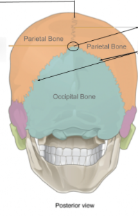
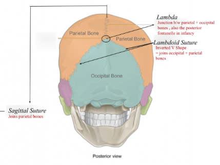
posterior view of sutures
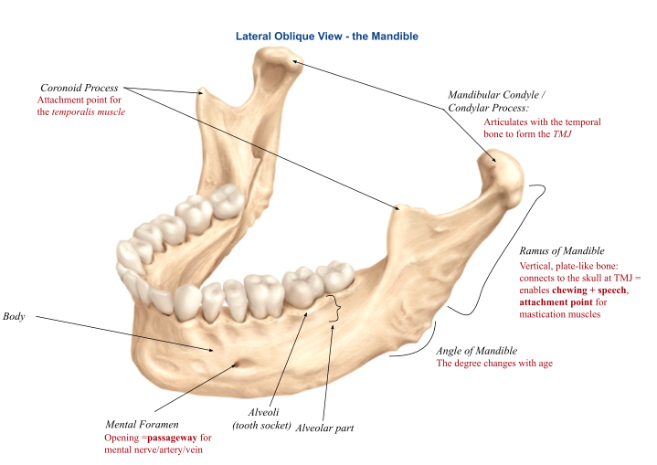
lateral oblique view of mandible
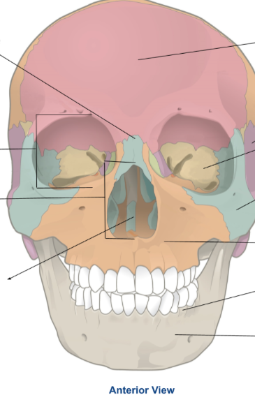
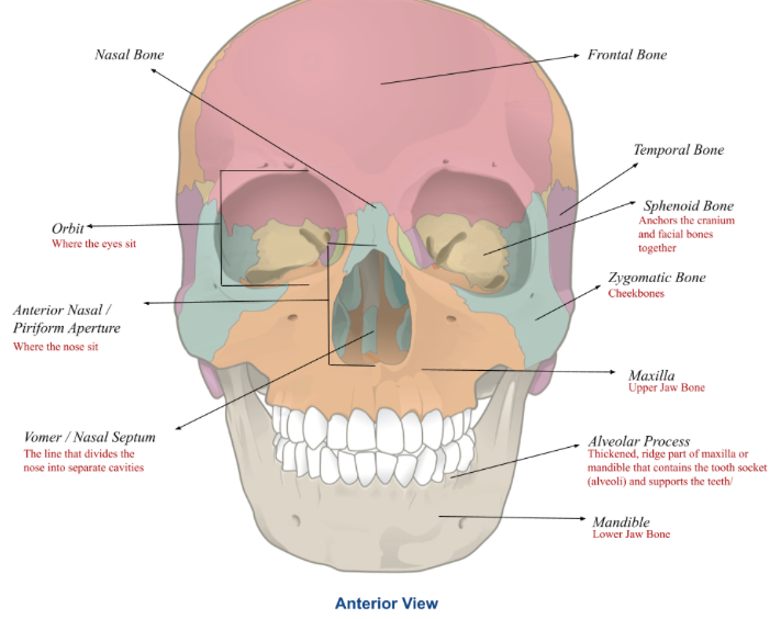
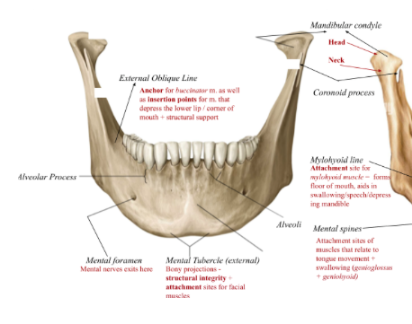
frontal view of mandible

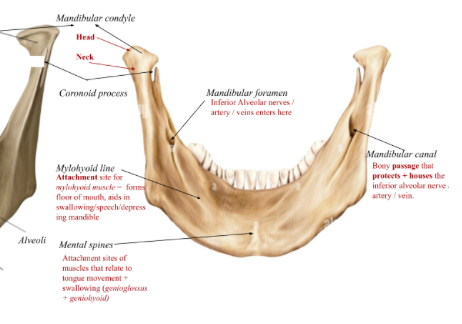
posterior view of mandible

name the paired and unpaired bones in the neurocranium:
frontal bones (1)
parietal bones (2)
occipital (1)
temporal (2)
sphenoid (1)
ethmoid (1)
key features of frontal bone:
houses frontal sinus
key features of parietal bone:
both bones joined medially by the saggittal suture
key feature sof the occipital bone
Foramen magnum: allows spinal nerves etc to go from brain > spinal cord
Articulates with the atlas (C1 vertebrae) = nodding / turning of head
Paired occipital condyles is joint to the atlas
key features of temporal bone:
5 distinct parts that fuse after birth (critical for hearing + balance structures)
Squamous = thin, flat-like part:
Incl zygomatic process > articulates with zygomatic bone to create cheekbone (zygomatic arch)
Attachment point for masseter m. (jaw movement)
Mastoid = posterior part
Mastoid process = attachment site for neck muscles
Petrous = bumpy, dense area (hardest bone)
Houses internal ear + internal acoustic meatus
Tympanic = u shaped plate
Forms the areas of the external acoustic meatus (ear canal)
Styloid Process = pointed protrusion that extends down
Anchor for muscles / ligaments of the tongue and larynx
key features of sphenoid bone:
“Bat-shaped”
Centre: contains sphenoidal sinuses
Has paired lesser wings + greater wings
Pterygoid process = medial pterygoid plate
Hamulus (of medial…): allows for muscle to be hooked around > controls soft palate tension + ear tension
Lateral pterygoid plate: vital for jaw movement + TMJ mechanics
key features of the ethmoid bone:
“Spongy” + perforated: olfactory nerves pass through = smell
Structure of nasal cavity
name the unpaired and paired bones in viscerocranium:
mandible (1)
vomer / nasal septum (1)
maxillae (2)
zygomatic bones (2)
nasal bones (2)
lacrimal bones (2)
palatine bones (2)
inferior nasal conchae (2)
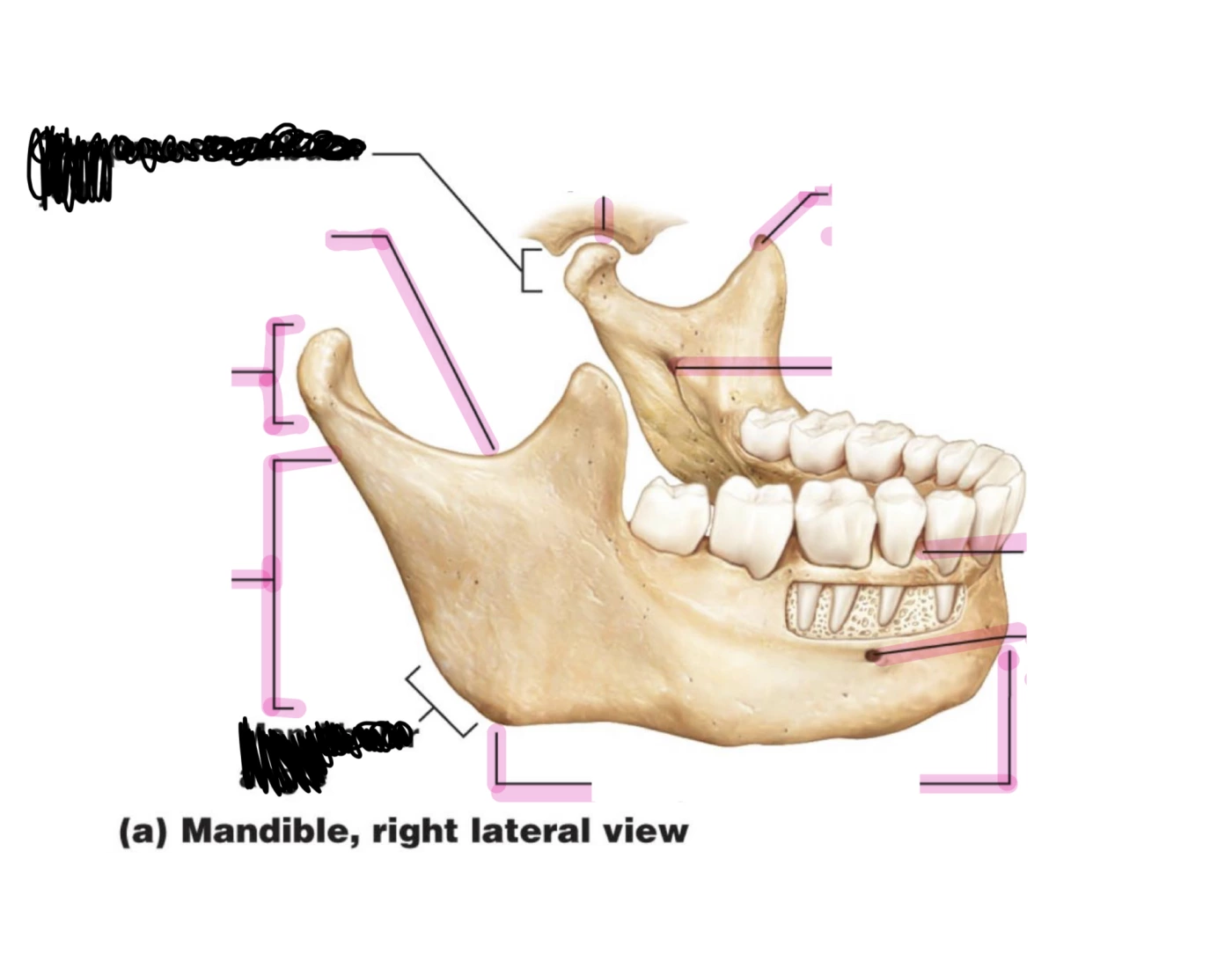
what are key features of the mandible:
mandibular condyle, ramus, cornoid process, external oblique, angle, mental foramen, mental tubercle, alveolar part, mandibular foramen, mandibular canal, mylohoid line, mental spine
U-shaped horizontal body:
Mandibular Condyle: rounded projection that articulates w temporal bone = Temporo-Mandibula Joint (TMJ)
Ramus of Mandible: extend upwards from mandible body
Coronoid Process: temporalis muscle attachment
External Oblique Line: anchor for buccinator m. + m that depress lip/mouth
Angle of Mandible
Mental Foramen: for passage of nerves / vessels
Mental Tubercle (external): structural integrity + m attachment sites
Alveolar Part: the area where the tooth sits, (Alveoli = tooth socket)
Mandibular Foramen: inferior alveolar nerves/artery/veins enter here
Mandibular Canal: passage that protects + houses inferior alveolar nerve/artery/vein
Mylohyoid line: attachment site of mylohyoid m > floor of mouth + swallowing/speech/depressing mandible
Mental Spine: attachment site of m that allow tongue movement + swallowing
what is the vomer?
Line that divides the nose into separate cavities
key features of the maxillae?
Pair of pyramid-shaped bones > fused at midline = upper jaw
Central part: maxillary sinus/paranasal sinus
Alveolar process: thickened ridge > holds upper teeth
Palatine process: horizontal plate > forms anterior ¾ of hard palate (roof of mouth)
Frontal Process: articulates with frontal / nasal / lacrimal bones
Zygomatic Process: articulates with zygomatic bone
It is immovable - does not move
zygomatic bones - key features?
Origin point for the masseter muscle (jaw closure)
Attachment point for temporal fascia
what is the nasal bones?
forms the bridge of the nose
what is the lacrimal bones?
smallest , most fragile bones in facial skeleton
Forms part of bony pathway that drains tears from the eye into the nasal cavity
what is the palatine bones?
Support key functions: separates oral + nasal cavities, facilitates speech, allows breathing + aids in chewing/swallowing
what is the inferior nasal conchae?
Filtres, heats and humidifies inhaled air due to its high surface area + vascularity
Sinus protection
what are the 5 cranial sutures and their locations?
Coronal | frontal bone + 2 parietal bones | ||
Sagittal | Two parietal bones (along the midline) | ||
Lambdoid | Parietal bones + occipital (at back of skull - inverted V shape) | ||
Squamous | Temporal + parietal (on each side) | ||
Metopic | Two frontal bones (from top of head to forehead) | ||
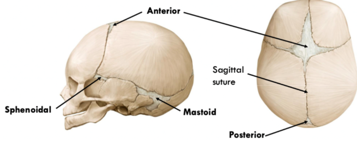
what are the suture junctions (4) and their locations?
Asterion | b/w temporal, parietal, occipital bones | ||
Pterion | b/w frontal, sphenoid, parietal and temporal (weakest part of the skull) | ||
Bregma | B/w frontal and parietal bones
| ||
Lambda | b/w parietal and occipital bones
| ||
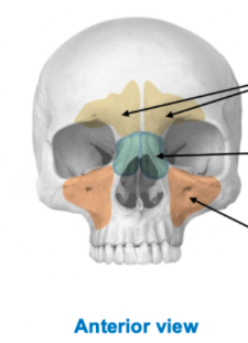
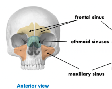
paranasal sinuses:
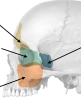
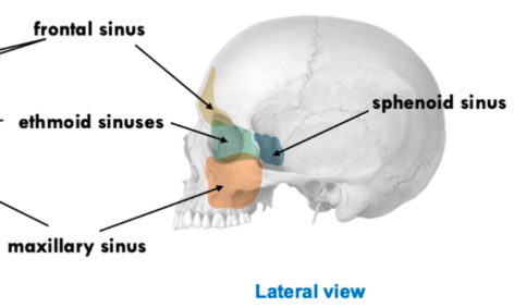
paranasal sinuses lateral view
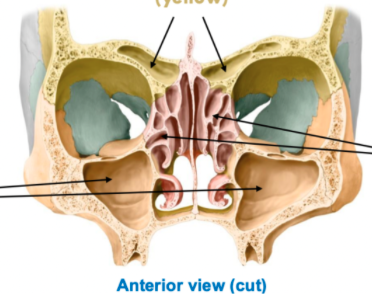
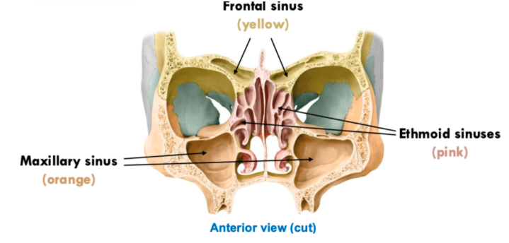
functions of sinuses:
Functions |
|
Infection | If the sinus cavities become infected = sinusitis
|
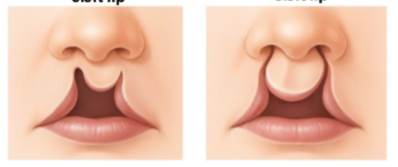
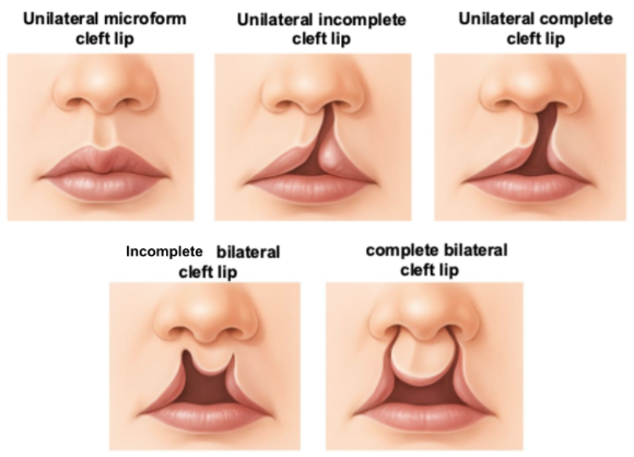
how is a cleft lip formed and its effect + treatment?
Cause | Tissues in baby’s face/ mouth don’t come together properly before birth
|
Effect | Physical malformations, Disrupts normal airflow + velopharyngeal closure, effects breathing, speech, swallowing Leads to:
|
Treatment/Management | Surgery, Speech Therapy, OT, ENT, Orthodontic care, neurologist, audiologist |
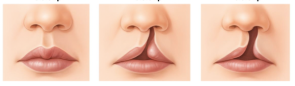
name the cleft lips shape
what does ossification mean?
development of bone
what is the endochondral ossificaiton and its examples?
Bone develops by replacing a pre-existing cartilage model
e.g cranial base, sphenoid, ethmoid, petrous temporal bone, basilar occipital region
what is membranous ossification?
Bone develops directly from mesenchyme without a cartilage scaffold
e.g flat bones of cranial vault (frontal, parietal, squamous temporal, parts of occipital, most of facial bones in viscerocranium
mesenchyme = A loosely organised tissue that develops into connective + skeletal tissues
what is appositional growth?
bone increases in size by adding new layers to its outer surface by bone-making cells (osteoblasts)
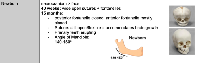
what are fontanelles?
a place b/w bone sin skull where ossification is NOT complete + sutures are not fully formed
Accommodates for the baby’s growth + fuses as it grows
name these fontanelles?
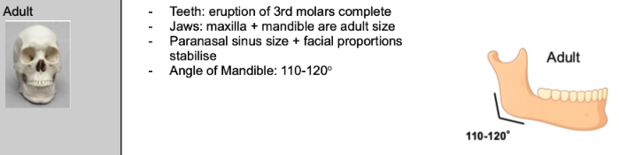
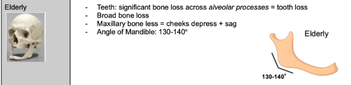
how does sinuses change with age?
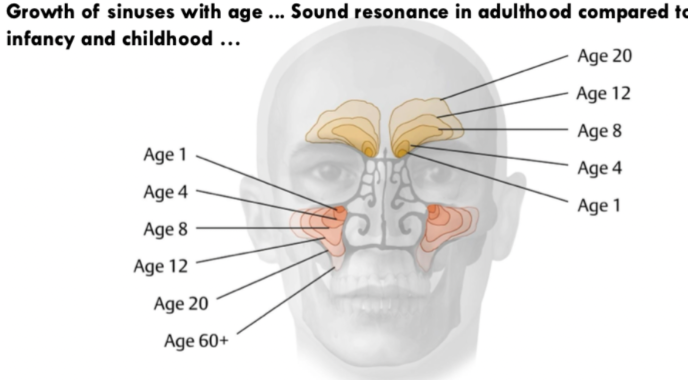
why does the angle of mandible change with age?
Masticatory forces from muscles (attached to the jaw) affect the angle
Angle is reduced when masticatory forces increase
how does the skull is as a newborn?
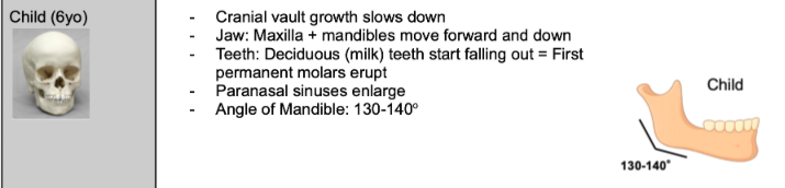
how is the skull as a child?
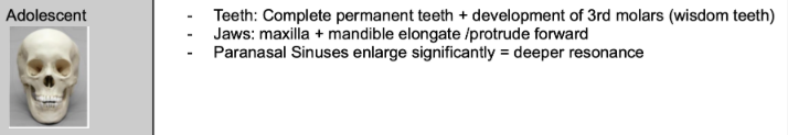
how is the skull as an adolecesent?
how is the skull as an adult?
how is the skull as an elderly?
what type of joint is the TMJ
synovial biaxial condylar joint
during the second stage of the mouth opening, what movement occurs?
anterior gliding of the condyle and disc onto the articular tubercle
which muscle primarily protrudes the mandible?
lateral pterygoid
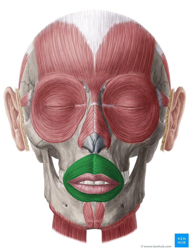
which muscle rounds the mouth for the /oo/ sound?
orbicularis oris
which fontanelle corresponds to what cranial suture junction?
Bregma → anterior fontanelle
Lambda → Posterior fontanelle
Asteroid→ Mastoid Fontanelle
Pterion→ Sphenoid Fontanelle
Describe the TMJ:
Synovial, biaxial condylar joint formed b/w the mandible and the temporal bone
Articular surfaces: articular tubercle + mandibular fossa with the condylar process (head)
Has a loose but strong joint capsule
what is the TMJ’s function?
Allows movement of the mandible in relation to the maxilla (which is immobile)
Mastication (chewing): approx jaw opens 50mm
Speech: small but important movements
Mandibular movement influences: lip posture, tongue position, oral cavity configuration, laryngeal height
what are the 4 key features of the TMJ and their description + functions:
Mandibular Condyle
Articular Tubercle + Mandibular Fossa
articular tubercle = prevents the mandible from moving further upwards
Articular Disc: A wedge of fibrocartilage
Has 2 synovial membranes that line each cavity
Function: Increases congruency, range of motion + absorbs shock, divides the TMJ into superior / inferior compartments
Shape: biconcave structure
Attachments: Lateral pterygoid (anteriorally), Head of Mandible (anteriorly), Joint capsule (circumferentially)
Retrodiscal Tissue:band of connective tissue that contains nerves that attaches the articular disc (posterior) → mandibular fossa + mandibular condyle
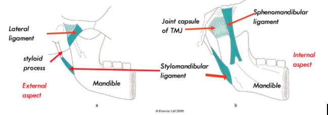
what are the two TMJ ligaments and its function?
Lateral (temporomandibular) ligament: strengthens TMJ joint capsule laterally + holds it taut in all movements
Prevents inferoposterior condylar displacement or excess lateral movement
Stylomandibular ligament + Sphenomandibular ligament = accessory ligaments >> prevents excessive mandibular opening
what are the 3 key positions of the TMJ and the mandible?
Resting Position: 3-4 mm teeth separation
Close-packed position: molars firmly clenched, overlap incisors, mouth closed
the most stable position >> the muscles will try to act + ligaments make It taut
Provides max congruency, condyle lies within the articular fossa = prevents further upward movement
Closed Position: the joint capsule / natural ligaments / muscles: medial pterygoid, masseter and temporalis help keep the mandible and TMJ closed
what allows for the stability of the TMJ?
Disc shape + attachments: Strongly attached, deformable pad improves mechanical fit, allows rotation + sliding movements to occur
Ligament: Lateral ligament prevents dislocation
Muscles: Temporalis, lateral pterygoid provide dynamic stability + temp & masseter elevate mandible = stable position
what muscles are key in moving the mandible?
temporalis, masseter, lateral pterygoid, medial pterygoid, suparhyoid muscles = geniohyoid / mylohyoid / ant. belly of diagastric
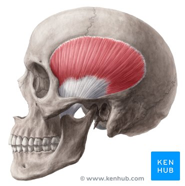
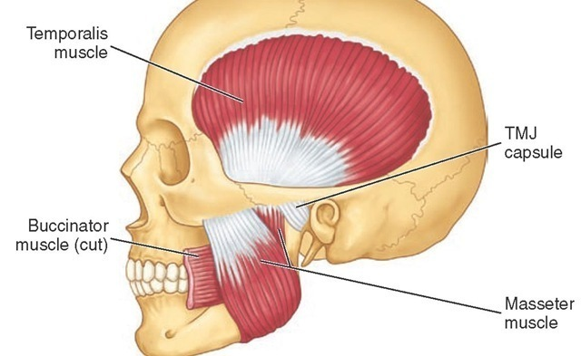
for the temporalis muscle: what is its location / shape / origin / insertion / actions when contracted / its fibres and their actions?
Temporalis (fan-shaped muscle): (retraction - pos / elevation - rest)
Origin: floor of temporal fossa/passes deep in the zygomatic arch/ insertion: coronoid process + ramus (ant.)
Actions: elevation + retraction (when muscles contract)
Posterior Fibre: retraction (more horizontal fibres - travel > arch > coronoid process)
Middle / Anterior Fibres: elevation (more vertical fibres)
for the masseter muscle: what is its location / shape / origin / insertion / actions when contracted / its fibres and their actions/ the two types?
Masseter (on the jaw) = stabilises the jaw (elevation, partially protrusion + retraction) > primary jaw closer + chewing muscle
Superficial:
Origin: maxillary process on zygomatic bone / insertion: lateral surface of mandibular ramus + angle
Action: Elevation + Protrusion
Deep
Origin: zygomatic arch / insertion: ramus of mandible
Action: Elevation + Retraction
= used for mastication
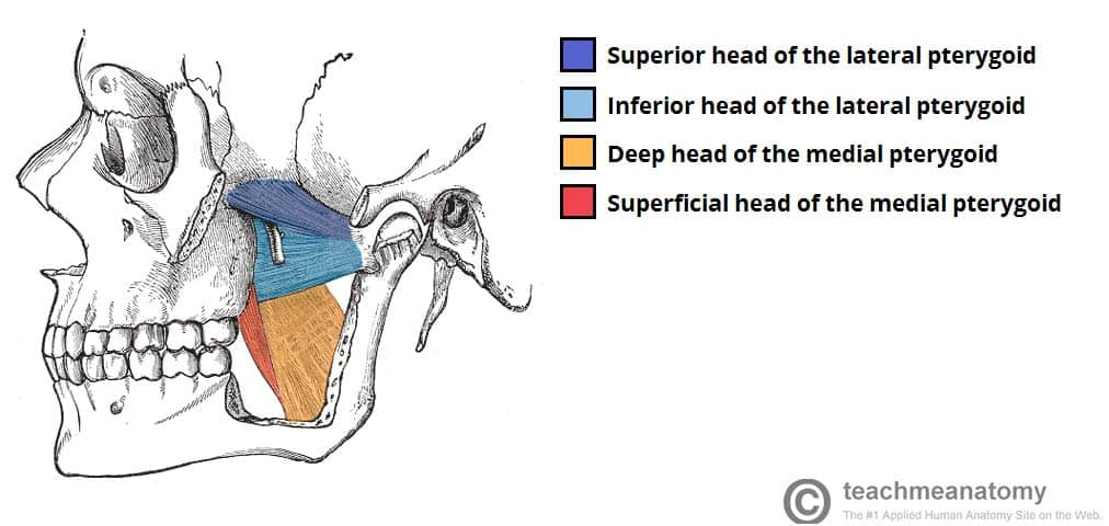
for the lateral pterygoid muscle: what is its location / shape / origin / insertion / actions when contracted / the two types?
Lateral Pterygoid (protrusion + depression)
Superior Head > Origin: greater wing of sphenoid / insertion: neck of mandible
Inferior Head > Origin: lateral pterygoid plate of sphenoid / insertion: neck of mandible
Actions: Protrusion, Depression
Purpose: controls slight forward movement for coordination with lips

for the medial pterygoid muscle: what is its location / shape / origin / insertion / actions when contracted / the two types?
Medial Pterygoid (elevation + protrusion) > has same direction as masseter so moves like it BUT a bit deeper, so not as impactful on mandible
Superficial Head > origin: maxilla / insertion: internal surface of angle of mandible
Deep Head > origin: lateral pterygoid plate of sphenoid / insertion: internal mandible angle
Actions: elevation + protrusion
helps close the jaw + stabilise side-to-side movement
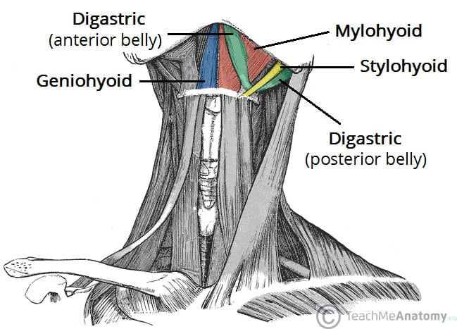
for the suprahyoid muscle: the types of muscles / what is its location / shape / origin / insertion / actions when contracted
Geniohyoid → superior to mylohyoid
Mylohyoid → covers floor of mylohyloid
Ant. Belly of Diagastric →inferior to mylohyoid
Joined from “chin” >> joined to diagnostic fossa
Main function: moves hyoid bone (which is above the larynx and is suspended within the body)
If hyoid bone is fixed, the muscles will help depress the mandible
If the mandible is fixed, the hyoid will be elevated
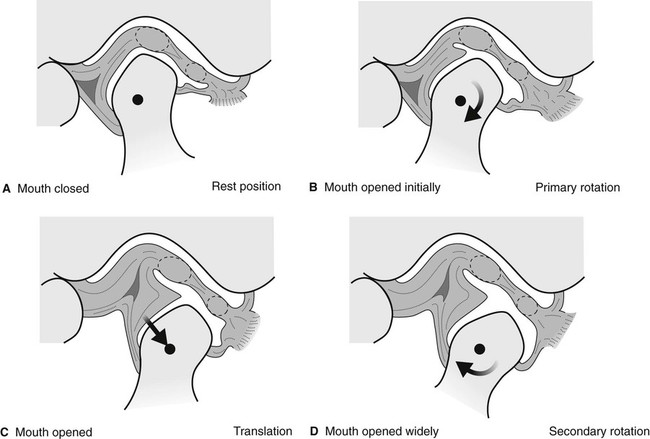
outline the process of the mouth opening and closing within the TMJ:
Resting Position: open packed jaw position, connective tissue is loose
Medial + lateral pterygoid muscles relax
Teeth not in contact, mouth slightly open
Isometric contraction of temporalis + masseter
Rotation: first half of mouth opening (20-25mm) >> inferior compartment of TMJ
Condylar process rolls anteriorly
Eccentric contraction of temporalis
Inferior portion of lateral pterygoid tenses
Superior lateral pterygoid relaxes
Translation: second half of mouth opening >> superior compartment of TMJ
Disc + condylar process glide anteriorly
Eccentric contraction of temporalis
Superior + inferior lateral pterygoid contracts concentrically (shortens)
Connective tissue taut posteriorly
Closure:
Disc + condylar process glide posteriorly
Masseter (+ post temporalis, eccentric lateral pterygoid)
Connective tissue returns to rest
name key facial muscles for speech
orbicularis oris, buccinator, lip muscles, upper lip elevators, lower lip depressors, mentalis
what is the orbicularis oris?
circular muscle that surrounds the mouth
For Speech: Closes the lips / rounds the lips / protrudes the lips
Bilabial sounds: /p/ /b/ /m/ = requires 2 lips to close together
Labiodental Sounds: /f/ /v/ > requires controlled lip positioning against teeth
Rounded Vowels: /u/ (food), /o/ (go) > requires lip rounding + protrusion
If weak / impaired = difficulty in closing or rounding lips
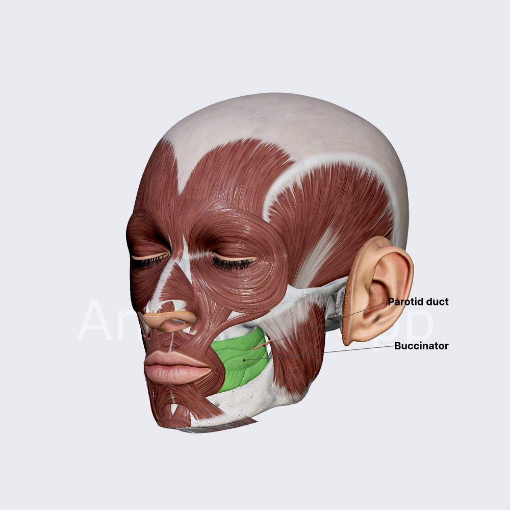
what is the buccinator?
Muscles in the cheek
For Speech: keeps cheeks tight against teeth / prevents air from collecting in cheeks while speaking / maintains inner mouth pressure = builds up air pressure then releases it
Plosives (stops): /p/ /b/ /t/ /k/ = need air pressure buildup b4 the sound
Fricatives: /s/ /f/ /v/ = controlled airflow thru narrow space
If weak / impaired = air may escape into the cheeks > reduces speech precision