Cell Cycle
1/36
There's no tags or description
Looks like no tags are added yet.
Name | Mastery | Learn | Test | Matching | Spaced | Call with Kai |
|---|
No analytics yet
Send a link to your students to track their progress
37 Terms
what are the stages of the cell cycle in eukaryotes
G1 phase
S phase
G2 phase
M phase
which stages are interphase
G1 + S + G2 = Interphase
What is G0 phase
some cells enter a resting phase where they aren’t in any stages of the cell cycle e.g neurones
explain the G1 phase and how much of the time it takes
around 45%
cell volume increases
protein synthesis
explain the G2 phase and how long it takes
30% of cell cycle time
rapid increase in volume
checking and repairing DNA before cell division
explain the S phase and how long it takes
20% of the time
DNA replication takes place
DNA helicase and DNA polymerase allow semi-conservative DNA replication to take place
dna is only replicated DNA content doubles but the number of chromosomes stays the same- at the end of the S phase chromosomes are in their replicated form
explain the M phase and how long it takes
5%
mitosis and cytokinesis
how does the DNA content of a cell change over the cell cycle
G1 stays the same
S it double
G2 stays at double
Mitosis stays at 2 then halves during mitosis
explain how the volume of the cell changes of the cell cycle
G1 it increases
S stays teh same
G2 increases again
Mitosis stays the same and then halves back to origional
What are the phases of mitosis
Prophase
Metaphase
Anaphase
Telophase
PMAT
Explain the process of interphase
the cell has 2 pairs of chromosomes (more in real life but just for this example)
individual chromosomes are not visible
g, S G2 takes place
explain the process of prophase-3
the chromosmes condense to become visible
the nuclear envelope breaks down
cetrioles migrate to opposite poles of the cell
explain the process of metaphase
spindle fibers made by the centrioles attach to the centromeres of each chromosome
the chromosomes are moved to the equator as a single line/layer
explain the process of anaphase
the spindle fibres are retracted , the centromeres split and the chromatids (now chromosmes)are pulled to opposite poles
explain the process of telophase-3
the chromosmes decondense
nuclear membrane reforms
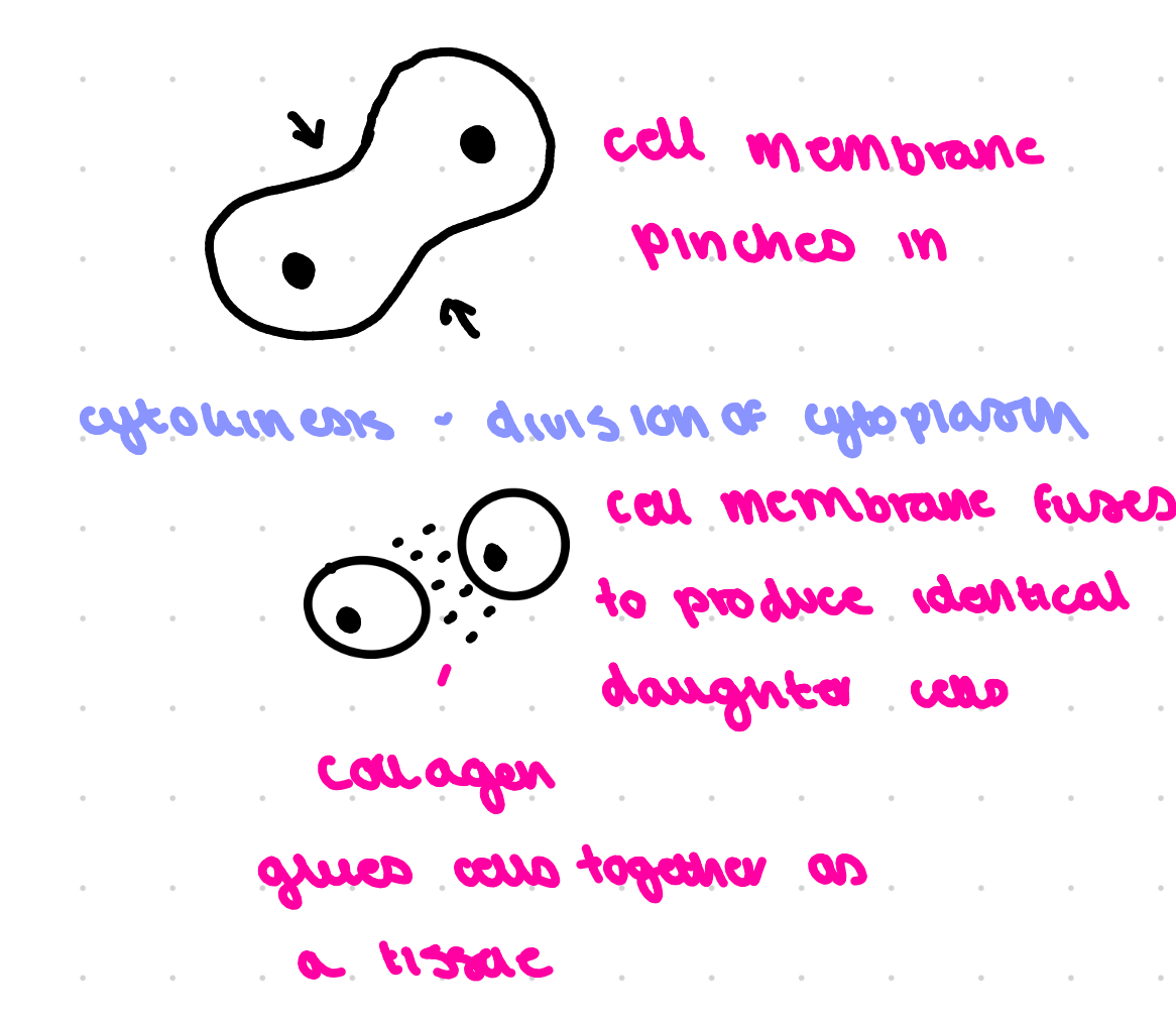
whilst telophase happens cytokinesis takes place to produce idenitcal daughter cell (splitting of the cytoplasm)
describe the strucutre of a replicated chromosme
2 identical sister chromatids held together by a centromere in the centre - this is one chromosome
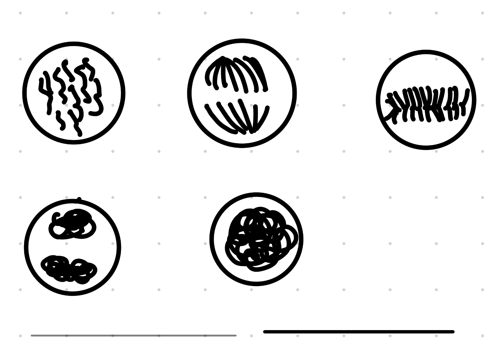
How can you identify the different stages of the cell cycle on a picture
Metaphase- lining up in the middle, should look like a big clump mascara eye
Anaphase- two clumps being split apart
Telophase-two clear sections with no connection
Prophase- can see individual chromosomes not just a black circle
Put these vents in order
A chromatids separate
B nuclear envelope disappears
C cytoplasm divides
D chromosomes condense and become visible
E chromosomes on the equator of the spindle
D B E A C
Name the phase in which DNA replication occurs
Interphase/ S phase
Describe the role of the spindle during mitosis
Attaches to the centromeres of chromosomes positions on the equator/ retract and separate chromatids
In which stages would the chromosomes be visible and would consist of a pair chromatids joined together
Prophase and metaphase
Give two reasons that when done on another day the garlic root tip has a different results
Warmer day/ different time of day
Different plant
Anaphase is a stage in mitosis describe how anaphase results in identical cells being produced- 3
Identical chromatids are pulled to their opposite poles by spindle fibres by their centromeres
Describe the changes that occur from early prophase to late prophase
nuclear membrane breaks down
Chromosomes condense Into visible chromosomes
Centrioles move to opposite poles
Spindle fibres form
Explain why the S-phase is faster at 25 degrees than at lower temperatures-3
Increased temp increases kinetic energy
DNA helicase and DNA polymerase have the required activation energy
Nucleotides are diffusing faster
DNA replication is faster
How do you calculate the mitotic index
(Number of cells in mitosis/total number of cells) X 100
Describe how you can tell from a micrograph what stage of mitosis cells are in
Anaphase- chromosomes are being pulled to opposite poles
There is a lack of DNA at the equator
prophase- the individual chromosomes can now be seen
Interphase- no visible chromosomes can be seen
Metaphase- mascara eye the chromosomes are trying to occupy the equator as a single line
Explain cell division in plant cells
A new cell wall forms down the centre
2 identical daughter cells are produced but the 2 cells stay attached
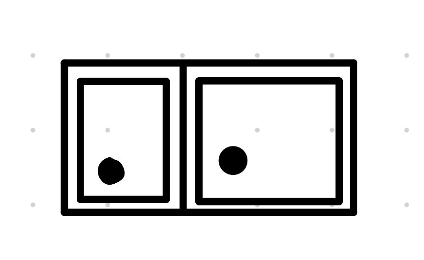
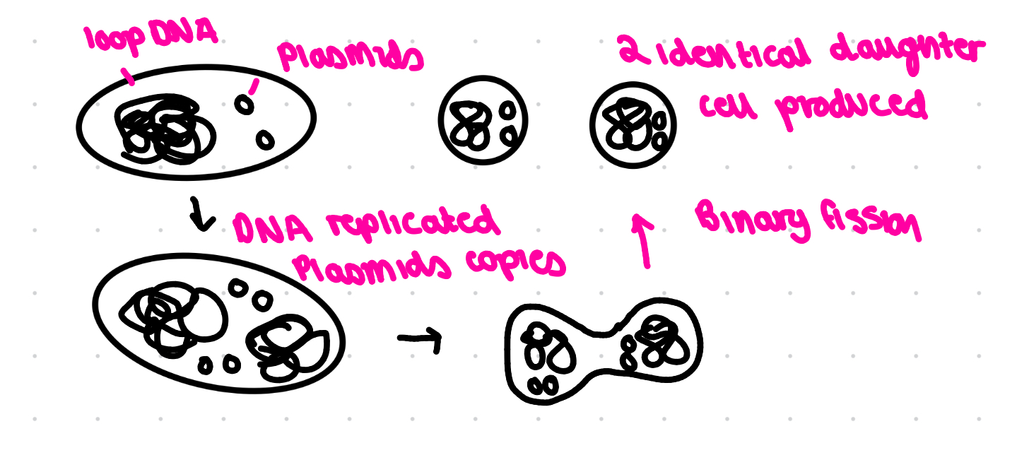
Binary fission
Label each diagram
Prophase anaphase metaphase telophase interphase
Explain animal cell division
Why is the prokaryotic cell cycle faster
They are smaller so have a larger surface area to volume ration so gain nutrients faster e.g make nucleotides for DNA replication
When observing stem cells that are dividing too quickly and therefore forming a tumour the cells often appear smaller than normal and large nuclei are common, suggest an explanation for these 2 features
Smaller as one of the growth phases is missed out therefore when cytokinesis halves the cell is it going to be smaller than it began as
Larger nuclei more likely to have been through s phase therefore dna is replicated
Explain why the s phase and the m phase take the same amount of time for a tumour cell and a normal cell
S phase is when dna is replicated, tumour/ normal cells have their same amount of DNA to replicate
M phase mitosis takes the same time.g same number of chromosomes to move in the cell
A drug has been developed to prevent anaphase , it is readily absorbed by cells in the ileum , evaluate the drug as a potential treatment for tumours forming in the human ileum
For:
It is absorbed by the cells we want to treat
Stops anaphase so prevents cell division
Against:
Data is for mice not human- human cells may react differently
Side effects as all cells in the ileum not just tumour cells would be effected
Suggest why the mitoitc index is a useful metric to record
The data can be compared to e.g normal range
The data can be plotted
Statistical work can be carried out
Explain a method to see root tips under the microscope
Cut 1 cm of a garlic root tip with scissors, make sure you know which end is the tip
Using a scalpel, cut around 2mm from the growing root tips on the tile and discard the rest as the tip is where most cell division occurs
Put 6 drops of 50:50 HCl and ethanol into a staining block, place the root tips i and cover with a watch glass for 10 minutes
Use a mounted needle to transfer the root tips to a second the root tip to a second staining block containing 6 drops of 45% glacial acetic acid and cover with a watch glass, eave for 5 mins
Put onto glass slide
Add one drop of toluidine blue stain make the DNA visible
Tap the tissue on the slide 20 times with a glass rod to form a single layer of cells allowing the light to pass through and then cover with a coverslip
Fold a paper towel over the slide and squash
Look under the microscope