Physiology II Exam 2 - Spring 2023
0.0(0)
Card Sorting
1/181
Earn XP
Last updated 1:25 PM on 5/10/23
Name | Mastery | Learn | Test | Matching | Spaced | Call with Kai |
|---|
No analytics yet
Send a link to your students to track their progress
182 Terms
1
New cards
2
New cards
What does sympathetic control?
“Fight or Flight”
3
New cards
What does parasympathetic control?
“Rest and Digest”
4
New cards
Autonomic Nervous System General Features?
Involuntary
\
Visceral
\
Moter/efferent control
\
If the somatic motor system influences skeletal muscle… what does the autonomic nervous system influence?
\
Visceral
\
Moter/efferent control
\
If the somatic motor system influences skeletal muscle… what does the autonomic nervous system influence?
5
New cards
Visceral Motor Effectors
Smooth Muscle
Smooth Muscle
Within vasculature
(vascular smooth muscle)
\
Within walls of GI tract
\
Sphincters (GI and GU)
\
Within walls of bladder
\
Within eye:
Iris (dilates and constrict pupil)
Ciliary (near/far accommodation)
(vascular smooth muscle)
\
Within walls of GI tract
\
Sphincters (GI and GU)
\
Within walls of bladder
\
Within eye:
Iris (dilates and constrict pupil)
Ciliary (near/far accommodation)
6
New cards
Visceral Motor Effectors
Heart
Heart
Cardiac muscle in walls of ventricles and atria
\
SA and AV nodes
\
SA and AV nodes
7
New cards
Visceral Motor Effectors
Glandular
Glandular
Sweat glands
\
Salivary glands
\
Pancreas
\
Stomach
\
Salivary glands
\
Pancreas
\
Stomach
8
New cards
Visceral Motor Effectors
Miscellaneous
Miscellaneous
Adipose Tissue
\
Liver
\
Kidney
\
Liver
\
Kidney
9
New cards
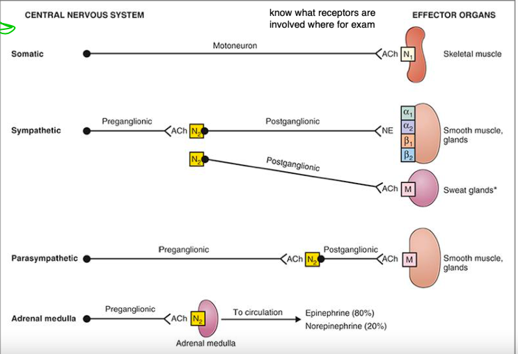
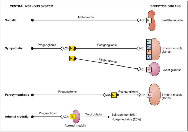
Autonomic Nervous System
Anatomical Features:
2 peripheral neuron system
Synapse on autonomic ganglia
Each neuron is differentiated by term:
*Pre-ganglionic (before ganglion)*
*Post-ganglionic (after the ganglion)* - goes to the target organ
\
2 peripheral neuron system
Synapse on autonomic ganglia
Each neuron is differentiated by term:
*Pre-ganglionic (before ganglion)*
*Post-ganglionic (after the ganglion)* - goes to the target organ
\
10
New cards
Central Nervous System
*Know this chart*
11
New cards
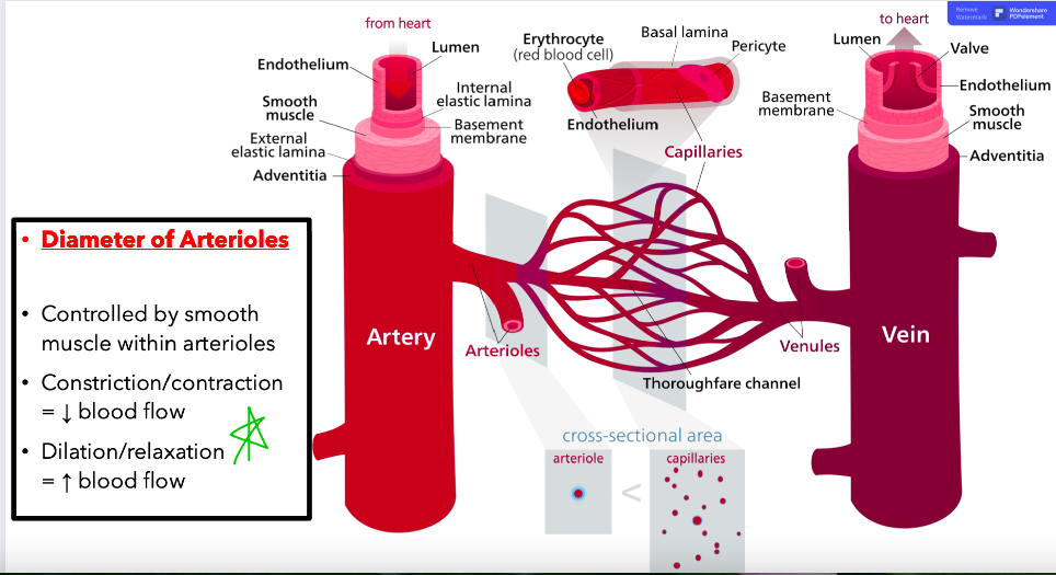
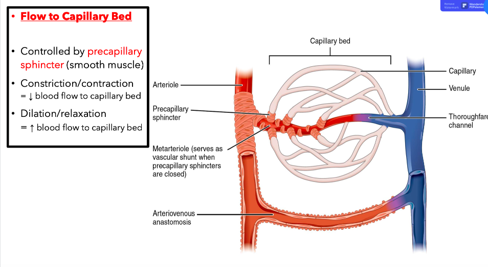
Control of Local Blood Flow: Sympathetics
Flow of Capillary Beds:
Precapillary sphincters restrict flow to capillary beds
Able to control the amount of blood within local capillary circulation (mesenteric circulation)
\
Diameter of Arterioles:
Vasoconstriction → decreased blood flow (ex. vasoconstriction of arterioles supplying GIT during times of stress/anger)
Vasodilation → increased blood flow (ex. vasodilation of arterioles supplying skeletal muscle during exercise)
12
New cards
Diameter of Arterioles
Image
Image
*See Image*
13
New cards
Flow to Capillary Bed
Image
Image
*See Image*
14
New cards
Neurotransmitter and Receptors Intro
Each division of the autonomic nervous system has its own set of neurotransmitters and receptors
\
The unique function of each division is directly related to these
\
The function depends entirely on which receptors are present on which organs/tissues
\
The unique function of each division is directly related to these
\
The function depends entirely on which receptors are present on which organs/tissues
15
New cards
Neurotransmitters and Receptors Continued…
Norepinephrine
NE
Sympathetic Post-ganglionic
*neurotransmitter released at target organ*
\
Acetylcholine
ACh
Pre-ganglionic (sympathetic and parasympathetic)
Parasympathetic post-ganglionic
NE
Sympathetic Post-ganglionic
*neurotransmitter released at target organ*
\
Acetylcholine
ACh
Pre-ganglionic (sympathetic and parasympathetic)
Parasympathetic post-ganglionic
16
New cards
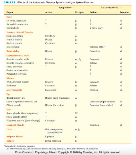
Receptor Chart Image
*See Image*
17
New cards
Receptors of Effector Organs
*Intro*
*Intro*
Adrenergic - NE
Sympathetic
Alpha-1
Alpha-2
Beta-1
Beta-2
\
Cholinergic - ACh
Parasympathetic
Muscarinic
Sympathetic
Alpha-1
Alpha-2
Beta-1
Beta-2
\
Cholinergic - ACh
Parasympathetic
Muscarinic
18
New cards
Receptor *EXCEPTION*
Sympathetic fibers going to sweat glands use the neurotransmitter ACh
\
Receptor is Muscarinic
\
Receptor is Muscarinic
19
New cards
Receptor Chart
*See Image*
20
New cards
Receptors Made Easy
In the *parasympathetic division*, effector organs have *muscarinic receptors*
\
In the sympathetic division there are multiple receptor types in effector organs including the four adrenoreceptors (alpha-1-2, beta 1-2); and in tissues with sympathetic cholinergic innervation, there are muscarinic receptors.
\
In the sympathetic division there are multiple receptor types in effector organs including the four adrenoreceptors (alpha-1-2, beta 1-2); and in tissues with sympathetic cholinergic innervation, there are muscarinic receptors.
21
New cards
Alpha Receptors Made Easy
“Among the sympathetic adrenoreceptors, receptor type is related to function.”
\
The alpha-1: increases smooth muscle activity
Receptors cause contraction of smooth muscle such as vascular smooth muscle, gastrointestinal, and urethral sphincters, pilomotor muscles, and in the radial muscle of the iris.
\
The alpha-2 receptors are rare. The main function is quite different than other receptors. Present on *autonomic nerve terminals and in GI tract*, these receptors inhibit activity of both sympathetic and parasympathetic pathways.
\
All other receptors are on the tissue itself, these are on post ganglionic neurons
\
The alpha-2 receptors are rare. There are 2 types
\
Autoreceptors: Located on sympathetic post-ganglionic neurons.
*Inhibits release of NE (conserves NE), not present in adrenal medulla*
\
Heteroreceptors: Located on parasympathetic post-ganglionic neurons
*Inhibits release of ACh (indirectly inhibits GI activity)*
\
The alpha-1: increases smooth muscle activity
Receptors cause contraction of smooth muscle such as vascular smooth muscle, gastrointestinal, and urethral sphincters, pilomotor muscles, and in the radial muscle of the iris.
\
The alpha-2 receptors are rare. The main function is quite different than other receptors. Present on *autonomic nerve terminals and in GI tract*, these receptors inhibit activity of both sympathetic and parasympathetic pathways.
\
All other receptors are on the tissue itself, these are on post ganglionic neurons
\
The alpha-2 receptors are rare. There are 2 types
\
Autoreceptors: Located on sympathetic post-ganglionic neurons.
*Inhibits release of NE (conserves NE), not present in adrenal medulla*
\
Heteroreceptors: Located on parasympathetic post-ganglionic neurons
*Inhibits release of ACh (indirectly inhibits GI activity)*
22
New cards
Beta Receptors Made Easy
Beta-1:
Activity of heart - All functions of the heart
Increase metabolism - Receptors are involved in homeostatic functions such as, lipolysis, renin secretion
\
Beta-2:
Decrease smooth muscle activity
Receptors cause relaxation of smooth muscle in bronchioles, wall of the bladder, and wall of the GIT
*+relaxation of vascular smooth muscle supplying skeletal muscle*
Inhibits urine
\
\
23
New cards
Sympathetic Nervous System
“Fight or Flight”
\
Originates from lateral gray horn from T1-L2
*Peripheral components of sympathetic nervous system*
\
Called thoracodorsal due to origin from thoracic and upper lumbar spine
\
Originates from lateral gray horn from T1-L2
*Peripheral components of sympathetic nervous system*
\
Called thoracodorsal due to origin from thoracic and upper lumbar spine
24
New cards
Sympathetic Anatomy
Pre-ganglionic fibers (short) originate in lateral gray horn of spinal cord between T1-L2
\
Fibers will travel to synapse on post-ganglionic fibers in one of many sympathetic ganglia
\
\
Fibers will travel to synapse on post-ganglionic fibers in one of many sympathetic ganglia
\
25
New cards
Sympathetic Ganglia
Near the spinal cord
*Paravertebral (sympathetic chain)* - located further from the spine, supply cast area from head down to feet
*Prevertebral* - Located right next to spine, supply visceral organs (celiac, superior mesenteric, inferior mesenteric)
\
Pre-ganglionic fibers can synapse at the same segmental level, or ascend/descend to another level before synapsing on ganglion
\
Reflects the diffuseness of sympathetic function
26
New cards
Sympathetic Ganglia: Adrenal Medulla
Adrenal Medulla: Unique “specialized ganglion”
One autonomic fiber travels directly to adrenal medulla
Activates and releases into blood:
*Epinephrine (80%)* - aka adrenaline, preferentially activates Beta-2 receptors
*Norepinephrine (20%)*
\
Sympathetic NS maintains vascular tons, the apha-1 receptors maintain that tone. If you lesson the constricting signal, the vasculature will dilate.
27
New cards
Sympathetic Anatomy
Post-ganglionic fibers (long) travel from ganglia to effector organ
\
Synapse on effector organ is a diffuse branching varicosity - *Not as specific of a synapse, like a vine growing up a tree, very widespread, wide effect*
\
Sympathetic ganglia: Near the spinal cord
\
Synapse on effector organ is a diffuse branching varicosity - *Not as specific of a synapse, like a vine growing up a tree, very widespread, wide effect*
\
Sympathetic ganglia: Near the spinal cord
28
New cards
Sympathetic Nervous System
Mobilizes for activity
*Danger, Stress, Fear*
\
How exactly does it accomplish this?
\
What needs to happen physiologically, when you are in danger?
*Danger, Stress, Fear*
\
How exactly does it accomplish this?
\
What needs to happen physiologically, when you are in danger?
29
New cards
How is blood pressure raised?
Sympathetic activity causes:
\
Norepinephrine to be released at post-ganglionic synapse
Heart: Beta-1 adrenergic receptors are stimulated by NE
= Cardiac muscle in ventricles increase contractile force.
*Minimal effect*
\
Norepinephrine (NE) to be released at post-ganglionic synapse
Vascular smooth muscle: Alpha-1 adrenergic receptors are stimulated by NE
= Smooth muscle constricts in walls of arterioles.
*Much more notable effect*
\
\
Norepinephrine to be released at post-ganglionic synapse
Heart: Beta-1 adrenergic receptors are stimulated by NE
= Cardiac muscle in ventricles increase contractile force.
*Minimal effect*
\
Norepinephrine (NE) to be released at post-ganglionic synapse
Vascular smooth muscle: Alpha-1 adrenergic receptors are stimulated by NE
= Smooth muscle constricts in walls of arterioles.
*Much more notable effect*
\
30
New cards
How is heart rate raised?
Sympathetic activity causes:
\
Norepinephrine (NE) to be released at post-ganglionic synapse
SA & AV nodes: Beta-1 adrenergic receptors are stimulated by NE
= Increased set rate of depolarization.
\
Norepinephrine (NE) to be released at post-ganglionic synapse
SA & AV nodes: Beta-1 adrenergic receptors are stimulated by NE
= Increased set rate of depolarization.
31
New cards
How is skeletal muscle blood flow increased?
Sympathetic activity causes:
\
Vascular smooth muscle and within skeletal muscles: Beta-2 adrenergic receptors are stimulated by NE
= Increased blood flow to skeletal muscles
\
Vascular smooth muscle and within skeletal muscles: Beta-2 adrenergic receptors are stimulated by NE
= Increased blood flow to skeletal muscles
32
New cards
How is blood glucose increased?
Sympathetic activity causes:
\
Liver: Beta-2 adrenergic receptors are stimulated by NE
= Increased Gluconeogenesis and Glycogenolysis
\
Liver: Beta-2 adrenergic receptors are stimulated by NE
= Increased Gluconeogenesis and Glycogenolysis
33
New cards
How is respiration increased?
Sympathetic activity causes:
\
Smooth muscle of Bronchioles: Beta-2 adrenergic receptors are stimulated by NE
= Dilation of bronchioles increases amount of O2/CO2 exchange
*Causes relaxation of muscle = dilation*
\
Smooth muscle of Bronchioles: Beta-2 adrenergic receptors are stimulated by NE
= Dilation of bronchioles increases amount of O2/CO2 exchange
*Causes relaxation of muscle = dilation*
34
New cards
How is GIT activity decreased?
We need glucose to got to more important things, so this is stopped
\
Sympathetic activity causes:
\
GI Smooth Muscle: Beta-2 adrenergic receptors are stimulated by NE - Muscles are slowed
*In Walls*
\
GI Sphincters: Alpha-1 adrenergic receptors are stimulated by NE - sphincters are activated to decrease the overall GIT activity
*In Walls*
\
GI Smooth Muscle Sphincters: Decreased peristalsis and sphincters close
= Net decrease in GIT activity and digestion
*Thus chronic stress causes GIT issues, especially constipation*
\
Sympathetic activity causes:
\
GI Smooth Muscle: Beta-2 adrenergic receptors are stimulated by NE - Muscles are slowed
*In Walls*
\
GI Sphincters: Alpha-1 adrenergic receptors are stimulated by NE - sphincters are activated to decrease the overall GIT activity
*In Walls*
\
GI Smooth Muscle Sphincters: Decreased peristalsis and sphincters close
= Net decrease in GIT activity and digestion
*Thus chronic stress causes GIT issues, especially constipation*
35
New cards
What is super important to keep fluid balance and blood pressure?
Kidneys!
36
New cards
How is urine formation decreased?
Sympathetic activity causes:
\
Kidney: Beta-1 adrenergic receptors are stimulated by NE
Increased renin secretion (RAAS)
= increase in NA+ resorption by kidney
= decreased urine formation (osmosis)
*Also: decreased blood flow to kidney*
\
Kidney: Beta-1 adrenergic receptors are stimulated by NE
Increased renin secretion (RAAS)
= increase in NA+ resorption by kidney
= decreased urine formation (osmosis)
*Also: decreased blood flow to kidney*
37
New cards
How is far vision improved?
Accommodation
38
New cards
Accommodation
Ciliary Muscle of Eye:
Sympathetic stimulation - relaxes ciliary muscle, puts increased tension on lens, flattens lens for far vision
\
Sympathetic activity causes:
Ciliary muscle of eye: Beta-2 adrenergic receptors are stimulated by NE
Relaxation of ciliary muscle, increased flatness of lens
= decreases refractive power of eye
39
New cards
How is pupillary diameter increased? - Mydriasis
Radial muscle of Iris:
Sympathetic stimulation: Alpha-1
Contraction leads to dilation of pupil
Increases light available to retina
Sympathetic stimulation: Alpha-1
Contraction leads to dilation of pupil
Increases light available to retina
40
New cards
Parasympathetic Nervous System
aka Craniosacral
aka Craniosacral
“Rest and Digest”
\
Originated from cranial nerves:
3, 7, 9, 10
Sacral nerves: S2-S4 (at cord level, not spinal level)
\
Called Cranio-sacral due to origin from cranial nerves and sacral nerves.
\
Originated from cranial nerves:
3, 7, 9, 10
Sacral nerves: S2-S4 (at cord level, not spinal level)
\
Called Cranio-sacral due to origin from cranial nerves and sacral nerves.
41
New cards
Parasympathetic Anatomy
Pre-ganglionic fibers (long) originate in cranial nerves or sacral nerves S2-S4
\
Fibers will travel long distances to synapse on post-ganglionic fibers in parasympathetic ganglion located near or inside effector organ
\
Fibers will travel long distances to synapse on post-ganglionic fibers in parasympathetic ganglion located near or inside effector organ
42
New cards
Parasympathetic Nervous System
Controls the body’s desire to conserve energy and digest
\
How exactly does it accomplish this?
\
What needs to happen physiologically, when you rest verses when you are in danger?
\
How exactly does it accomplish this?
\
What needs to happen physiologically, when you rest verses when you are in danger?
43
New cards
Physiological Processes
Maintains resting:
Heart rate
Respiration
\
Increases GI activity
Increases secretion from accessory digestive organs/glands
*saliva, gastric acid, pancreatic enzymes*
Increases lacrimal secretions
Improves near vision (accommodation) & constricts pupil
\
Heart rate
Respiration
\
Increases GI activity
Increases secretion from accessory digestive organs/glands
*saliva, gastric acid, pancreatic enzymes*
Increases lacrimal secretions
Improves near vision (accommodation) & constricts pupil
\
44
New cards
How is heart rate managed?
Parasympathetic activity causes:
\
Acetylcholine (ACh) to be released at post-ganglionic synapse
Heart: Muscarinic receptors are stimulated by ACh
= SA & AV nodes decrease rate of depolarization - up to resting heart rate
= Cardiac muscle in atria decreases contractile force
\
Acetylcholine (ACh) to be released at post-ganglionic synapse
Heart: Muscarinic receptors are stimulated by ACh
= SA & AV nodes decrease rate of depolarization - up to resting heart rate
= Cardiac muscle in atria decreases contractile force
45
New cards
How is respiration managed?
Parasympathetic activity causes:
\
Acetylcholine (ACh) to be released at post-ganglionic synapse
Lungs: Muscarinic receptors are stimulated by ACh
= Constriction of smooth muscle of bronchioles.
\
Acetylcholine (ACh) to be released at post-ganglionic synapse
Lungs: Muscarinic receptors are stimulated by ACh
= Constriction of smooth muscle of bronchioles.
46
New cards
How is GIT activity increased?
Parasympathetic activity causes:
\
Acetylcholine (ACh) to be released at post-ganglionic synapse
Smooth muscle in walls of GIT: Muscarinic receptors are stimulated by ACh
= Increased activity of smooth muscle peristalsis
\
Acetylcholine (ACh) to be released at post-ganglionic synapse
Smooth muscle in walls of GIT: Muscarinic receptors are stimulated by ACh
= Increased activity of smooth muscle peristalsis
47
New cards
What is Peristalsis?
Circular and longitudinal layers of smooth muscle alternating contractions to propel food.
48
New cards
How is GI activity increased?
Parasympathetic activity causes:
\
Acetylcholine (ACh) to be released at post-ganglionic synapse
Sphincters of GIT: Muscarinic receptors are stimulated by ACh
= Relaxation of smooth muscle sphincters
\
Acetylcholine (ACh) to be released at post-ganglionic synapse
Sphincters of GIT: Muscarinic receptors are stimulated by ACh
= Relaxation of smooth muscle sphincters
49
New cards
How is digestion increased?
Parasympathetic activity causes:
\
Salivary glands, pancreas, stomach: Muscarinic receptors are stimulated by ACh
= Increased rate of digestive enzyme secretions from accessory digestive organs
\
Salivary glands, pancreas, stomach: Muscarinic receptors are stimulated by ACh
= Increased rate of digestive enzyme secretions from accessory digestive organs
50
New cards
How is urination increased?
aka micturation
aka micturation
Parasympathetic activity causes:
\
Detruser Muscle of Bladder: Muscarinic receptors are stimulated by ACh
*detruiser the juicer*
Internal Urethral Sphincter: Muscarinic receptors are stimulated by ACh
*not under voluntary control, we will also relax in response to parasympathetic activity*
\
= Urination is not exclusively controlled by the voluntary external urethral sphincter.
\
Detruser Muscle of Bladder: Muscarinic receptors are stimulated by ACh
*detruiser the juicer*
Internal Urethral Sphincter: Muscarinic receptors are stimulated by ACh
*not under voluntary control, we will also relax in response to parasympathetic activity*
\
= Urination is not exclusively controlled by the voluntary external urethral sphincter.
51
New cards
How is near vision improved? → Accommodation
Parasympathetic activity causes:
\
Ciliary Muscle of Eye: Muscarinic receptors are stimulated by ACh
Contraction of ciliary muscle
increased roundness of lens
= Increases refractive power of eye → improves near vision
\
Ciliary Muscle of Eye: Muscarinic receptors are stimulated by ACh
Contraction of ciliary muscle
increased roundness of lens
= Increases refractive power of eye → improves near vision
52
New cards
Accommodation
Default is distance vision
\
Accommodation allows for near vision
\
Far vision: Refracted mostly by cornea
\
Near vision: increases refraction needed by lens
Increases lens thickness = increased refraction
\
*Our default is distance vision, when we need to see close, accommodation occurs for that to happen*
\
Accommodation allows for near vision
\
Far vision: Refracted mostly by cornea
\
Near vision: increases refraction needed by lens
Increases lens thickness = increased refraction
\
*Our default is distance vision, when we need to see close, accommodation occurs for that to happen*
53
New cards
How is pupillary diameter decreased? → Miosis
Circular sphincter muscle of Iris:
Parasympathetic stimulation →
Contraction leads to constriction of pupil
Makes less light available to retina
\
*What mediates response to constrict in the eye? → Parasympathetic*
\
This is helpful to remember for people with headaches!
Parasympathetic stimulation →
Contraction leads to constriction of pupil
Makes less light available to retina
\
*What mediates response to constrict in the eye? → Parasympathetic*
\
This is helpful to remember for people with headaches!
54
New cards
Autonomic Control Centers → CNS Discussed more with Exam 3, but know this info…
Hypothalamus →
Temperature Regulation
Thirst
Food Intake
\
Brain Stem →
Micturition Center
Pneumotaxic Center (respiratory inhibition)
Vasomotor Center
Respiratory Center
Swallowing, coughing, vomiting
Temperature Regulation
Thirst
Food Intake
\
Brain Stem →
Micturition Center
Pneumotaxic Center (respiratory inhibition)
Vasomotor Center
Respiratory Center
Swallowing, coughing, vomiting
55
New cards
Case #1 → Patient with motion sickness is treated with a muscarinic receptor antagonist. What are pros and cons?
Pros →
Effectively treats motion sickness
\
Cons →
What would you expect side effects to be?
Dry mouth, dilation of pupils, increased heart rate, difficulty voiding urine, constipation
Effectively treats motion sickness
\
Cons →
What would you expect side effects to be?
Dry mouth, dilation of pupils, increased heart rate, difficulty voiding urine, constipation
56
New cards
Case #2 → Horner Syndrome… what is it and what symptoms would you expect?
Loss of sympathetic tone to 1/2 of face
*What provides sympathetic tone to the face? T1 (technically T1-L2)*
\
What symptoms would you expect?
Drooping eyelid to 1/2 of face → Superior tarsal muscle is actually under autonomic control (ptosis of the lid)
Lack of sweating to 1/2 of face
Constriction of pupil to 1 eye
\
*What provides sympathetic tone to the face? T1 (technically T1-L2)*
\
What symptoms would you expect?
Drooping eyelid to 1/2 of face → Superior tarsal muscle is actually under autonomic control (ptosis of the lid)
Lack of sweating to 1/2 of face
Constriction of pupil to 1 eye
\
57
New cards
Case #3 → Pheochromocytoma
Tumor of Adrenal Medulla
\
What symptoms would you expect?
Panic attacks, racing heart, increased blood pressure, throbbing headaches, cold hands, cold feet, feeling hot, nausea, vomiting
\
*They’re not being chased by a tiger, but they feel like they are. Just massive, constant, adrenaline response*
\
What symptoms would you expect?
Panic attacks, racing heart, increased blood pressure, throbbing headaches, cold hands, cold feet, feeling hot, nausea, vomiting
\
*They’re not being chased by a tiger, but they feel like they are. Just massive, constant, adrenaline response*
58
New cards
Vasovagal Syncope
Increase in parasympathetic tone in response to increase in sympathetic tone
\
Triggers:
Sight of blood, fear, injury, heat
\
Symptoms:
Decreased heart rate, decreased blood pressure, lightheaded, vision problems, warm sensation
\
Rule out serious cardiac pathology!
via EKG, Echo
\
Triggers:
Sight of blood, fear, injury, heat
\
Symptoms:
Decreased heart rate, decreased blood pressure, lightheaded, vision problems, warm sensation
\
Rule out serious cardiac pathology!
via EKG, Echo
59
New cards
All reflexes (visceral and somatic) need what?
Receptor
Sensory Nerve
CNS Center
Efferent Nerve
Effector
Sensory Nerve
CNS Center
Efferent Nerve
Effector
60
New cards
Somatic Reflex vs Visceral Reflex
Somatic Reflexes:
Receptors are somatic sensory nerve endings
*nociceptors, muscle spindles, golgi tendon organs*
Effectors are skeletal muscles
Protective in nature
\
Visceral Reflexes:
Receptors are in visceral organs & mediated by autonomic nerves
Effectors are smooth muscle, cardiac muscle, glands
Regulatory in nature (help maintain homeostasis)
61
New cards
Autonomic (Visceral) Reflexes
Occur in smooth muscle (vascular & GI), cardiac muscle, & glands
\
Baroreceptor Reflex
Pupillary Light Reflex
Vagovagal Reflex
Blushing
Salivary
\
Baroreceptor Reflex
Pupillary Light Reflex
Vagovagal Reflex
Blushing
Salivary
62
New cards
Autonomic (Visceral) Reflexes → Blood Pressure
Baroreceptor Reflex → Sensory Component
Baroreceptor Reflex → Sensory Component
Mechanoreceptors (Baroreceptos) detect changed in blood pressure
\
Frequency of firing (strength) is:
Enhanced with increased pressure/stretch = stimulates signal to decrease BP
Decreased with decreased pressure/stretch = stimulates signal to increase BP
\
Carotid Sinus →
Innervated by carotid sinus nerve → glossopharyngeal nerve → nucleus of solitary tract (medulla)
Transduce pressures between \~50 - 180 mm Hg
\
Aortic Arch →
Innervated by Vagus Nerve → nucleus of solitary tract (medulla)
\
Frequency of firing (strength) is:
Enhanced with increased pressure/stretch = stimulates signal to decrease BP
Decreased with decreased pressure/stretch = stimulates signal to increase BP
\
Carotid Sinus →
Innervated by carotid sinus nerve → glossopharyngeal nerve → nucleus of solitary tract (medulla)
Transduce pressures between \~50 - 180 mm Hg
\
Aortic Arch →
Innervated by Vagus Nerve → nucleus of solitary tract (medulla)
63
New cards
Autonomic (Visceral) Reflexes → Blood Pressure
Baroreceptor Reflex → Motor Component: Sympathetics
Baroreceptor Reflex → Motor Component: Sympathetics
In self-defence → Karate chop thing stimulates this(video)
\
When blood pressure INCREASES: Sympathetics
When blood pressure increases, sympathetics will reduce rate of firing
Origin of signal = vasomotor (vasoconstrictor) center in medulla
*decrease rate of firing - signal sent from medulla to spinal cord where sympathetic fibers originate*
Same pathway: only difference is reduced rate of firing
Result is reduced vasoconstrictor activity to lower BP
\
When blood pressure DROPS: Sympathetics will act to increase BP
Origin of signal = Vasomotor (vasoconstrictor) center in medulla
*Increase rate of firing - signal sent from medulla to spinal cord where sympathetic fibers originate*
Origin of preganglionic fibers = Intermediolateral gray column from T1-L2
Destination of postganglionic fibers = smooth muscle of arterioles
*Case vasoconstriction via Alpha-1 adrenergic receptors*
\
\
When blood pressure INCREASES: Sympathetics
When blood pressure increases, sympathetics will reduce rate of firing
Origin of signal = vasomotor (vasoconstrictor) center in medulla
*decrease rate of firing - signal sent from medulla to spinal cord where sympathetic fibers originate*
Same pathway: only difference is reduced rate of firing
Result is reduced vasoconstrictor activity to lower BP
\
When blood pressure DROPS: Sympathetics will act to increase BP
Origin of signal = Vasomotor (vasoconstrictor) center in medulla
*Increase rate of firing - signal sent from medulla to spinal cord where sympathetic fibers originate*
Origin of preganglionic fibers = Intermediolateral gray column from T1-L2
Destination of postganglionic fibers = smooth muscle of arterioles
*Case vasoconstriction via Alpha-1 adrenergic receptors*
\
64
New cards
Autonomic (Visceral) Reflexes → Blood Pressure
Orthostatic Hypotension
Orthostatic Hypotension
Diminished ability to vasoconstrict when patient takes upright position/stands up after sitting/laying down.
65
New cards
Autonomic (Visceral) Reflexes → Digestion
Control of GI Activity
Control of GI Activity
Extrinsic (autonomic nervous system)
*Nerves that innervate have cell bodies outside gut wall*
\
Intrinsic (enteric nervous system)
*Nerves that innervate have cell bodies inside gut wall*
Submucosal & Myenteric Plexuses contained here.
*Nerves that innervate have cell bodies outside gut wall*
\
Intrinsic (enteric nervous system)
*Nerves that innervate have cell bodies inside gut wall*
Submucosal & Myenteric Plexuses contained here.
66
New cards
Autonomic (Visceral) Reflexes → Digestion
Vagovagal GI Reflex → Sensory Component
Vagovagal GI Reflex → Sensory Component
Parasympathetic Innervation:
Vagus nerve innervated esophagus, stomach, gallbladder, pancreas, & intestines (ending at proximal colon)
*stretch detected in stomach*
Pelvic/sacral nerves S2-S3 innervate the distal colon and anorectal region
\
Sympathetic Innervation:
Afferents from prevertebral ganglia (celiac, superior an inferior mesenteric)
These follow similar course as spinal somatic sensory neurons
\
Sensory fibers return to nucleus of solitary tract (Vagus)
Synapse in interneuron
Interneuron communicated with efferent fibers (vagus)
\
67
New cards
Autonomic (Visceral) Reflexes → Digestion
Vagovagal GI Reflex → Motor Component
Vagovagal GI Reflex → Motor Component
Efferent fibers from vagus (dorsal vagal complex) return to GIT
Stomach relaxes to accommodate large amounts of food in response to stretch stimulus
Parietal cels in stomach release gastric acid
Efferent fibers also communicate with enteric nervous system to regulate peristalsis throughout entire GIT
Stomach relaxes to accommodate large amounts of food in response to stretch stimulus
Parietal cels in stomach release gastric acid
Efferent fibers also communicate with enteric nervous system to regulate peristalsis throughout entire GIT
68
New cards
Autonomic (Visceral) Reflexes → Eyes
Pupillary Light Reflexes
Pupillary Light Reflexes
Diameter of pupil reacts to amount of light hitting retina
\
Large stimulation of retinal ganglia cells results in pupil constriction (miosis)
*decreased light hitting retina*
\
Minimal stimulation of retinal ganglia cells results in pupil dilation (mydriasis)
*increased available light for retina*
\
\
\
Large stimulation of retinal ganglia cells results in pupil constriction (miosis)
*decreased light hitting retina*
\
Minimal stimulation of retinal ganglia cells results in pupil dilation (mydriasis)
*increased available light for retina*
\
\
69
New cards
Autonomic (Visceral) Reflexes → Eyes
Pupillary Light Reflexes → Sensory Component
Pupillary Light Reflexes → Sensory Component
Retinal ganglia cells (optic nerve) return to pretectal region of midbrain
\
Bright light stimulates parasympathetic pathway
\
Low light stimulates sympathetic pathway
\
Bright light stimulates parasympathetic pathway
\
Low light stimulates sympathetic pathway
70
New cards
Autonomic (Visceral) Reflexes → Eyes
Pupillary Light Reflexes → Motor Component
Pupillary Light Reflexes → Motor Component
*Parasympathetics*
\
Bright light
Efferent fiber from pretectal region of midbrain communicated with Edinger Westphal Nucleus (CN3)
Edinger Westphal nucleus is origin of parasympathetic fibers of oculomotor nerve
Efferent fibers of CN3 cause pupillary constriction (stimulates circular sphincter muscle of iris)
\
*Sympathetics*
\
Low light
Efferent fiber from pretectal region of midbrain communicated with sympathetic preganglionic fibers in upper thoracics
Sympathetic pre-ganglionic fibers synapse in superior cervical ganglion
Post ganglionic fibers cause pupillary dilation (stimulates radial dilator) muscle of Iris
\
Bright light
Efferent fiber from pretectal region of midbrain communicated with Edinger Westphal Nucleus (CN3)
Edinger Westphal nucleus is origin of parasympathetic fibers of oculomotor nerve
Efferent fibers of CN3 cause pupillary constriction (stimulates circular sphincter muscle of iris)
\
*Sympathetics*
\
Low light
Efferent fiber from pretectal region of midbrain communicated with sympathetic preganglionic fibers in upper thoracics
Sympathetic pre-ganglionic fibers synapse in superior cervical ganglion
Post ganglionic fibers cause pupillary dilation (stimulates radial dilator) muscle of Iris
71
New cards
Layers of Motor Control
Voluntary Actions →
Conscious control by higher brain centers (primary motor cortex)
*Fine detailed movement especially in hands/finger and facial muscles*
\
Involuntary/Subconscious Action →
Learned patterns of movement controlled by basal nuclei (ganglia)
Maintenance of posture and muscle tone by extrapyramidal tracts and muscle spindles
Involuntary reflexes coordinated by the spinal cord
Coordinated actions that are reliant in the sensory/motor integration of the cerebellum
72
New cards
Primary Motor Cortex
Origin of the Corticospinal (pyramidal) Tracts
Direct pathway to spinal cord
Produces voluntary movements of skeletal muscle
“Upper Motor Neurons” → first neuron in motor pathway
Direct pathway to spinal cord
Produces voluntary movements of skeletal muscle
“Upper Motor Neurons” → first neuron in motor pathway
73
New cards
Brain Stem Nuclei
Origin of the Extrapyramidal Tracts
Indirect.reflexive
Postural
Indirect.reflexive
Postural
74
New cards
Cerebellum
Coordinates smooth motor activity
75
New cards
Basal Nuclei
Coordinates the inhibition/activation of motor cortex
76
New cards
Pre-Motor & Supplementary Motor Cortex
Prepares patterns of movement for the primary motor cortex
\
Receives information from basal nuclei and cerebellum vis thalamus
\
Receives information from basal nuclei and cerebellum vis thalamus
77
New cards
Spinal Cord
Houses descending tracts:
Pyramidal
Extrapyramidal
\
Serves to relay signal to lower motor neuron
Located in the anterior gray horn
\
Reflexes → Pre-programmed responses
Pyramidal
Extrapyramidal
\
Serves to relay signal to lower motor neuron
Located in the anterior gray horn
\
Reflexes → Pre-programmed responses
78
New cards
Primary Motor Cortex
Located in the precentral gyrus of the frontal lobe (Brodmann’s area 4)
\
Mapped in a similar fashion as the primary somatosensory cortex
→ Topographical representation is seen in the motor homunculus
→ 1/2 of the primary motor cortex is devoted to muscles controlling the hands and muscles of speech
\
3 Categories →
Functional Motor Cortex - Somatotopic Mapping - Motor Homunculus
\
Mapped in a similar fashion as the primary somatosensory cortex
→ Topographical representation is seen in the motor homunculus
→ 1/2 of the primary motor cortex is devoted to muscles controlling the hands and muscles of speech
\
3 Categories →
Functional Motor Cortex - Somatotopic Mapping - Motor Homunculus
79
New cards
Primary Motor Cortex
Output cells are called pyramidal cells
→ Very large neurons
→ Make up corticospinal (pyramidal) tract
\
50-100 pyramidal cells needed to cause excitation of muscle
Importantly: Electrical stimulation will result in muscle contraction
\
Surgical removal = loss of discrete control of movement in distal extremities (especially the hands)
\
Gross (without fine control) movement is spared even without primary motor cortex
\
Underscores complexity of motor control
→ Approx. 30% of Corticospinal tract fibers originate in pre/supplementary motor cortex
→ Approx. 40% of Corticospinal tract fibers originate in Primary Sensory Cortex
80
New cards
Primary motor Cortex - Main Output
Corticospinal (Pyramidal) Tracts → Key focus of output vis primary motor cortex
\
Giant Bets Cells → LMN lesions
Very large myelinated fibers (70m/sec transmission)
3% of fibers in Corticospinal (pyramidal) tracts
97% of fibers contribute to “background tonic signals”
\
Giant Bets Cells → LMN lesions
Very large myelinated fibers (70m/sec transmission)
3% of fibers in Corticospinal (pyramidal) tracts
97% of fibers contribute to “background tonic signals”
81
New cards
Primary Motor Cortex - Additional Output
In addition to pyramidal tracts:
→ Corticobulbar (cranial nerves)
→ Inferior olivary (to cerebellum)
→ Extrapyramidal tracts (below)
\
Primary motor cortex outputs to extrapyramidal (brain stem) nuclei
→ Rubrospinal (brainstem - posture)
→ Pontine-reticulospinal (brainstem - posture)
→ Medullary-reticulospinal (brainstem-posture)
→ Vestibulospinal (brainstem-posture)
\
Discussed later in the section
82
New cards
Incoming Sensory Pathways To Motor Cortex
Subcortical fibers from:
→ Adjacent areas of the cortex
→ Somatic sensory areas (via Thalamus)
→ Visual & Auditory cortex (via Thalamus)
\
Subcortical fibers from:
→ Opposite hemisphere which pass through corpus collosum
\
*Most of sensory motor is processed in cerebellum*
→ Adjacent areas of the cortex
→ Somatic sensory areas (via Thalamus)
→ Visual & Auditory cortex (via Thalamus)
\
Subcortical fibers from:
→ Opposite hemisphere which pass through corpus collosum
\
*Most of sensory motor is processed in cerebellum*
83
New cards
Incoming Sensory Pathways to Motor Cortex
Nuclei of Thalamus coordinate function between motor cortex, basal ganglia, and cerebellum
\
Fibers come from the intralaminar nuclei of Thalamus (control level of excitability of the motor cortex), some of these may be pain fibers
\
Fibers come from the intralaminar nuclei of Thalamus (control level of excitability of the motor cortex), some of these may be pain fibers
84
New cards
Motor Cortex and Conscious Control
The motor cortex is the focus for the start point of voluntary motor control pathways
\
The corticospinal (pyramidal) tract is the major pathway for controlled/precise output from the motor cortex
\
Corticospinal (pyramidal) tracts must pass from the cortex, all the way to the ventral gray horn of the cord within the white matter. Let’s look at the path that these tracts take…
*See Image*
\
The corticospinal (pyramidal) tract is the major pathway for controlled/precise output from the motor cortex
\
Corticospinal (pyramidal) tracts must pass from the cortex, all the way to the ventral gray horn of the cord within the white matter. Let’s look at the path that these tracts take…
*See Image*
85
New cards
Premotor and Supplementary Motor Cortex
Premotor Area:
Guided more by external stimuli (sensory)
→ Mirror neurons (mimicking movement)
*Like a dad doing it and the son mimicking. It happens, not as well*
\
Topographical organization similar to primary motor cortex
\
Work in concert with other motor areas.
Guided more by external stimuli (sensory)
→ Mirror neurons (mimicking movement)
*Like a dad doing it and the son mimicking. It happens, not as well*
\
Topographical organization similar to primary motor cortex
\
Work in concert with other motor areas.
86
New cards
Supplementary Motor Area
Learning and planning (mental rehearsal)
\
Topographically organized
\
Functions in concert with premotor area to provide positional movement for the body.
\
It provides the background for fine motor control of the arms and hands by primary motor cortex
\
\
Topographically organized
\
Functions in concert with premotor area to provide positional movement for the body.
\
It provides the background for fine motor control of the arms and hands by primary motor cortex
\
87
New cards
Motor Cortex
\
Supplementary vs Premotor Areas
\
Supplementary vs Premotor Areas
Case of a stroke victim
→ Could not smile evenly when asked
*Supplemental area damaged*
\
→ But - Could smile when told a joke
*Premotor area intact*
→ Could not smile evenly when asked
*Supplemental area damaged*
\
→ But - Could smile when told a joke
*Premotor area intact*
88
New cards
Specialized Areas of Motor Cortex
Broca’s Area →
Motor area, producing sounds that will become words
Damage causes impaired *motor* production of speech
Closely associated area controls appropriate respiratory function for speech.
\
*Spastic paralysis is hallmark of UMN lesion → Holding arm*
\
Eye Fixation and Head Rotation Area →
For coordinated head and eye movements
\
Hand Skills Area →
Can move hands, but struggles with intentional movements, figuring out how to do what they are trying to/asked to do.
Damage causes motor apraxia - the inability to perform fine hand movements if the hand skills area is damaged.
\
Fluent Aphasia → speech is effortless, but meaning is impaired. Issue is not in motor area
Likely writing is impaired too, but not sure on that.
89
New cards
Wernicke’s Area (NOT in Motor Cortex)
Damage causes →
Decreases speech comprehension
Incorrect choice of words
\
Could have fluent aphasia
Decreases speech comprehension
Incorrect choice of words
\
Could have fluent aphasia
90
New cards
Outgoing Cortical Motor Signals
*Remember motor cortex stimulates and inhibits*
\
Direct pathway (Pyramidal) →
Corticospinal tract
For discrete detailed movement
Modulate (inhibit) unintended movement
\
Indirect pathway (Extrapyramidal) →
Signals to basal ganglia, cerebellum, brainstem nuclei
Modulate (inhibit) unintended movement
\
Direct pathway (Pyramidal) →
Corticospinal tract
For discrete detailed movement
Modulate (inhibit) unintended movement
\
Indirect pathway (Extrapyramidal) →
Signals to basal ganglia, cerebellum, brainstem nuclei
Modulate (inhibit) unintended movement
91
New cards
Corticospinal (Pyramidal) Tracts
Descending Projection Fibers
→ Going from motor cortex, through internal capsule, down through pyramids, down the cord
\
Extend from topographical region associated with each particular muscle/group (motor homunculus)
\
Pass by the basal nuclei and thalamus (within posterior limb of internal capsule)
\
Extend through brain stem (cerebral peduncles) & cross (decussation of pyramids)
\
*Point where tracts cross marks the point where the spinal cord begins and medulla ends*
92
New cards
Corticospinal (Pyramidal) Tracts
Lateral Corticospinal →
*Main One*
1st Order/Upper Motor Neuron
Appendicular muscle control - limbs
Eventually synapses with 2nd order neuron that controls appendicular muscles (muscles that we have fine control over)
All fibers cross
\
\
Ventral Corticospinal →
1st Order/Upper Motor Neuron
Axial muscle control - End in mid thoracics
Eventually synapses with 2nd order neuron that controls axial muscles (muscles that we have gross control over)
Approx. 50% of fibers cross.
93
New cards
Motor Cortex - Effect on All Descending Tracts
Primary Motor Cortex →
*Stimulatory Function*
Should normally activate corticospinal tracts
Damage results in loss of voluntary movement
\
Primary motor Cortex →
*Inhibitory Function*
Should normally modulate (inhibit) unintended movement
Damage results in loss of inhibition of unintended movement
*When it can’t inhibit, we get things like spastic paralysis*
94
New cards
Lesions of the Motor Cortex - Effect on Pyramidal Tracts
Loss of voluntary control and loss of inhibition
\
Result is *Spastic Paralysis*
\
\
\
Result is *Spastic Paralysis*
\
\
95
New cards
Note on Stroke
Immediate Effect → Weakness
Thus we look for FAST
\
Over Time → Spasticity is long term effect
Thus we look for FAST
\
Over Time → Spasticity is long term effect
96
New cards
Outgoing Cortical Motor Signals
Direct Pathway (Pyramidal)
→ Corticospinal tract
→ For discrete detailed movement
→ Modulate (Inhibit) unintended movement
\
Indirect Pathway (not synapsing on LMN → Skeletal Muscle)
→ To Brainstem Nuclei (Extrapyramidal)
→ To Basal Ganglia, Cerebellum
→ Modulate (inhibit) unintended movement
→ Corticospinal tract
→ For discrete detailed movement
→ Modulate (Inhibit) unintended movement
\
Indirect Pathway (not synapsing on LMN → Skeletal Muscle)
→ To Brainstem Nuclei (Extrapyramidal)
→ To Basal Ganglia, Cerebellum
→ Modulate (inhibit) unintended movement
97
New cards
Brainstem: Extrapyramidal Control of Motor Function by the Brainstem
Posture:
→ Gross Extensors
→ Gross Flexors
\
Contains centers for repetitive movement and equilibrium (CN 8 plays role in equilibrium)
→ Gross Extensors
→ Gross Flexors
\
Contains centers for repetitive movement and equilibrium (CN 8 plays role in equilibrium)
98
New cards
Orientation of the Pontine and Medullary Reticular Nuclei
*See Image*
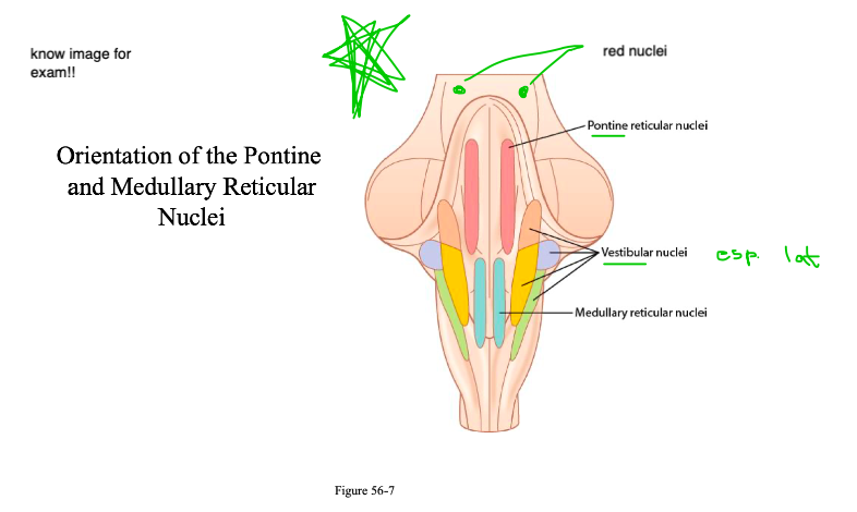
99
New cards
Brainstem → Extrapyramidal Control of Motor Function by the Brainstem
Vestibular Nuclei & Tracts - Anti-gravity/extensors (uncrossed)
Pontine-reticular Nuclei and Tract - Anti-gravity/extensors (Bilateral)
\
Red Nuclei & Rubrospinal Tract - Anti-extensors (flexor dominant) -- Crosses
Medullary-reticular Nuclei & Tract - Anti-extensors (flexor dominant) -- Bilateral
Pontine-reticular Nuclei and Tract - Anti-gravity/extensors (Bilateral)
\
Red Nuclei & Rubrospinal Tract - Anti-extensors (flexor dominant) -- Crosses
Medullary-reticular Nuclei & Tract - Anti-extensors (flexor dominant) -- Bilateral
100
New cards
Vestibular and Pontine Reticular Nuclei
Powerful Excitatory/Anti-Gravity Tone
→ High degree of natural excitability
→ When unopposed, they produce extension
\
Centers for control of extensors/antigravity muscle activity
\
Involved in activating and sending signals down cord via:
→ Lateral Vestibulospinal Tract - descends ipsilaterally
→ Pontine-reticulospinal Tract (medial reticulospinal) - descends bilaterally