Lecture 13: The Superior Colliculus and Primary Visual Cortex
1/30
There's no tags or description
Looks like no tags are added yet.
Name | Mastery | Learn | Test | Matching | Spaced | Call with Kai |
|---|
No analytics yet
Send a link to your students to track their progress
31 Terms
What is the role of the superior colliculus in the visual system?
Superior colliculus acts as a tracking mechanism (it points the eyes and head toward a visual object)
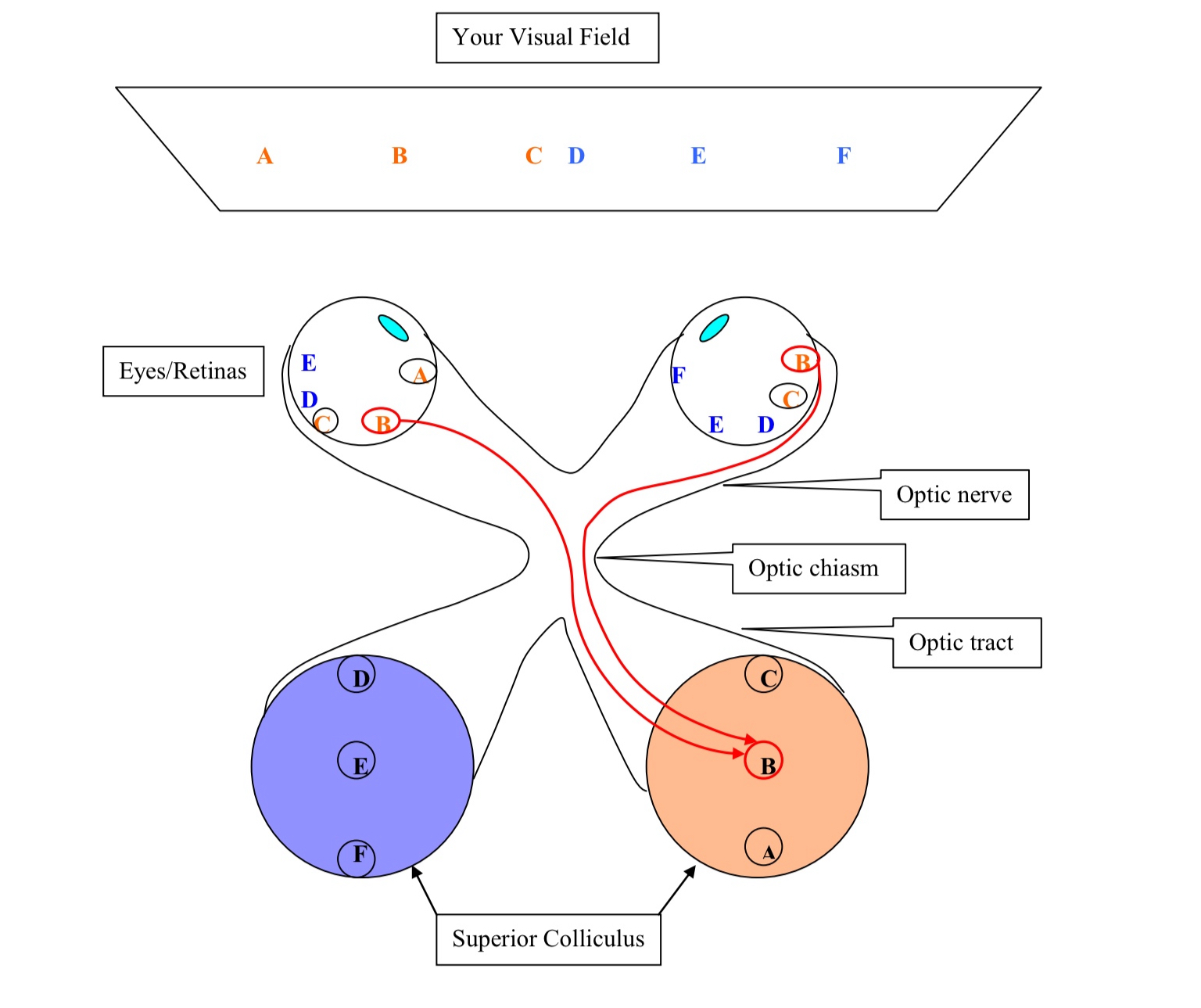
What does this diagram depict?
the superior colliculus and its inputs from the retina
What happens to neurons in the anterior portion of the superior colliculus ?
respond to a visual image on the center of gaze from both eyes (but only from the contralateral visual field)
What do the neurons in the posterior portion of the superior colliculus?
respond to visual image in the periphery of the visual field on the contralateral side
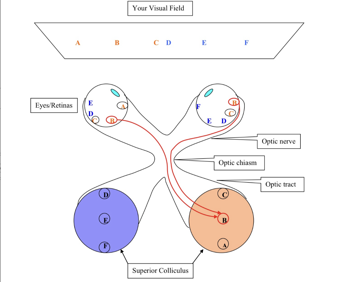
What do the neurons in the center portion (B) in the superior colliculus do ?
receive synaptic input from retinal ganglion cells (RGCs) from both eyes that respond to light at position B in the contralateral visual field
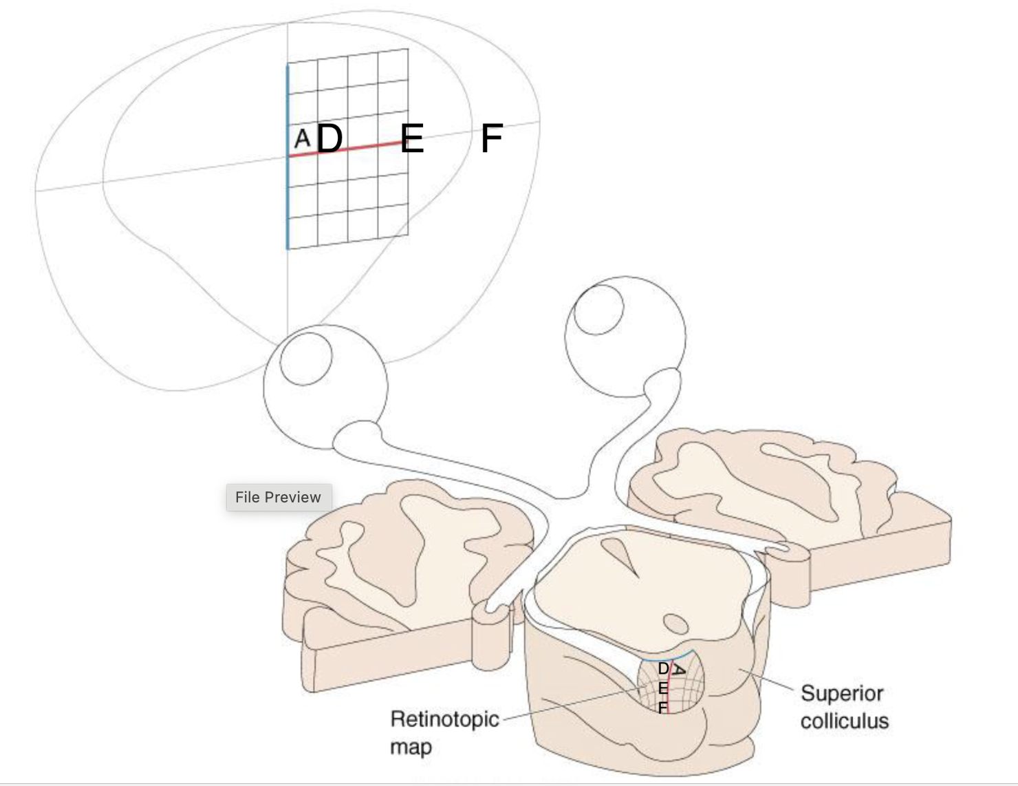
What does this figure present?
anatomical pattern of connections in the mechanism for generation of retinotopic maps in the superior colliculus
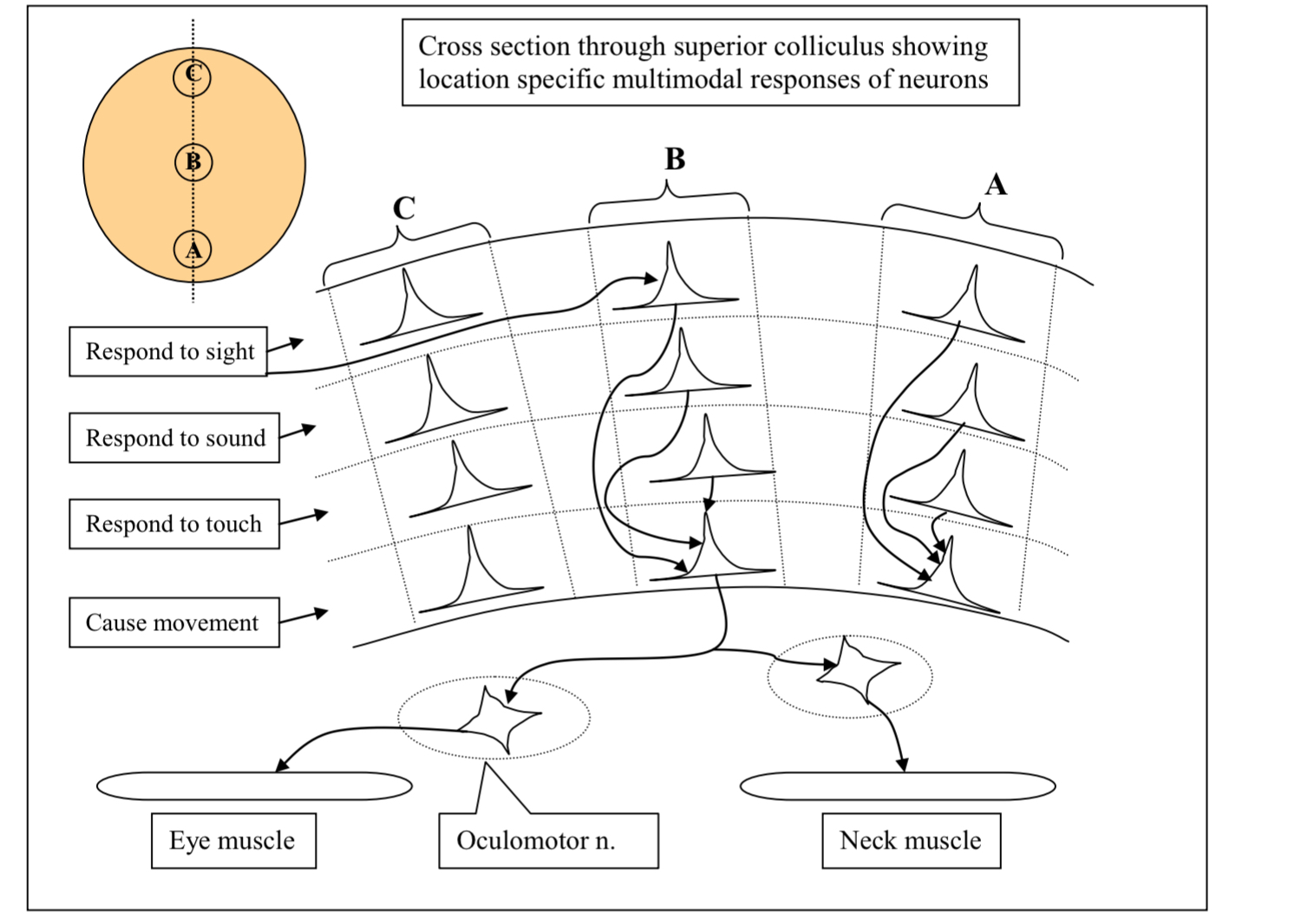
What does this diagram show?
cross section of the superior colliculus
showing how visual, auditory, somatosensory and motor maps are aligned with each other
What do the neurons in different layers in the superior colliculus do?
respond input from different sensory modalities: visual, auditory, and somatosensory
What does each layer contain?
contains a map of a particular sensory modality
these maps are all aligned with each other
What does the motor layer contain?
contains motor neurons not sensory neurons

What would happen if you stimulate neurons at position B in the motor layer of the superior colliculus?
causes the head eyes to move to look towards location B in the visual field
What do axons from the motor layer of the SC do?
terminate in the oculomotor nucleus
neurons from the oculomotor nucleus project to muscles that move the eye
also terminate onto the motor neurons which project to the muscles of the neck
Why is sensory tracking, eye pointing, and subsystem important for survival?
if you hear something unfamiliar this system will cause your head and eyes to orient towards the sound to visually identify what is it you heard
This occurs without conscious awareness
explains the phenomena called blind sight
What is blind sight?
sight that remains when a person is cortically blind
The eyes and the optic nerve work properly but the visual cortex is damaged so that person cannot perceive visual images
What can a person cortically blind do?
cannot see or identify visual objects
can localize and track objects in visual space and they do this without awareness
What is the superior colliculus homologous to and what are its characteristics?
homologous to the optic tectum (main visual brain area in non-mammalian vertebrates)
SC became specialized for tracking and lost the ability to contribute to object identification as that function expanded in visual cortex
What are the 2 visual subsystems discussed so far?
Superior Colliculus
involved in object tracking
Suprachiasmatic nucleus
brain’s clock
located dorsal (above) the optic chiasm
reason for jet lag and getting over jet lag: spontaneous daily patterns of activity of neurons in this region
What is another visual subsystem?
cortical portion of the visual system
part used in the perception of a visual stimulus

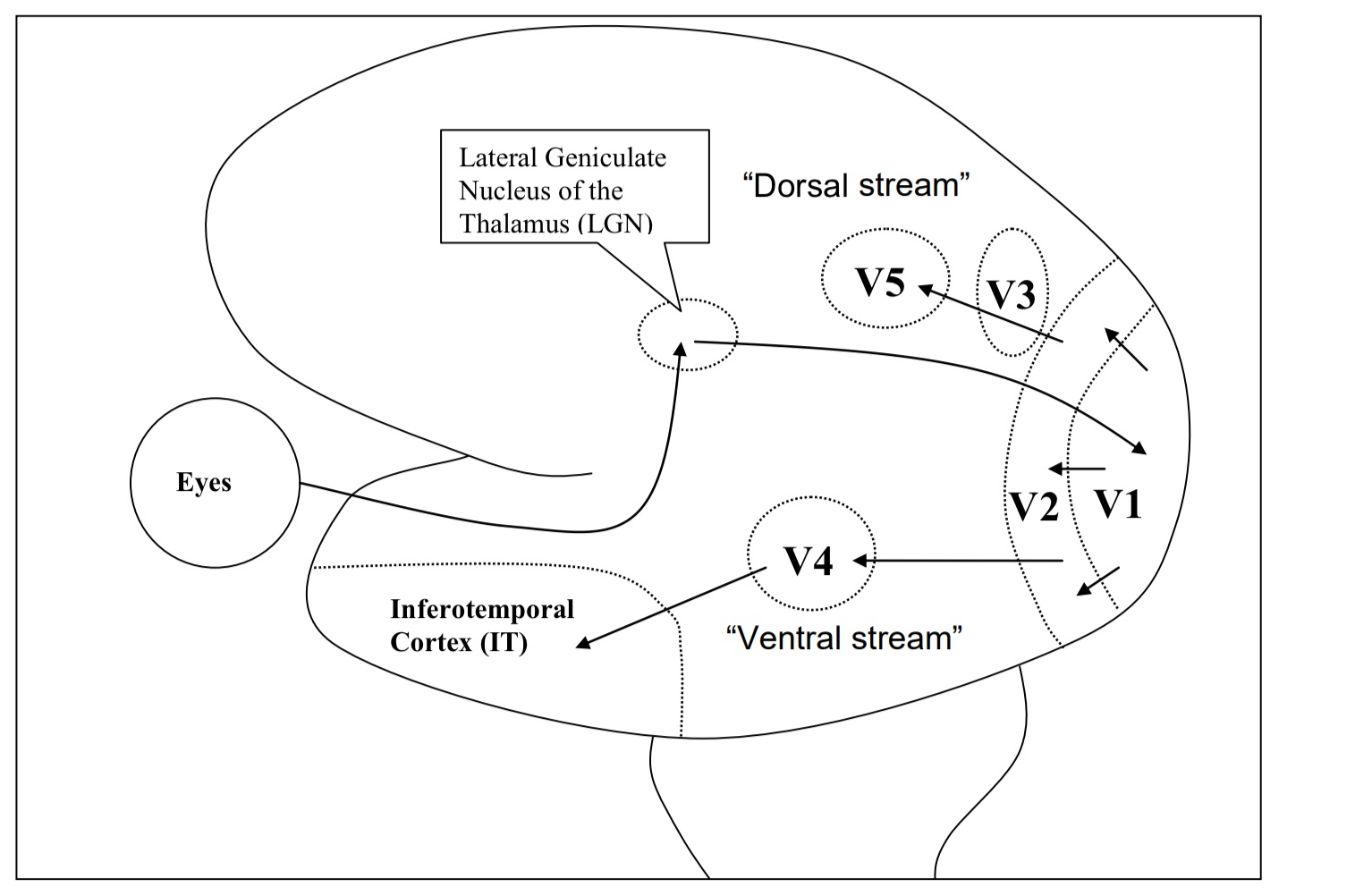
What does this diagram show?
diagram of a side view of a human brain depicting some of the major pathways in the cortical subsystem of the visual system in simplified form
Each of these cortical areas is retinotopically mapped (V1,V2…)


Explain in more detail whats ocurring in each point of the diagram?
Neurons in different regions of the visual cortex (V) have different functions
Neurons in V5: highly sensitive to movement but not color of objects in visual field
Neurons in V4: highly sensitive to color but not movement of objects in the visual field
What is perception of visual objects dependent on?
dependent on the activity of neurons in these cortical areas (V3, V4, V5)
Example: if a person’s V4 is destroyed, then that person’s visual world will be black and white, and memories are also in black or white

What is the area in the brain that is not retinotopically mapped?
Inferotemporal cortex (IT)
neurons in the IT cortex are not retinotopically mapped and don’t respond to color or movement
They respond to complex forms such as faces, hands, chairs, etc

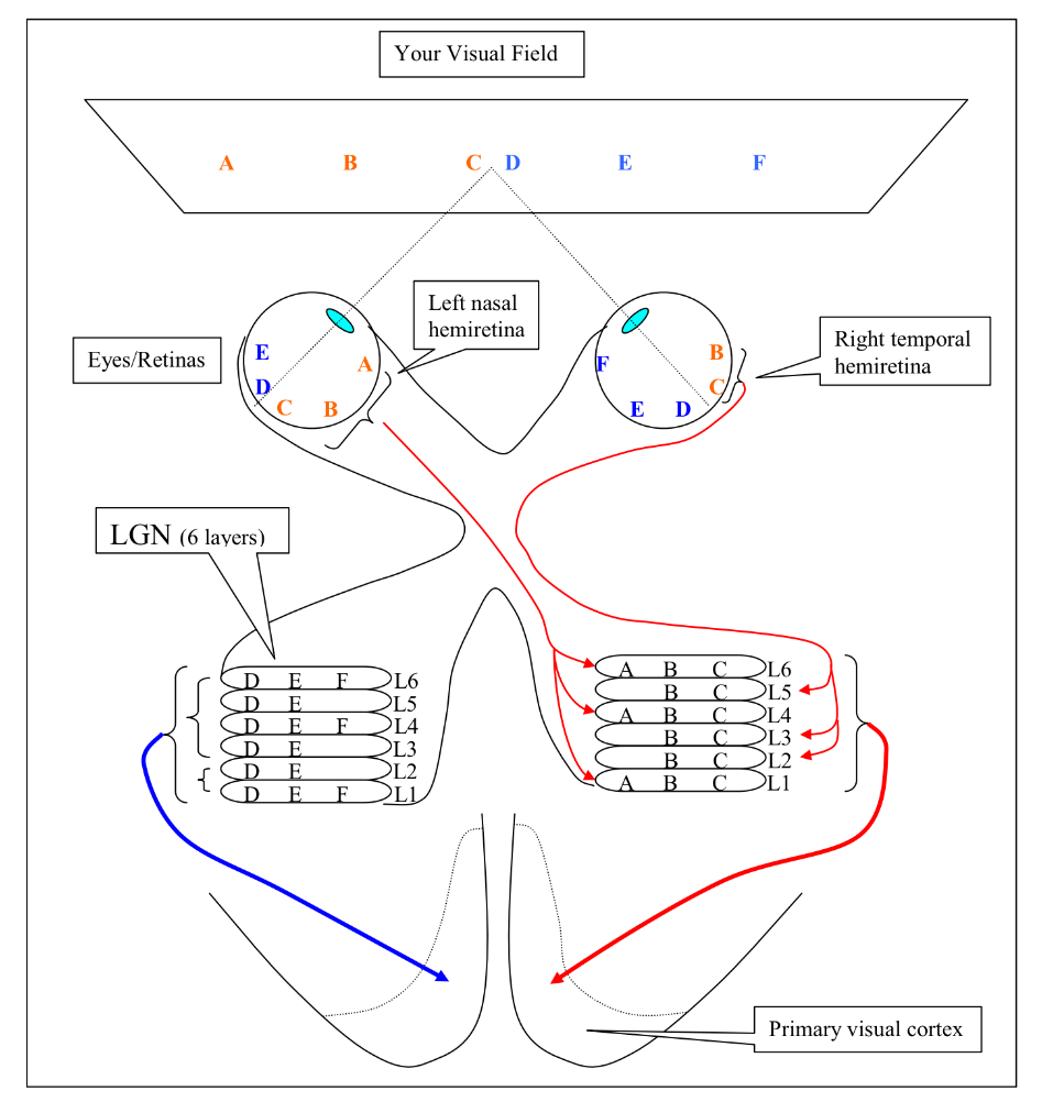
What does this diagram show?
the pattern of nasal and temporal hemiretina projections to the 6 layers of the lateral geniculate nucleus of the thalamus (LGN)
What are the basic form of projections from the retinal ganglion cells to the LGN similar to?
similar to that of the retinal ganglion cells to the superior colliculus
Neurons in the LGN respond only to visual images in the contralateral visual field
What are the characteristics of the LGN?
has 6 layers and each layer is retinotopically mapped
neurons in each layer receive input from the RGCs from only one hemiretina
the neurons in the LGN are monoculary (respond to input from one eye) driven
each of these maps are aligned with each other
Why is there repetition of visuotopic maps in the LGN?
cells in the different layers are processing different aspects of the visual field
What are the physical differences in the cells of the different layers?
cells in layers 1 and 2 are larger and are called magnocellular
cells here respond to movement
they are achromatic (don’t respond to color)
The cells in layers 3 through 6 are smaller, they’re called parvocellular
cells here are color sensitive
more sensitive to details

What is parallel processing?
the idea that different aspects of visual objects (aspects like color, movement, shape) are coded by different parallel pathways

What can projections to the primary visual cortex (V1) from the LGN be described as and what does that mean?
projections are ipsilateral
neurons in V1 respond exclusively to visual input from the contralateral visual field
Means if V1 is destroyed, you would be blind to input in the left visual field
there will still be “blind sight” due to an intact Superior Colliculus

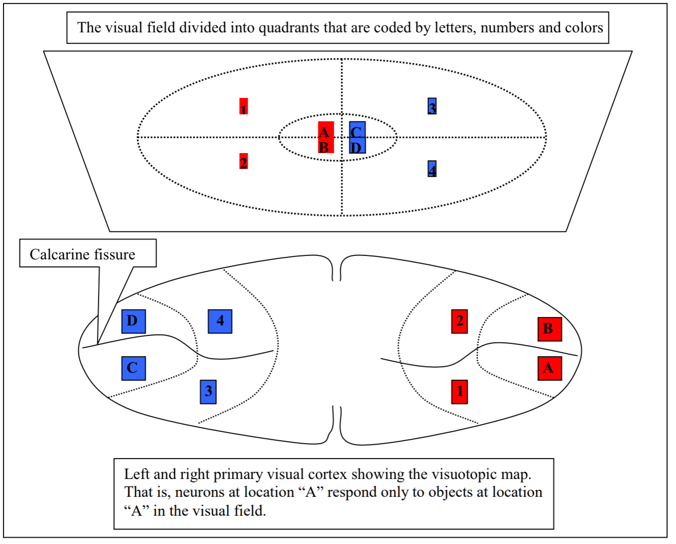
What does this diagram illustrate and describe it?
illustrates the visuotopic/retinotopic map in the primary visual cortex
½ of the neurons in the V1 code for the fovea region. even though this is only a very tiny portion of the visual field
this is b/c it takes a lot of neural machinery to decode the complex
Also, detailed info we can perceive from this area of highest visual acuity
