Facial Bones and Orbits
1/19
There's no tags or description
Looks like no tags are added yet.
Name | Mastery | Learn | Test | Matching | Spaced |
|---|
No study sessions yet.
20 Terms
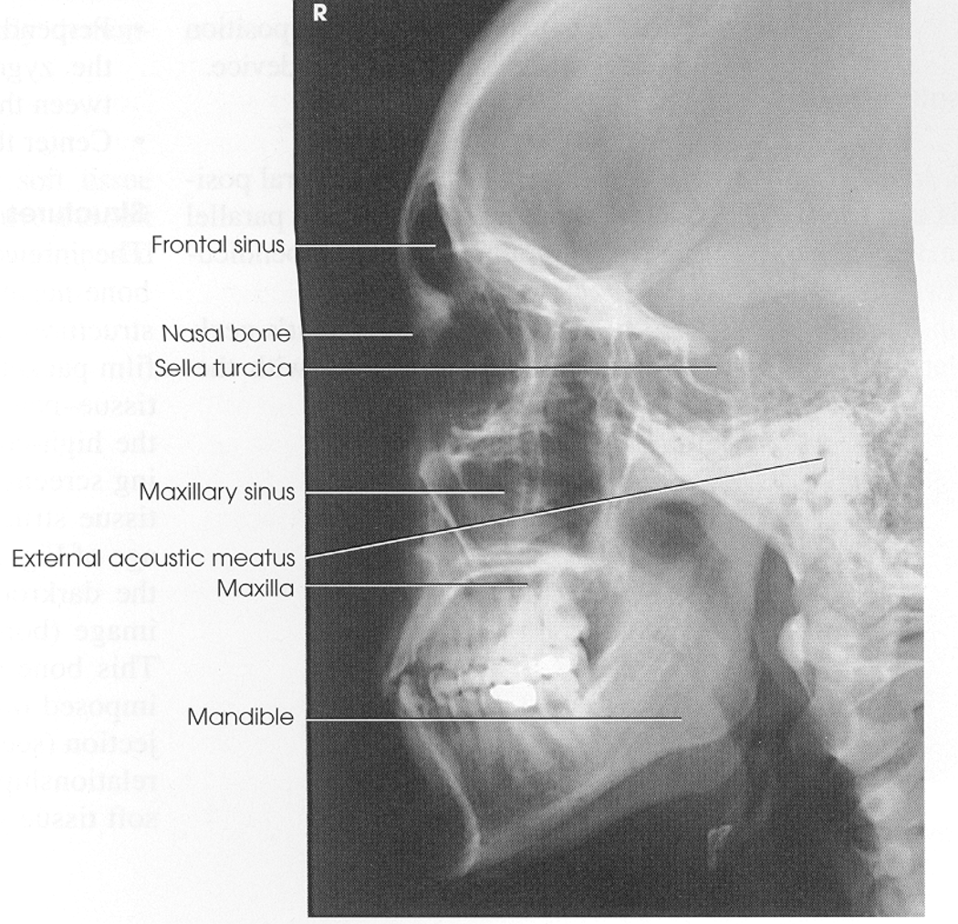
Lateral Facial Bones
Position: patient semi prone or oblique seated upright with MSP and IOML parallel to IR and interpupillary line perpendicular
CR: enter at lateral surface of zygomatic arch halfway between outer canthus and EAM
SS: lateral facial bones
EC: superimposed mandibular rami and superimposed orbital roofs and sella turcica
Lateral Facial Bones Image
xray
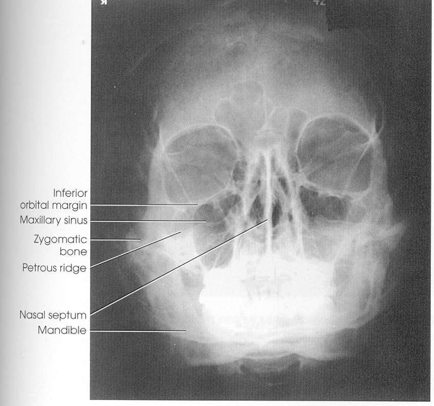
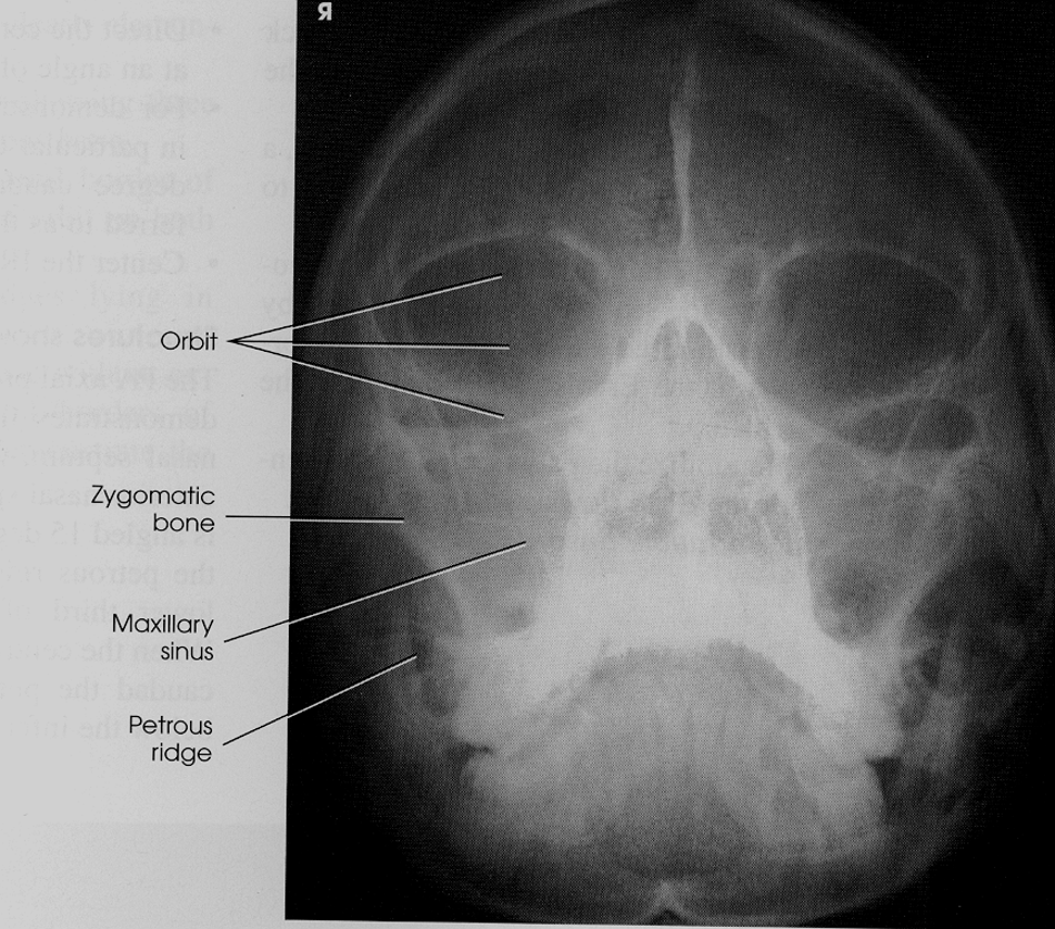
Parietocanthial Waters Method
Position: patient prone or seated upright with MSP perpendicular and OML 37 degrees with IR and MML perpendicular to IR
CR: perpendicular to acanthion
SS: orbits, maxillae, zygomatic arches
EC: equal distance b/w lateral border of skull and orbit on each side, petrous ridges below maxillary sinuses
Parietoacanthial Waters Method
xray
Modified Parietocanthial Waters Method
Position: patient prone or seated upright with OML 55 degrees with IR(neck extended less than waters)
CR: perpendicular to acanthion
SS: orbital floor perpendicular to IR, show blow out fx with inferior displacement of orbital floor
EC: petrous pyramids below orbital floors
Modified Parietocanthial Waters
xray
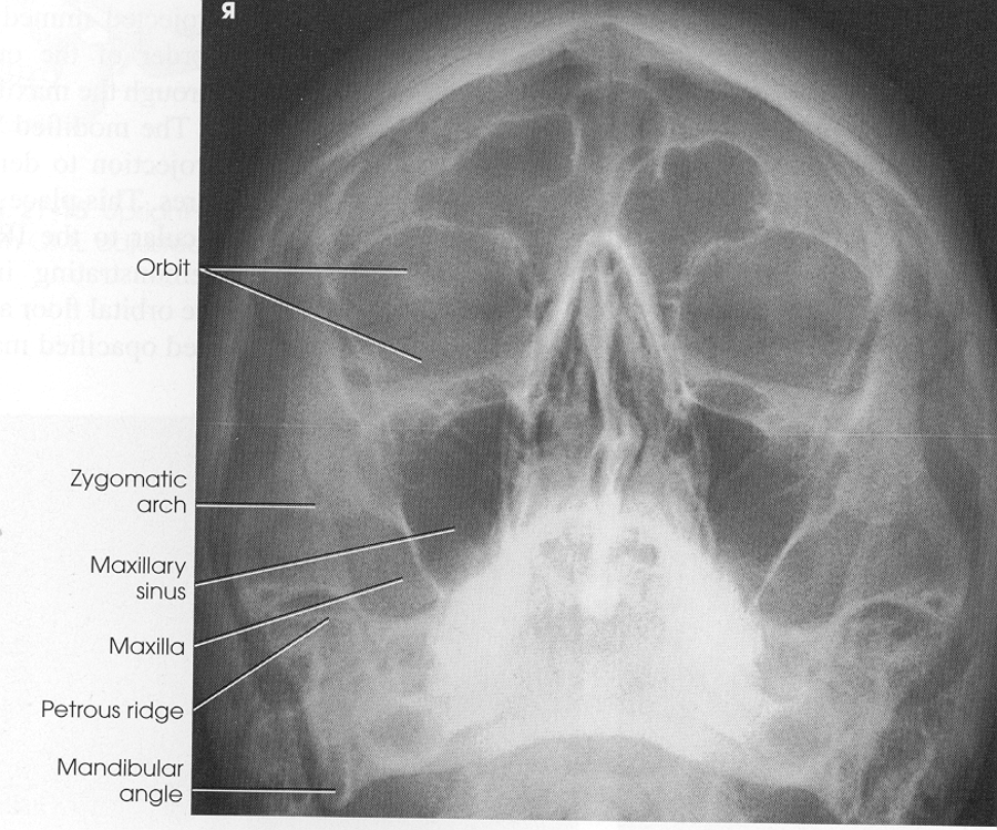
Acanthioparietal Reverse Waters
Position: patient supine with OML 37 degrees with IR and MML and MSP perpendicular to IR
CR: enters at acanthion
SS: magnified facial bones, superior facial bones
EC: equal distance b/w lateral border of skull and orbit on each side, petrous ridges below maxillary sinuses
Trauma angle cephalic 30 degrees to IOML
Acanthioparietal Reverse Waters
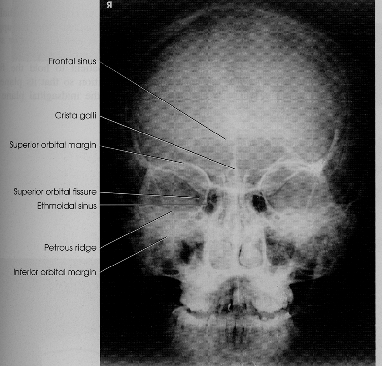
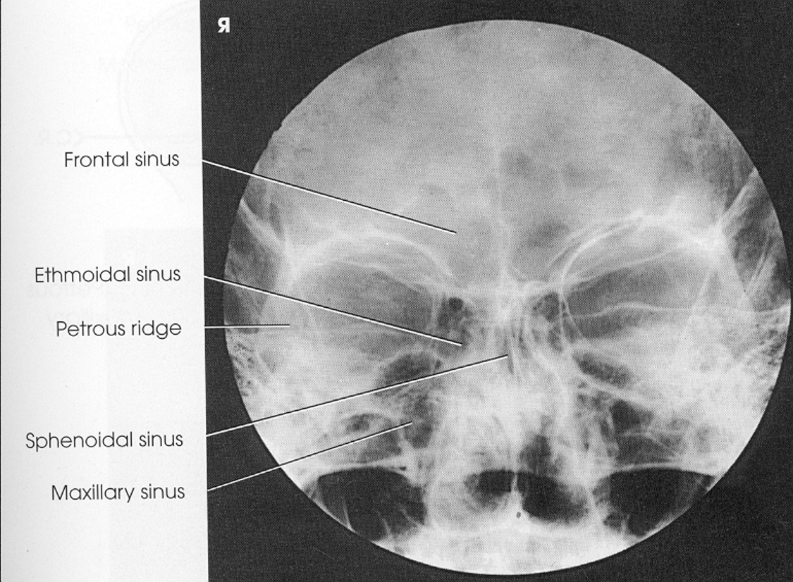
PA Axial Caldwell Skull
Position: patient prone or seated with OML and MSP perpendicular to IR, place sponge under forehead for obese
CR: angle 15 degrees caudad to nasion
SS: orbital rims, maxillae, nasal septum, zygomatic bones, petrous pyramids in lower 1/3rd of orbits, frontal bone, frontal and ethmoidal sinuses, crista galli
EC: equal distance b/w lateral border of skull and lateral orbit
PA Axial Caldwell Skull
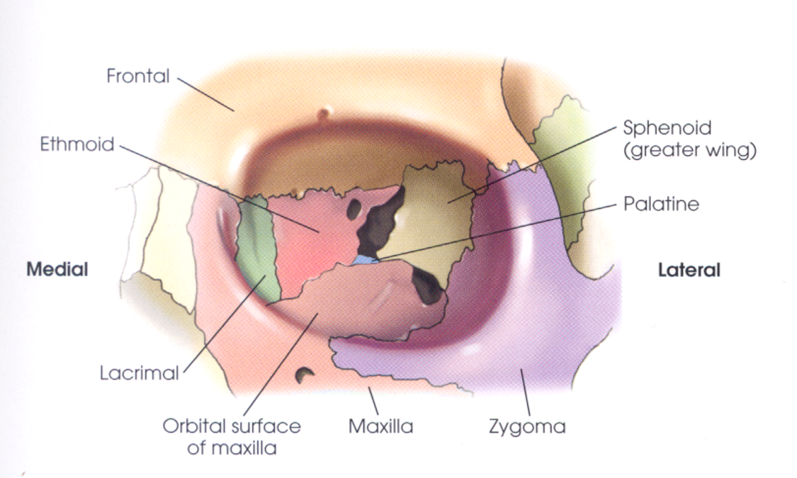
7 Orbit Bones
cranial: frontal, ethmoid, sphenoid
facial: lacrimal, palatine, maxillary, zygomatic
Orbit Openings
1.optic foramen(optic nerve)
2.superior orbital fissure(cleft between greater and lesser sphenoid wing)
3.inferior orbital fissure(cleft at lower anteriorlateral sphenoid bone)
Orbits
image
Lateral Orbits
Position: patient erect in RAO/LAO with MSP and IOML parallel to IR
CR: enter at outer canthus
SS: orbit closest to IR
EC: density and contrast to visualize orbital rims, superimposed orbital roofs, close beam restriction
Lateral Facial Bones
xray
PA Axial Caldwell Orbits
Position: patient seated upright facing IR with nose and forehead on grid and MSP and OML perpendicular to IR
CR: angle 15 degrees caudad to nasion
SS: frontal and ethmoidal sinuses, sphenoidal sinuses projected in nasal fossa, orbits
EC: equal distance b/w lateral skull borders and orbits, symmetrical petrous ridges in lower 1/3rd of orbits, close beam restriction
PA Axial Caldwell Orbits
Parietocanthial Waters Orbits
Position: patient seated upright with MSP and MML perpendicular to IR and OML forms 37 degree angle with IR
CR: perpendicular to acanthion
SS: both orbits, maxillary sinuses above petrous ridges, frontal and ethmoidal sinuses distorted
EC: petrous ridges below floor of maxillary sinuses, equal distance from lateral skull borders to lateral orbits, maxillary sinuses and orbits shown with close beam restriction
Parietocanthial Modified Waters Orbits
Position: patient seated upright with MSP perpendicular to IR and OML forms 50 degree angle with IR
CR: perpendicular to mid-orbits
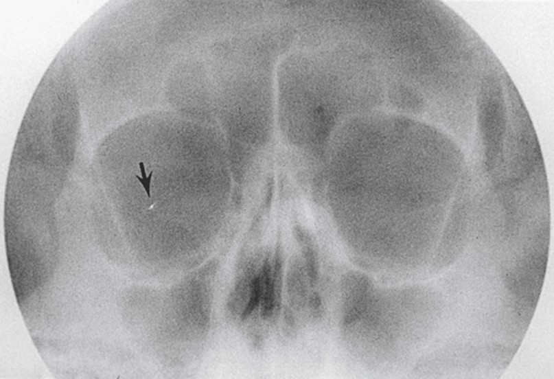
EC: entire orbits, petrous ridges in orbital shadows, close beam restriction centered on orbital region, bony orbit and soft tissues of eye (foreign body localization)
Parietocanthial Modified Waters Orbits
xray