Skull Osteology
1/109
Earn XP
Name | Mastery | Learn | Test | Matching | Spaced | Call with Kai |
|---|
No analytics yet
Send a link to your students to track their progress
110 Terms
Frontal description
Forms forehead and part of each orbit
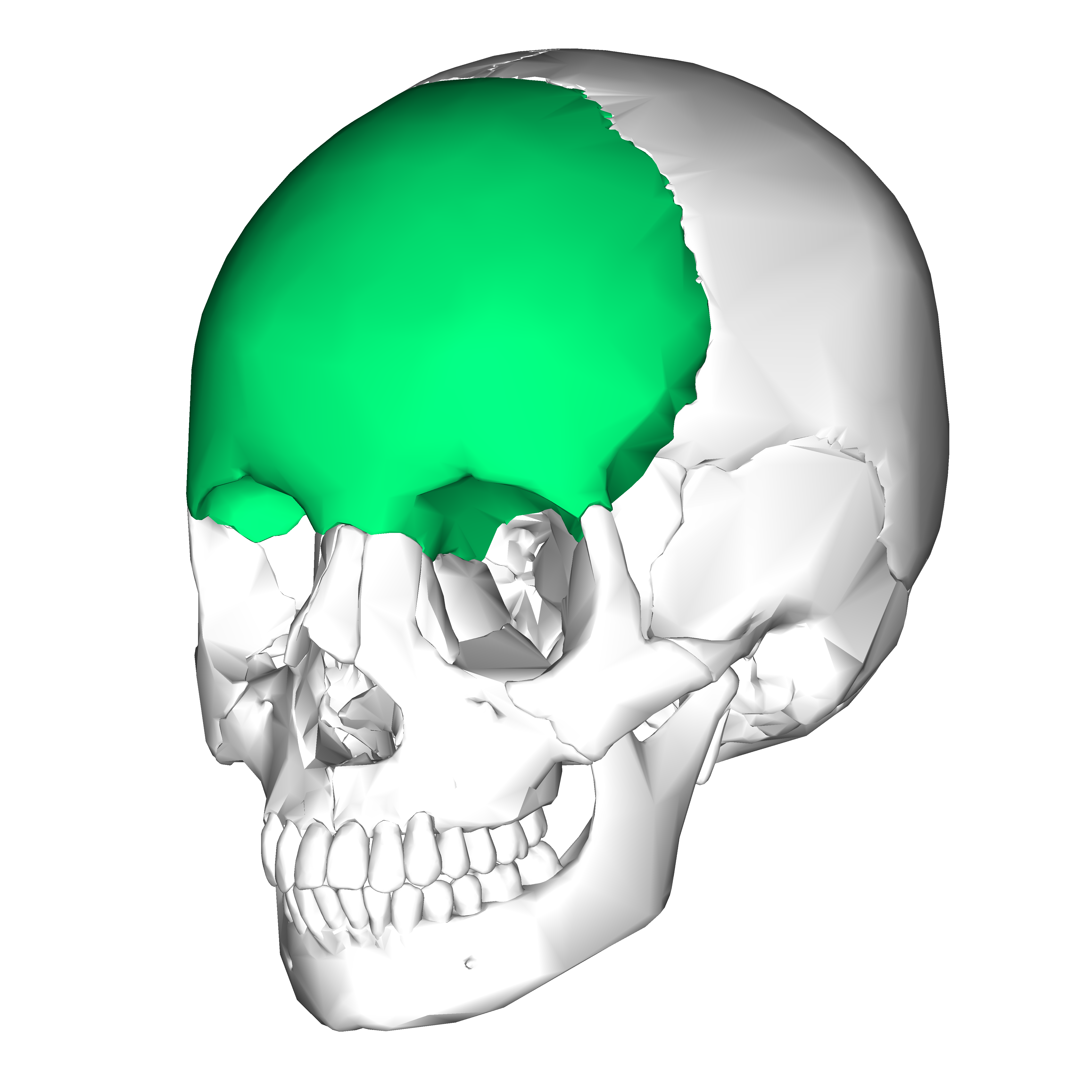
Frontal function
Protects frontal lobe, forms a portion of the orbital complex
Supraorbital Foramen (ext) description
Hole above eye sockets
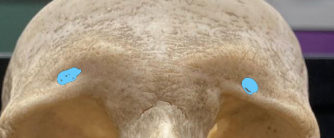
Supraorbital Foramen (ext) function
Passage for veins/arteries and CN V1
Occipital description
Forms back/base of skull
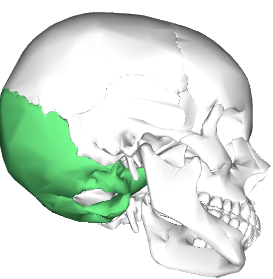
Occipital function
Protects cerebellum and occipital lobe
Foramen Magnum (ext/int) description
Large opening in occipital bone
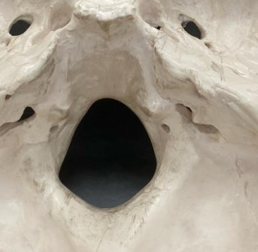
Foramen Magnum (ext/int) function
Spinal cord and CN XI passage
Hypoglossal Canal (int) description
Holes that look like they are inside of the Foramen magnum
Hypoglossal Canal (int) function
CN XII passage
Occipital Condyle (ext) description
Big bone bulges lateral to the Foramen magnum (inferior view only)
Occipital Condyle (ext) function
Articulates with the C1 vertebrae called the Atlas
Nuchal Line (superior and inferior)(ext) description
Line on occipital
Nuchal Line (superior and inferior)(ext) function
Muscle attachment
Parietal description
Posterior to frontal bone and on either side of midline
Parietal function
Protects parietal lobe
Temporal description
Anterior to occipital and inferior to parietal
Temporal function
Protects temporal lobe
Jugular foramen (int) description
Opening between occipital and temporal bone
Jugular foramen (int) function
Cranial nerve passage for CN IX, X, XI, and Jugular vein
External Acoustic Meatus (ext) description
Ear hole connecting to the internal acoustic meatus
External Acoustic Meatus (ext) function
Opening for initial sound waves to travel through before reaching the processing centers in the inner ear
Internal Acoustic Meatus (int) description
Tunnel in temporal bone
Internal Acoustic Meatus (int) function
Cranial nerve passage for CN VII and VIII
Carotid Canal (ext) description
Canal in temporal bone
Carotid Canal (ext) function
Vessel passage
Stylomastoid Foramen (ext) description
Hole between mastoid and styloid processes
Stylomastoid Foramen (ext) function
CN VII exits here
Styloid process (ext) description
Slender projection protruding from inferior of temporal bone
Styloid process (ext) function
Attachment site for hyoid ligaments and other neck muscles
Mastoid Process (ext) description
Projects inferiorly from temporal bone
Mastoid Process (ext) function
Attachment of diagastric and sternocleidomastoid muscles
Mandibular Fossa (ext) description
Bole like feature directly posterior to the zygomatic arch on the inferior view of the skull
Mandibular Fossa (ext) function
Articulates with the mandible, where people can get Temporomandibular joint issues
Zygomatic Arch (ext) description
Arch formed from zygomatic and temporal bones
Zygomatic Arch (ext) function
Attachment of masseter muscle
Zygomatic description
Forms prominences of cheeks and part of each orbit
Zygomatic funciton
Articulates with the frontal, sphenoid, temporal, and maxilla bones, forms portion of the orbital complex
Maxilla description
Forms upper jaw, part of hard palate, part of each orbit
Maxilla function
Helps form portion of orbital complex and roof of oral cavity
Infraorbital Foramen (ext) description
Inferior to each orbit
Infraorbital Foramen (ext) function
Passageway for blood vessels and CN V2
Incisive Foramen description
Singular hole in the anterior portion of the maxilla (roof of mouth behind the big front teeth)
Incisive Foramen function
Passage for nasopalatine nerve
Palatine Process of Maxilla (ext) description
Forms hard palate
Palatine Process of Maxilla (ext) function
Floor of nasal cavity and roof of mouth
Mandible description
Forms lower jaw
Mandible function
Holds teeth, allows mastication, and articulates with temporal bone to form joint
Alveolar Processes (ext) (on both Mandible and Maxilla) description
Oral margins of the maxilla and mandible
Alveolar Processes (ext) (on both Mandible and Maxilla) function
Contain tooth roots and alveolar nerves
Mental Protuberance (ext) description
Bottom of chin
Mental Protuberance (ext) function
Forms the chin
Mental Foramen (ext) description
Opening in mandible lateral to chin
Mental Foramen (ext) funciton
Vessel and nerve passage, CN V3
Mandibular Body (ext) description
Horizontal, curved portion
Mandibular Body (ext) function
Forms lower jaw
Mandibular Ramus (ext) description
Vertical portion of mandible
Mandibular Ramus (ext) function
Condyles articulate with temporal bone (allows movement in jaw)
Coronoid Process (ext) description
Projection on mandible that is anterior to condylar process
Coronoid Process (ext) function
Attachment of temporalis muscle
Condylar Process (ext) description
Rounded projection on mandible
Condylar Process (ext) function
Articulates with mandibular fossa
Mandibular Head (ext) description
Located on Condylar process
Mandibular Head (ext) function
Provides surface for articulation
Mandibular Foramen (int) description
Located on the inner surface of the mandibular ramus
Mandibular Foramen (int) function
Passage of CN V3
Nasal description
Forms bridge of nose
Nasal function
Be able to identify
Lacrimal description
Next to the zygomatic portion/nasal portion of the orbit, where the lacrimal gland sits (produces tears)
Lacrimal function
Forms part of the medial portion of the orbital complex
Palatine description
Forms part of hard palate
Palatine function
It contributes to floor of nasal cavity, roof of mouth, adn a small portion of the orbital complex
Greater Palatine Foramen (ext) description
Paired holes in hard palate (back towards molars on the roof of the mouth)
Greater Palatine Foramen (ext) function
Passage for nerves/vessels, CN V2
Vomer (ext) description
Forms part of nasal septum
Vomer (ext) function
Be able to identify
Sphenoid description
Posterior to face, extends from one temple ot the other, forms part of orbit
Sphenoid function
Articulates with frontal, parietal, and temporal bones, part of the orbital complex
Superior Orbital Fissure (ext) description
Top part of the C shape in the eye socket
Superior Orbital Fissure (ext) function
Passageway for CN III, CN IV, CN V1, CN VI
Inferior Orbital Fissure (ext) description
Separates the lower wall and floor of orbit (lower part of C shape in eye socket)
Inferior Orbital Fissure (ext) function
Passageway for CN V2
Optic Canal (ext/int) description
Round pening in sphenoid bone in posterior part of orbit
Optic Canal (ext/int) function
Cranial nerve passage CN II
Greater wings of the Sphenoid (int/ext) description
A process of the sphenoid bone, mainly located behind the eyes in the middle of the cranial vault, Foramen Rotundum, Ovale, and Spinosum can be found on this portion of the sphenoid
Greater wings of the Sphenoid (int/ext) function
Aids in forming the base and a little of the sides of the skull, aids in forming the inferior and lower lateral portion of the eye socket
Lesser wings of the Sphenoid (int/ext) description
A triangular shaped region of the sphenoid located superiorly to the greater wings of the sphenoid, the optic canal the superior orbital fissure can be found in this region
Lesser wings of the Sphenoid (int/ext) function
Supports portions of the frontal lobe and forms the posterior, superior aspect of the eye socket, and helps form the superior orbital fissure
Foramen Lacerum (in association with Temporal and Occipital bones, the marking belongs to the Sphenoid, but it lies smack between all three bones) (ext/int) description
Most medial foramen is seemingly composed of the Sphenoid, Temporal, and Occipital bone, slightly posterior and lateral to the vomer
Foramen Lacerum (in association with Temporal and Occipital bones, the marking belongs to the Sphenoid, but it lies smack between all three bones) (ext/int) funciton
Nerve/artery/vein passageway for function
Foramen Rotundum (int) description
Located on the greater wing of the sphenoid, this opening. is inferior to the superior orbital fissure, and both superior and medial to foramen ovale, often almost a perfect circule in shape
Foramen Rotundum (int) function
Passage for Cn V2 to exit skull
Foramen Ovale (ext/int) description
Oval opening in the greater wing of the sphenoid bone, inferior to foramen Foramen Rotundum but superior to the Foramen Spinosum, lateral to the Sella Turcica
Foramen Ovale (ext/int) function
Passageway for Cn V3 to exit skull
Foramen Spinosum (ext/int) description
Smallest foramen, almost shaped like a perfect circle, located inferior to foramen ovale, lateral to Sella Turcica
Foramen Spinosum (ext/int) function
Nerves and vessels pass through here
Sella Turcica (int) description
Depression in sphenoid bone, saddle shaped
Sella Turcica (int) function
Location of pituitary gland
Dorsum Sellae (int) description
A bony ridge which forms posterior wall of Sella Turcica
Dorsum Sellae (int) function
Location of pituitary gland