3.2 Cells
1/198
There's no tags or description
Looks like no tags are added yet.
Name | Mastery | Learn | Test | Matching | Spaced |
|---|
No study sessions yet.
199 Terms
What are the distinguishing features of eukaryotic cells?
● Cytoplasm containing membrane-bound organelles
● So DNA enclosed in a nucleus
Nucleus function
● Holds / stores genetic information which codes for polypeptides (proteins)
● Site of DNA replication
● Site of transcription (part of protein synthesis), producing mRNA
● Nucleolus makes ribosomes / rRNA
Structure of of nucleus
Nuclear envelope: double membrane surrounding nucleus, outer membrane continuous with the (R)ER of the cell.
Nuclear pores: allow the passage of larger molecules, such as mRNA, out of the nucleus.
Nucleoplasm: granular, jelly-like material making up the bulk of the nucleus.
Chromosomes: protein-bound, linear DNA.
Nucleolus: small spherical region(s) in nucleoplasm. Manufactures ribosomal RNA and assembles ribosomes.

Rough Endoplasmic Reticulum (RER) structure
-A network of flattened sacs enclosed by a membrane.
-Surface is studded with ribosomes, giving it a "rough" appearance.
Rough Endoplasmic Reticulum function
● Ribosomes on surface synthesise proteins
● Proteins processed / folded / transported inside rER
● Proteins packaged into vesicles for transport eg. to Golgi apparatus
Smooth Endoplasmic Reticulum (SER) structure
A network of membrane-bound sacs without ribosomes.
Smooth Endoplasmic Reticulum (SER) Function
● Synthesises and processes lipids
● Eg. cholesterol and steroid hormones
Golgi Apparatus structure
A stack of fluid-filled, flattened, curved sacs, with vesicles at the edges.
Golgi Apparatus function
● Modifies protein, eg. adds carbohydrates to produce glycoproteins
● Modifies lipids, eg. adds carbohydrates to make glycolipids
● Packages proteins / lipids into Golgi vesicles
● Produces lysosomes (a type of Golgi vesicle)
Golgi vesicles function
● Transports proteins / lipids to their required destination
● Eg. moves to and fuses with cell-surface membrane
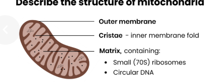
Mitochondria structure
-Oval-shaped, surrounded by a double membrane.
-The inner membrane is folded into cristae, which increase the surface area for respiration.
-The internal space, called the matrix, contains enzymes needed for aerobic respiration to produce ATP.
Mitochondria function
● Site of aerobic respiration
● To produce ATP for energy release
● Eg. for protein synthesis / vesicle movement / active transport
Ribosomes structure
-Made of two subunits (large and small).
-Found either free in the cytoplasm or attached to the RER.
Ribosomes Function
Site of protein synthesis (translation)
Lysosomes structure
Small vesicles bound by a single membrane, containing digestive enzymes.
Lysosomes Function
● Release hydrolytic enzymes (lysozymes)
● To break down / hydrolyse pathogens or worn-out cell components
In complex multicellular organisms, eukaryotic cells become specialised for specific functions, What is cell specialisation?
Cell specialisation is the process by which cells develop specific structures and functions to perform particular roles within an organism.
Differentiation
Differentiation is the process through which unspecialised cells (e.g., stem cells) become specialised.
Describe the Specialised cells organisation
Specialised cells are organised into tissues, tissues into organs and organs into organ systems.
What are the distinguishing features of prokaryotic cells?
● Cytoplasm lacking membrane-bound organelles
● So genetic material not enclosed in a nucleus
What do prokaryotic cells contain
Cell wall, capsule, plasmid, flagellum, pili,
Cell Wall
-A rigid outer layer made of peptidoglycan (also known as murein), a polymer of sugars and amino acids.
-Provides structural support and protection against osmotic pressure.
Capsule
-A protective slimy layer outside the cell wall.
-Offers protection against the immune response of host organisms.
Plasmid
-A small, circular piece of DNA separate from the chromosomal DNA.
-Contains genes for survival traits, such as antibiotic resistance.
Flagellum
A tail-like structure that rotates to propel the cell through its environment.
Pili
-Thin, hair-like projections on the surface of the cell.
-Help to attach to other bacterial cells or surfaces.
Ribosomes
-The site of protein synthesis.
-Smaller than eukaryotic ribosomes, identified as 70S ribosomes.
Compare the prokaryotic and eukaryotic cells
•Size: E is larger than P
•Nucleus: E has nucleus, P has no nucleus.
•DNA Structure: E is Linear DNA associated with histones, P is Circular DNA without histones
•Organelles: E has Many membrane-bound organelles (e.g., mitochondria), P has Few (non-membrane-bound, e.g., ribosomes)
•Ribosomes- E has 80s while P has 70s
•Cell Wall- E: In plants: cellulose; in fungi: chitin; absent in animals. P made of murein
•Histones- E present while P absent
What are viruses
-Viruses are non-living particles much smaller than bacteria.
-They lack cellular structures and are completely dependent on a host cell to reproduce.
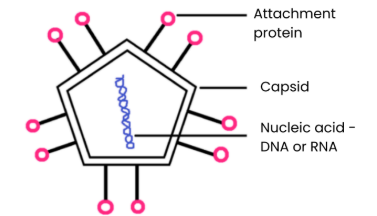
What is the structure of viruses?
1. Nucleic acids surrounded by a capsid (protein coat)
2. Attachment proteins allow attachment to specific host cells
3. No cytoplasm, ribosomes, cell wall, cell-surface membrane etc.
4. Some also surrounded by a lipid envelope eg. HIV
Explain why viruses are described as acellular and non-living
● Acellular - not made of cells, no cell membrane / cytoplasm / organelles
● Non-living - have no metabolism, cannot independently move / respire / replicate / excrete
What is reverse transcriptase
-an enzyme that catalyzes the formation of DNA from an RNA template in reverse transcription.
-This incorporate their genetic material into the host genome
Differences Between Viruses and Bacteria:
•Cell Type:
B: Prokaryotic cells with cell membranes and organelles.
V: Non-cellular particles with no organelles or membranes.
•Genetic material:
B: Circular strand of DNA.
V: Can be DNA or RNA, single or double-stranded.
•Reproduction:
B: Divide by binary fission.
V: Replicate only inside a host cell.
•Size:
B- Larger
V- Smaller
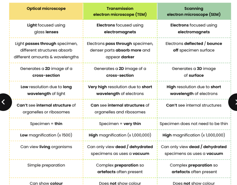
How do optical microscope work
-Use a pair of convex glass lenses to magnify specimens.
-Rely on visible light, limiting their resolution to about 0.2 μm.
Resolution
Resolution is the minimum distance at which two objects can be distinguished as separate entities.
Factors Affecting Resolution:
1.) Type of microscope:
-Light microscopes have limited resolution.
-Electron microscopes (e.g., TEM, SEM) provide higher resolution.
2.) Wavelength of light or electrons:
-Shorter wavelengths give better resolution.
Magnification
Magnification refers to how many times larger an image appears compared to the actual size of the object.
Describe the difference between magnification and resolution
● Magnification = number of times greater image is than size of the real (actual) object
○ Magnification = size of image / size of real object
● Resolution = minimum distance apart 2 objects can be to be distinguished as separate objects
Wavelength of light
The wavelength of light limits resolution, which cannot resolve structures smaller than 0.2 μm.
Formula for magnification
Magnification = image size / real size
How do electron microscope works?
-Use a beam of electrons focused by electromagnets.
-Operate in a vacuum to prevent air particles from scattering the electrons.
-Can resolve objects as small as 0.1 nm, providing a much greater resolution than light microscopes.
Two type of electron microscopes
transmission electron microscope and scanning electron microscope
Transmission Electron Microscope (TEM)
-A beam of electrons passes through a thin specimen.
-Produces a detailed 2D image of the internal structure.
-Areas absorbing more electrons appear darker.
Scanning Electron Microscope (SEM):
-A beam of electrons scans the surface of the specimen.
-Produces a detailed 3D image of the specimen's surface.
Advantages of each
-TEM: Extremely high resolution for internal details.
-SEM: Produces 3D images, useful for understanding surface structures.
Limitations of each
-Require a vacuum, so living specimens cannot be observed.
-A complex staining process is necessary, which may introduce artefacts (false structures not present in the specimen).
-Specimens must be thin (especially for TEM).
-SEM has a lower resolving power than TEM, but both are far superior to light microscopes.
Compare the principles and limitations of optical microscopes,
transmission electron microscopes and scanning electron microscopes
How to prepare a specimen
-A thin section of the plant tissue is prepared and placed on a slide.
-A drop of iodine solution is added to the tissue to stain the starch grains.
-The slide is covered with a coverslip and observed under a light microscope.
Describe how the size of an object viewed with an optical microscope can be
measured
1. Line up (scale of) eyepiece graticule with (scale of) stage micrometre
2. Calibrate eyepiece graticule - use stage micrometre to calculate size of divisions on eyepiece graticule
3. Take micrometre away and use graticule to measure how many divisions make up the object
4. Calculate size of object by multiplying number of divisions by size of division
5. Recalibrate eyepiece graticule at different magnifications
Guidelines for Scientific Diagrams:
1.) Guidelines for Drawing Biological Diagrams:
-Use of Pencil:
Sharp pencil for clear lines and precision.
-Shading & Colouring:
Avoid shading or colouring to maintain simplicity and clarity.
-Diagram Size:
Ensure diagrams are large and clear for easy identification.
-Labelling:
Label structures with straight lines that don't cross each other.
Include the name of the specimen and the magnification used.
Labels should be:
Horizontal: Use specific terms (e.g., nucleus, mitochondrion).
-Proportions:
Accurately represent the relative size of structures.
Use grid paper if necessary to help maintain proportions.
-Scale Bars:
Include a scale bar to indicate the actual size of structures.
Suggest how the scientific community distinguished between artefacts (eg.
dust, air bubbles occurring during preparation) and cell organelles
● Scientists prepared specimens in different ways
● If an object was seen with one technique but not another, it was more likely to be an artefact than an organelle
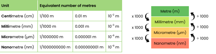
Describe how to convert between different units
Cell fractionation
Cell fractionation is a technique used to separate and isolate different organelles within a cell so that their structure and function can be studied in detail. The process involves homogenisation and ultracentrifugation.
Homogenisation
-Cells are broken apart using a homogeniser (a blender or similar device).
-This forms a liquid called the homogenate, which contains a mixture of cell organelles.
-The homogenate is then filtered to remove debris and unbroken cells.
Filtration
● Remove large, unwanted debris eg. whole cells, connective tissue
Ultracentrifugation
-The homogenate is placed in a centrifuge and spun at different speeds to separate the organelles based on their density.
Initial Spin
-The tube is spun at a low speed.
-The heaviest organelles (e.g., nuclei) settle at the bottom of the tube as a pellet.
-The remaining liquid, called the supernatant, is carefully removed.
Subsequent Spins
-The supernatant is transferred to a new tube and spun at a higher speed.
-The next heaviest organelles (e.g., mitochondria) form a pellet at the bottom.
-This process is repeated, increasing the speed each time to separate lighter organelles like lysosomes and ribosomes.
Conditions for Successful Cell Fractionation:
● Cold to reduce enzyme activity
○ So organelles not broken down / damaged
● Isotonic so water doesn’t move in or out of organelles by osmosis
○ So they don’t burst
● Buffered to keep pH constant
○ So enzymes don’t denature
Order of Organelle Separation
Nuclei (heaviest).
Mitochondria (and chloroplasts in plant cells).
Lysosomes and endoplasmic reticulum.
Ribosomes (lightest).
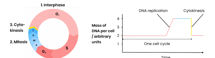
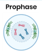
Summarise the stages of the cell cycle in eukaryotic cells
Stage 1 Interphase
● DNA replicates semi-conservatively (S phase)
○ Leading to 2 chromatids (identical copies) joined at a centromere
● Number of organelles & volume of cytoplasm increases, protein synthesis (G1 / G2)
Stage 2 Mitosis
● Nucleus divides
● To produce 2 nuclei with identical copies of DNA produced by parent cell
Stage 3 Cytokinesis
● Cytoplasm and cell membrane (normally) divide
● To form 2 new genetically identical daughter cells
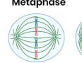
Stage 1 Prophase
● Chromosomes condense, becoming shorter / thicker so visible
○ Appear as 2 sister chromatids joined by a centromere
● Nuclear envelope breaks down
● Centrioles move to opposite poles forming spindle network
● Spindle fibres start to attach to chromosomes by their centromeres
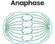
Stage 2 Metaphase
● Spindle fibres attach to chromosomes by their centromeres
● Chromosomes align along equator
Stage 3 Anaphase
● Spindle fibres shorten / contract
● Centromere divides
● Pulling chromatids (from each pair) to opposite poles of cell
Stage 4 Telophase
● Chromosomes uncoil, becoming longer / thinner
● Nuclear envelopes reform = 2 nuclei
● Spindle fibres / centrioles break down

Why do some eukaryotic cells not undergo the cell cycle?
● Within multicellular organisms, not all cells retain the ability to divide (eg. neurons)
● Only cells that do retain this ability go through a cell cycle
Explain the importance of mitosis in the life of an organism
Parent cell divides to produce 2 genetically identical daughter cells for:
● Growth of multicellular organisms by increasing cell number
● Replacing cells to repair damaged tissues
● Asexual reproduction
Describe how tumours and cancers form
● Mutations in DNA / genes controlling mitosis can lead to uncontrolled cell division
● Tumour formed if this results in mass of abnormal cells
○ Malignant tumour = cancerous, can spread (metastasis)
○ Benign tumour = non-cancerous
Suggest how cancer treatments control rate of cell division
● Some disrupt spindle fibre activity / formation
○ So chromosomes can’t attach to spindle by their centromere
○ So chromatids can’t be separated to opposite poles (no anaphase)
○ So prevents / slows mitosis
● Some prevent DNA replication during interphase
○ So can’t make 2 copies of each chromosome (chromatids)
○ So prevents / slows mitosis
Describe the fluid-mosaic model of membrane structure
● Molecules free to move laterally in phospholipid bilayer
● Many components - phospholipids, proteins, glycoproteins and glycolipids
Structure of phospholipid bilayer
-Hydrophilic heads face outward, interacting with water.
-Hydrophobic tails point inward, avoiding water.
Explain the arrangement of phospholipids in a cell membrane
● Bilayer, with water present on either side
● Hydrophobic fatty acid tails repelled from water so point away from water / to interior
● Hydrophilic phosphate heads attracted to water so point to water
Intrinsic proteins
Proteins of the cell-surface membrane that completely span the phospholipid bilayer from one side to the other.
Extrinsic proteins
Embedded in the bilayer on one side but don't extend through
Cholesterol function
● Restricts movement of other molecules making up membrane
● So decreases fluidity (and permeability) / increases rigidity
Glycolipids structure
-Made of carbohydrates bound to lipids.
-Extend from the surface of the cell membrane.
Glycolipids function
-Act as cell-surface receptors for specific molecules.
-Facilitate cell adhesion to form tissues.
Glycoproteins structure
Made of carbohydrates attached to extrinsic proteins.
Glycoproteins function
-Act as receptors for signalling molecules like neurotransmitters.
-Enable cell recognition.
-Aid cell adhesion to form tissues.
Suggest how cell membranes are adapted for other functions
● Phospholipid bilayer is fluid → membrane can bend for vesicle formation / phagocytosis
● Glycoproteins / glycolipids act as receptors / antigens → involved in cell signalling / recognition
What is the importance of the membrane
1.) Selective Permeability:
-Controls the entry and exit of substances such as ions, nutrients, and waste.
-Maintains the internal environment of the cell or organelle.
2.) Communication and Interaction:
-Contains receptors for hormones, neurotransmitters, and signalling molecules.
-Enables cells to interact and adhere to form tissues.
3.) Flexibility:
-The fluid nature allows membranes to change shape, important for processes like endocytosis and exocytosis
What are the causes of cancers?
1.) Genetic Mutations:
-Changes in DNA that affect proto-oncogenes or tumour suppressor genes.
2.) Carcinogens:
-Substances that increase the risk of cancer by causing DNA damage.
-Examples include tobacco smoke, UV radiation, and asbestos.
Describe how movement across membranes occurs by simple diffusion
● Lipid-soluble (non-polar) or very small substances eg. O2
, steroid hormones
● Move from an area of higher concentration to an area of lower conc., down a conc. gradient
● Across phospholipid bilayer
● Passive - doesn’t require energy from ATP / respiration (only kinetic energy of substances)
Explain the limitations imposed by the nature of the phospholipid bilayer
● Restricts movement of water soluble (polar) & larger substances eg. Na+/ glucose
● Due to hydrophobic fatty acid tails in interior of bilayer
Describe how movement across membranes occurs by facilitated diffusion
● Water-soluble / polar / charged (or slightly larger) substances eg. glucose, amino acids
● Move down a concentration gradient
● Through specific channel / carrier proteins
● Passive - doesn’t require energy from ATP / respiration (only kinetic energy of substances)
Explain the role of carrier and channel proteins in facilitated diffusion
● Shape / charge of protein determines which substances move
● Channel proteins facilitate diffusion of water-soluble substances
○ Hydrophilic pore filled with water
○ May be gated - can open / close
● Carrier proteins facilitate diffusion of (slightly larger) substances
○ Complementary substance attaches to binding site
○ Protein changes shape to transport substance

Describe how movement across membranes occurs by osmosis
● Water diffuses / moves
● From an area of high to low water potential (ψ) / down a water potential gradient
● Through a partially permeable membrane
● Passive - doesn’t require energy from ATP / respiration (only kinetic energy of substances)
What is water potential
Water potential is a measure of how likely water molecules are to move out of a solution - pure (distilled) water has the maximum possible ψ (0 kPA), increasing solute concentration decreases ψ
Describe how movement across membranes occurs by active transport
● Substances move from area of lower to higher concentration / against a concentration gradient
● Requiring hydrolysis of ATP and specific carrier proteins
Describe the role of carrier proteins and the importance of the hydrolysis of
ATP in active transport
1. Complementary substance binds to specific carrier protein
2. ATP binds, hydrolysed into ADP + Pi, releasing energy
3. Carrier protein changes shape, releasing substance on side
of higher concentration
4. Pi released → protein returns to original shape

Describe how movement across membranes occurs by co-transport
● Two different substances bind to and move simultaneously via a
co-transporter protein (type of carrier protein)
● Movement of one substance against its concentration gradient is often
coupled with the movement of another down its concentration gradient
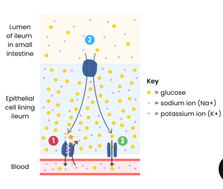
Describe an example that illustrates co-transport (Absorption of sodium ions and glucose (or amino acids) by cells lining the mammalian ileum)
1 ● Na+ actively transported from epithelial cells to blood (by Na+/K+ pump)
● Establishing a conc. gradient of Na+ (higher in lumen than epithelial cell)
2 ● Na+ enters epithelial cell down its concentration gradient with glucose against its concentration gradient
● Via a co-transporter protein
3 ● Glucose moves down a conc. gradient into blood via facilitated diffusion
Describe how surface area, number of channel or carrier proteins and
differences in gradients of concentration or water potential affect the rate of
movement across cell membranes
● Increasing surface area of membrane increases rate of movement
● Increasing number of channel / carrier proteins increases rate of facilitated diffusion / active transport
● Increasing concentration gradient increases rate of simple diffusion
● Increasing concentration gradient increases rate of facilitated diffusion
○ Until number of channel / carrier proteins becomes a limiting factor as all in use / saturated
● Increasing water potential gradient increases rate of osmosis
Explain the adaptations of some specialised cells in relation to the rate of
transport across their internal and external membranes
● Cell membrane folded eg. microvilli in ileum → increase in surface area
● More protein channels / carriers → for facilitated diffusion (or active transport - carrier proteins only)
● Large number of mitochondria → make more ATP by aerobic respiration for active transport
Explain the changes in plant tissue mass when placed in different
concentrations of solute
Increase in mass:
● Water moved into cells by osmosis
● As water potential of solution higher than inside cells
Decrease in mass
● Water moved out of cells by osmosis
● As water potential of solution lower than inside cells
No change
● No net gain/loss of water by osmosis
● As water potential of solution = water potential of cells
Binary fission
The process by which prokaryotic cells divide to produce two genetically identical daughter cells.
Steps of Binary Fission:
1. Replication of circular DNA
2. Replication of plasmids
3. Division of cytoplasm to produce 2 daughter cells
○ Single copy of circular DNA
○ Variable number of copies of plasmids
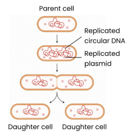
Is binary fission slow or fast
-It is a fast and efficient process, allowing bacteria to reproduce rapidly under ideal conditions.
-Daughter cells are genetically identical except for potential differences in the number of plasmids.
Describe how viruses replicate
1. Attachment proteins attach to complementary receptors on host cell
2. Inject viral nucleic acid (DNA/RNA) into host cell
3. Infected host cell replicates virus particles:
a. Nucleic acid replicated
b. Cell produces viral protein / capsid / enzymes
c. Virus assembled then released
Why do viruses have Viral Replication
-Viruses lack cellular structures such as ribosomes and enzymes, so they depend entirely on the host cell's metabolic machinery for replication.
-The process can destroy the host cell, leading to tissue damage and disease.