Nerve, Muscle, and Synapse Physiology
1/208
There's no tags or description
Looks like no tags are added yet.
Name | Mastery | Learn | Test | Matching | Spaced |
|---|
No study sessions yet.
209 Terms
Neuroscience
The study of the nervous system.
The human body contains roughly 100 million neurons.
Consists of two main parts: The Central Nervous System (CNS) and Peripheral Nervous System (PNS).
Research includes genes and other molecules that are the basis for the nervous system, individual neurons, and ensembles of neurons that make up systems and behaviour.
Central Nervous System (CNS)
Comprised on the cerebral cortex, cerebellum, brainstem, and spinal cord.
Peripheral Nervous System (PNS)
Peripheral nerves.
For example, the axons of nerve cells which connect the spinal cord to the muscles are located here.
The Stretch Reflex - Mallet
If going to the doctor for a physical examination, the doctor will hit the tendon with a mallet.
The tendon is connected to the quadriceps muscle. Tapping the tendon causes the muscle to stretch.
∴ Activation of nerve impulses in special receptors located in the quadriceps.
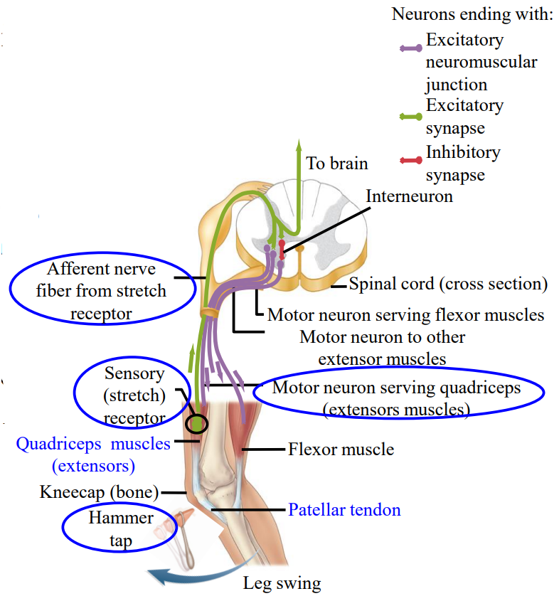
The Stretch Reflex - Impulses
The activation of nerve impulses in special receptors located in the quadriceps, these special receptors are called stretch receptors.
The nerve impulses are sent back to the spinal cord along the sensory neurons.
They activate another nerve cell which feeds back out into the quadriceps.
This activates another muscle causing contraction.
“A knee-jerk” reaction.

Natural Reflex - Biceps
The example is holding a bag.
The bicep muscle will be stretched based on the activation.
If something is added to the bag, there is a slight shift downwards, causing the biceps to stretch.
The degree of activation for the biceps feeds back onto the muscle and allows the arm to stay at 90°.
This stretch reflex is used often and controls posture and muscle tone.
Withdrawal Reflex
Touching a hot stove would activate this reflex.
It is a standard reflex, slightly more complex than the stretch reflex as more cells are involved.
The main process is the same. Your get activation of nerve fibers in the periphery with the withdrawal reflex.
Sends a nerve impulse from the affected area to the spinal cord, where interactions with other neurons occur.
Neurons
A type of cell that receives and sends messages from the body to the brain and back to the body.
10% of cells in the CNS, but they occupy 50% of the volume.
∴ much bigger than glia.
Three different types: afferent, efferent, and interneurons.
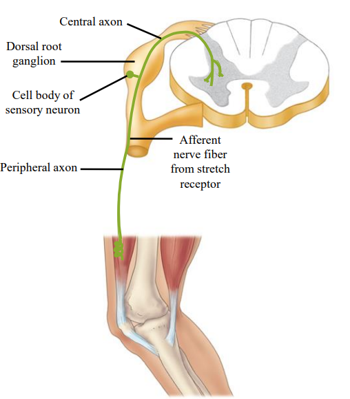
Afferent Neurons
Carry information from periphery to the spinal cord via the dorsal roots.
Make synaptic contact onto either efferent neurons directly, or the make excitatory contact onto interneurons.
Afferent neurons always use the excitatory neurotransmitter glutamate.
Efferent Neurons
Carry information from the spinal cord to the periphery via the ventral roots.
They receive input from either afferent neurons directly or from interneurons to make contact onto muscle.
Uses a excitatory neurotransmitter called acetylcholine.
Interneurons
Carry information between neurons.
Can excite or inhibit neurons.
Located only in the CNS, either the spinal cord or the brain.
Can receive input from afferent neurons, efferent neurons, or other interneurons.
They output information onto other interneurons or onto efferent neurons.
Glia
Acts as the glue, very numerous and tiny.
Another type of neuron. 90% of the cells in the NS and ½ the volume in the NS.
Three different types:
Oligodendrocytes, astrocytes, and microglia.
Oligodendrocytes
Makes myelin which is important in action potential conduction in the CNS. Schwann Cells do this in the PNS.
Crucial for myelin production.
Astrocytes
Physically and metabolically support neurons by buffering extracellular K⁺, removing neurotransmitter, helping maintain the blood brain barrier, and plays a role in signalling and information processing.
Microglia
Serve immune functions in CNS.
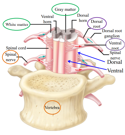
Intertwining Afferent and Efferent Neurons
When afferent and efferent neurons innervate a muscle, they are separated around the muscle.
As they travel toward the CNS, they intertwine (bundle) and travel within a mixed peripheral or spinal nerve.
Before entering the spinal cord, they split: afferent fibers enter dorsally, while efferent fibers exit ventrally.
Information goes in via afferent fibers to the CNS and leaves the CNS via efferent fibers.
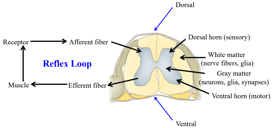
Reflex Loop
The activation of a receptor activates a afferent fiber. The information comes through the dorsal horn.
The efferent neurons, located in the ventral spinal cord, leaves the ventral area.
Afferent comes through dorsal part of the cord.
Efferent comes through the ventral part of the cord.
The efferent fiber then activates a muscle.

White Matter
Within the white matter of the spinal cord, myelinated axons of neurons which are travelling up or down the spinal cord.
This is why white matter appears white.
It contains myelinated axons, giving a white appearance.

Grey Matter
Contains cell bodies of interneurons and unmyelinated processes.
The lack of myelin makes it look grey.
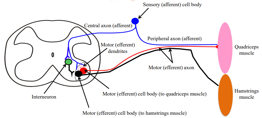
The Stretch Reflex - Afferent Neurons
Tendon tap of the patellar tendon results in stretching of the quadriceps muscle.
Stretching of the muscle activates stretch receptors attached to afferent neurons.
The afferent neurons will travel together with the efferent neurons in the mixed peripheral nerve until it is outside the spinal cord.
Step 1.
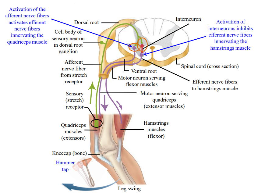
The Stretch Reflex - Interneurons
Branches of the green, afferent nerve fiber also end on the red inhibitory interneurons (in this specific reflex, the interneurons are inhibitory).
When activated, the interneurons inhibit efferent neurons which innervate the hamstrings.
This inhibits contraction of the hamstrings muscle which would otherwise interfere in reflex response.
Step 2.

The Stretch Reflex - Efferent Neurons
The afferent neuron takes a separate pathway from the efferent neuron and enters through the dorsal root of the spinal cord.
Connects with a efferent neuron in the ventral part of the spinal cord and sends a synapse.
Efferent neurons coils through the ventral root of the spinal cord, travels the mixed peripheral nerve to the quadriceps, resulting in contraction.
Step 3.

Agonistic and Antagonistic Pair
All muscles have a opposing-function pair.
i.e. biceps (agonistic) → lifts forearm, triceps (antagonistic) → extends forearm.
Coordinated movement is controlled by innervation and activation of agonist/antagonist muscles.

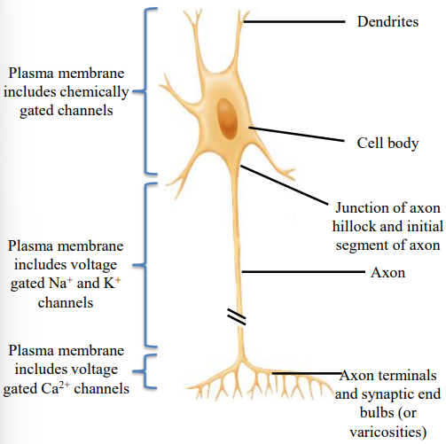
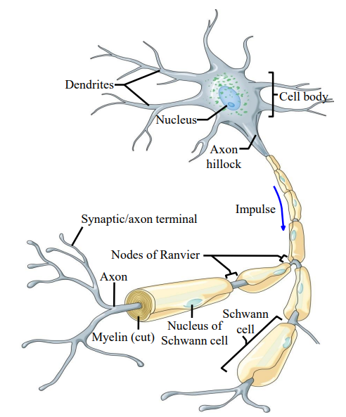
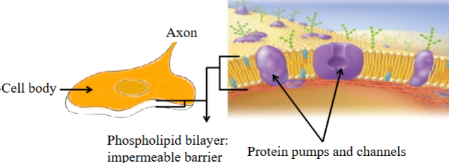
Dendrites
Receive information either from the periphery or from other cells.
Receive stimuli through activation of chemically or mechanically gated ion channels; in sensory neurons, produce generator or receptor potentials; in motor neurons and association neurons, produce excitatory and inhibitory postsynaptic potentials (EPSPs and IPSPs).

Nucleus
Genetic material factory.
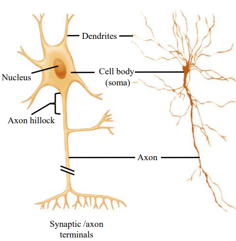
Axon
Propagates (conducts) nerve impulses from initial segment (or from dendrites of sensory neurons) to axon terminals in a self-reinforcing manner
Impulse amplitude does not change as it propagates along the axon.
Axon Hillock
Input coming into the dendrites is integrated in the axon.
An impulse is initiated here if there is enough input for a nerve impulse.
Trigger zone; integrates EPSPs and IPSPs and, if sum is a depolarization that reaches threshold, initiates action potentials (nerve impulse).

Synaptic/Axon Terminals
Contains neurotransmitters.
The neurotransmitter will be released and cause other cells/muscle to be activated or inhibited.
Inflow of Ca2+ caused by depolarizing phase of nerve impulse triggers neurotransmitter release by exocytosis of synaptic vesicles.

Cell Body (soma)
Contains ribosomes.
Has all the information and machinery to manufacture proteins.
Receives stimuli and produces EPSPs and IPSPs through activation of chemically or mechanically gated ion channels

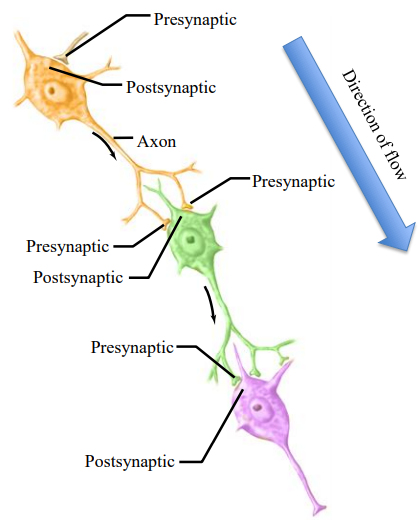
Direction of Flow
Information in the nervous system flows in one direction.
It goes from the dendrites to the axon, ending at the synaptic terminal.
It will never flow in the opposite direction.

Presynaptic & Postsynaptic Neuron
The presynaptic neuron sends the signal.
The postsynaptic neuron receives the signal.
A neuron can be postsynaptic to one cell and presynaptic to another cell.
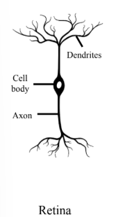
Bipolar Cell
A specialized type of nerve cell characterized by having one dendrite and one axon extending from the cell body.
It has two distinct processes, emerging from the cell body.
These extensions are one axon and one dendrite.
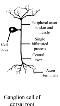
Pseudo-Unipolar Cell
Has one axon that splits into two branches.
These branches act as both dendrites and axons, with one branch carrying signals to the periphery and the other to the CNS.
Afferent neurons of the stretch reflex fall into this category.
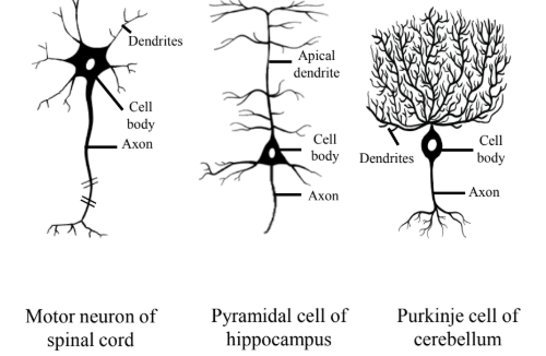
Multipolar Cells
Characterized by a single axon and multiple dendrites extending from their cell body.
This structure allows them to receive and integrate a large amount of information from other neurons.
All of them have a axon terminal that terminate in the muscle, affecting activity in the muscle.
Both interneurons and efferent neurons are this type.
Pseudo-Unipolar Function
Peripheral Axon
The peripheral axon extends from the cell body to the muscle. Acts like the dendrites.
Central Axon
Extends from the cell body to the CNS.
Acts like a normal axon and makes contact with other axons on interneurons.
Dorsal Root Ganglion
The cell body of the neuron is located here.
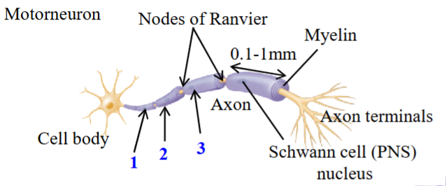
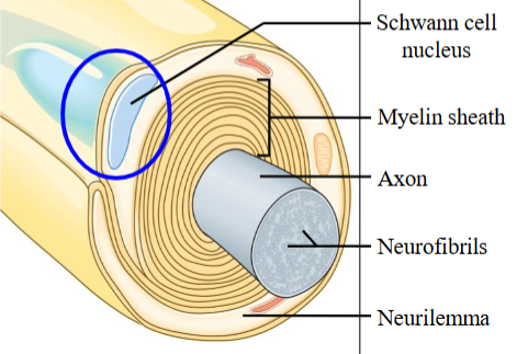
Myelin
A fatty, insulating sheath that surrounds the axons of neurons.
A lipid-protein mix; 80% lipid and 20% protein.
The white fatty myelin is made of either oligodendrocytes or Schwann cells.
Ingredients: myelin basic protein, proteolipid protein, and glycolipids.
Increases the speed of electrotonic conduction.
The Stretch Reflex - Pseudo-Unipolar (Afferent)
The afferent neuron is a pseudo-unipolar cell, receiving input via the peripheral axon and providing output to the CNS via the central axon.
There are two synapses that the pseudo-unipolar neuron makes.
The Stretch Reflex - Multipolar Neuron (Efferent)
During the stretch reflex, the afferent neuron makes direct monosynaptic contact onto a efferent neuron (red)
The efferent neuron innervates the quadriceps.
The activation of this excitatory efferent neuron results in the quadriceps contracting.
The Stretch Reflex - Multipolar Neuron (Inter.)
The afferent neuron is excitatory and activates this inhibitory interneuron (green).
Makes contact onto a efferent neuron (black).
The efferent neuron innervates the antagonistic muscle, for quadriceps its the hamstring.
The inhibitory interneuron inhibits the efferent neuron to the hamstring, preventing the hamstrings from contracting.
Neuronal Membrane
A phospholipid bilayer, impermeable on it own, but has protein pumps and channels embedded in it.
The most important protein pumps/channels for setting resting membrane potential are the Na⁺/K⁺ pump and the Na⁺ and K⁺ leak channels.
Neuronal Pumps and Channels
Controls movement of ions through membrane.
Pumps: active transport.
Ion channels.
Passive channels (leak channels).
Gated channels: ligand-gated and voltage-gated.
Active Transport
Use an energy source (ATP) and is not driven by a concentration gradient.
Neurons specifically.
Ion Channels
Does not use a energy source (ATP).
Acts as a door which is either open or closed.
Allows the flow of ions down its concentration gradient.
Pumps and Channels Characteristics
There is many different types of protein pumps and channels, all with different characteristics.
Due to the presence of pumps & channels, there is a uneven distribution of different ions inside and outside the cell.
Gives the cell a resting membrane potential (RMP).
Charge separation b/w the ECF and ICF.
Leak Channels (Passive Channels)
A channel that is always open. Allows the the passive flow of ions into/out of the neuron.
Allow diffusion of ions down their electrical and chemical concentration gradients in an attempt to reach equilibrium.
Selective: each ion has its own channels which they can pass.
Two forces acting on each ion: chemical and electrical gradient.
Ligand-Gated Channel
A neurotransmitter binds to its receptors, leading to conformation change (close → open), allowing the flow of ions through them.
The ligand is the neurotransmitter.
Into/out of the cell.
Voltage-Gated Channels
Open and close in response to changes in the electrical potential across the cell membrane.
These channels are crucial for generating and propagating action potentials.
Resting Membrane Potential (Em or RMP)
Steady-state condition determined by relative permeability of membrane to K⁺ and Na⁺.
Measure of electrical potential different between ICF and ECF.
Due to the activity of the Na⁺/K⁺ pump, there is an uneven distribution of Na⁺ and K⁺ across the membrane.
∴ Charge separation, approx. -70mV.
Ions involved in setting RMP:
K⁺, Na⁺, [Cl⁻, organic anions (-)].
Pumps/channels involved in setting RMP:
Na⁺/K⁺ pump, Na⁺ leak channels, K⁺ leak channels, [Cl⁻ pump].
![<ul><li><p>Steady-state condition determined by relative permeability of membrane to K⁺ and Na⁺.</p></li><li><p>Measure of electrical potential different between ICF and ECF.</p></li><li><p>Due to the activity of the Na⁺/K⁺ pump, there is an uneven distribution of Na⁺ and K⁺ across the membrane.</p><ul><li><p>∴ Charge separation, approx. -70mV.</p></li></ul></li><li><p>Ions involved in setting RMP:</p><ul><li><p>K⁺, Na⁺, [Cl⁻, organic anions (-)].</p></li></ul></li><li><p>Pumps/channels involved in setting RMP:</p><ul><li><p>Na⁺/K⁺ pump, Na⁺ leak channels, K⁺ leak channels, [Cl⁻ pump].</p></li></ul></li></ul><p></p>](https://knowt-user-attachments.s3.amazonaws.com/fc4eaf25-8bd6-45c0-a773-618761a3949c.png)
Net Negative Charge
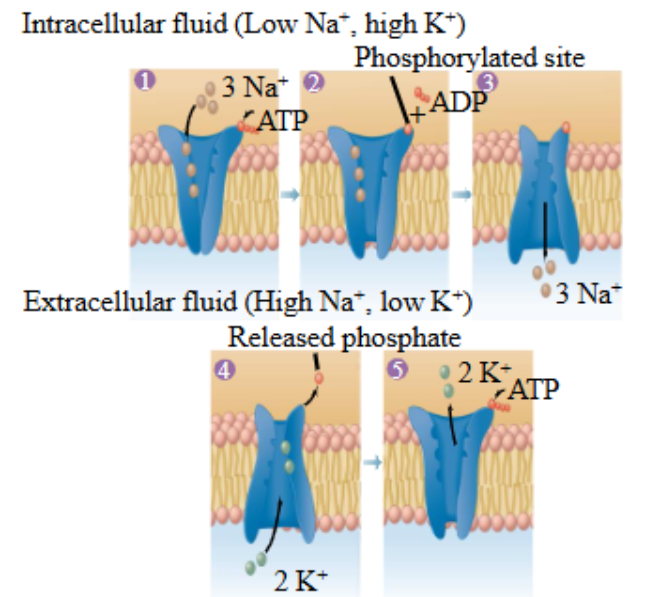
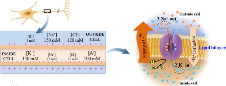
Na⁺/K⁺ pump is electrogenic, moving charge across the membrane.
Requires energy obtained from the hydrolysis of ATP (an energy carrying molecule) to ADP + Pi.
3 Na⁺ molecules move out of the cell, 2 K⁺ molecules move into the cell.
Results in a net negative charge inside the cell.
Moving a net +1 ion charge out of the cell for every cycle in the Na⁺/K⁺ pump, resulting in a net negative charge.
Na+/K+ ATPase
3 Na⁺ bind to the Na⁺/K⁺ pump.
ATP binds to the pump causing conformation change in the protein, leaving a phosphate group attached to the protein and ADP.
The energy that comes from hydrolysis results in a change of conformation of the change Na⁺/K⁺ pump.
The phosphate group leaves, resulting in conformation change, taking in 2 K⁺.
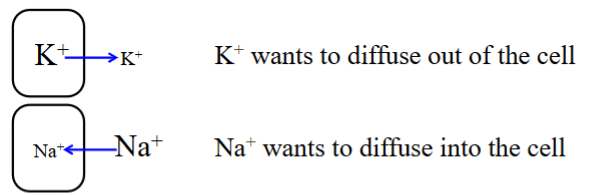
Na+/K+ Pump Creates Gradients
Both chemical and electrical gradients are present.
For the electrical gradient, the intracellular environment wants to become more positive.
The Na⁺/K⁺ pump pumps K⁺ into the cell, and the K⁺ then wants to move back out of the cell.
The Na⁺/K⁺ pump pumps Na⁺ out of the cell, which then wants to move back into the cell.
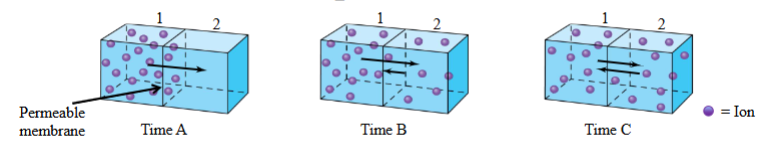
Equilibrium
At any time you have a gradient for a ion across a membrane, you will have more ions on side of the permeable/semi-permeable membrane.
The cell will try to reach a state of equilibrium; equal distribution.
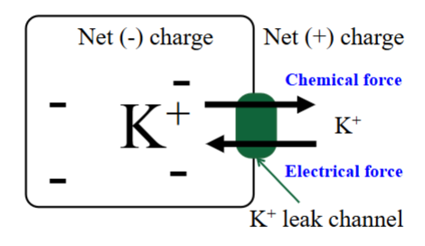
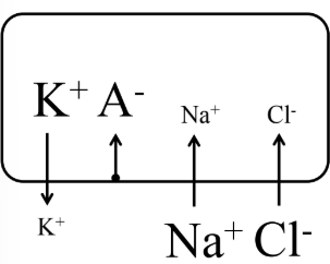
Potassium Leak Channel
There is more K⁺ in the cell than outside the cell.
The chemical force pushes K⁺ outside the cell through the leak channel.
The electrical force pushes the K⁺ into the cell (as a positively charged ion).
Both of these forces act on K⁺ at the same time.
Net charge in the ICF is negative, ECF is positive.

Equilibrium Potential
The membrane potential at which the chemical concentration gradient is balanced.
No net movement of any ions into or out of the cell because the chemical and electrical force are equal and acting in opposite directions.
Cancels out.
Na⁺ and K⁺ attempt to reach an ideal potential, +55mV and -90mV respectively.
Can be calculated using Nernst equation.
Equilibrium Potential for Potassium
No net movement of K⁺ into or out of the cell.
The chemical force and the electrical force are equal and acting in opposite directions.
With the Nernst equation, you can calculate the equilibrium potential of an ion, based on the amount present, temperature, etc.
Potassium at Resting Potential
K⁺ will be moving out of the cell.
K⁺ is a positively charged ion and the Em is -70mV, K⁺ will move to try to bring the Em of the cell to -90mV.
-90mV is equilibrium potential that K⁺ wants to maintain.
∴ Net movement is out of the cell, taking with it the positive charge.
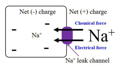
Sodium Leak Channel
There is more Na⁺ outside the cell than the inside.
The chemical force acts to push Na⁺ into the cell, to achieve a state of equilibrium.
The electrical force acts to push Na⁺ into the cell.
Net charge inside the cell is negative, and the net charge outside the cell is positive.
Sodium at Resting Potential
Na⁺ will be moving into the cell.
At a RMP of -70mV, Na⁺ will move to make it equal to its equilibrium potential of +55mV.
Neither K⁺ or Na⁺ are successful in shifting the membrane potential to its equilibrium membrane potential.
Equilibrium Channel for Sodium
If the inside of the cell was +55mV, the electrical force would push positively charged Na⁺ out of the cell while the chemical force would push Na⁺ into the cell.
The forces acting on Na⁺ would be equal and opposite, resulting in no movement of Na⁺ into or out of the cell.
Na⁺ would be at equilibrium.
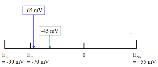
Leak Channels and Em Rule
Na⁺ alone would force RMP to +55mV.
K⁺ alone would force RMP to -90mV.
The more permeant the ion, the greater its ability to force Em towards its own equilibrium potential.
The more permeable a membrane is to a specific ion, the closer the cell’s RMP will be to that ion’s equilibrium potential.
Leak Channels and Em Ions
There is more K⁺ leak channels in the membrane, so the RMP is closer to the equilibrium potential for K⁺.
Permeability is 50-100x times greater to K⁺ than Na⁺.
Em is therefore closer to EK+ → more leak channels for K⁺.
Em Variability
The variable values for RMP is because there are different amounts of leak channels in different cells.
The specific ratio of Na⁺ and K⁺ determines the RMP of a given cell.
∴ Subject to variability.
RMP Summary
Activity of Na⁺/K⁺ ATPase causes intracellular environment to be negatively charged with respect to extracellular environment (3 Na⁺ out/ 2 K⁺ in).
At RMP passive ionic fluxes are balanced so that there is charge separation and Em remains constant.
Value of RMP (-70mV) is closest to equilibrium potential of the ion with greatest membrane permeability (K⁺).
RMP Ion Movement
At RMP, there is movement of ions (K⁺ and Na⁺) across the PM.
K⁺ is pumped into the cell by the Na⁺/K⁺ pump and is leaving through K⁺ leak channels.
Na⁺ is pumped out of the cell via the Na⁺/K⁺ pump and coming back into the cell via Na⁺ leak channels.
Less Na⁺ is coming out of the leak channels.
End result of ion movement = -70mV.
Action Potential
A large change in membrane potential from -70mV to +30mV and back to resting over a period of a few milliseconds.
An electrical signal is generated due to activity of voltage-gated Na⁺ and voltage-gated K⁺ channels.
Opening of these channels results in a ion flow and membrane potential changes.
Occurs when specific stimuli (physical or chemical) disrupt RMP, causing ion-selective channels to open.
Afferent Activation
Muscle stretch (stretch reflex) or other sensory stimuli increase opening of specialized Na⁺ receptors.
Allows Na⁺ entry into the afferent fiber and depolarizes the neuron.
If depolarization reaches threshold (~ -50 mV), voltage-gated Na⁺ channels open and an action potential is generated.
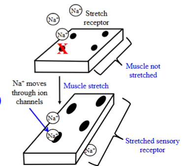
Stretch Receptor
Embedded in the muscle. There is a receptor at the tip of the afferent neuron.
It senses muscle stretch (i.e. stretch in the quadriceps muscle).
Stretched/Unstretched Sensory Receptor
When the muscle is not stretched, the Na⁺ cannot casually flow inside as it is too large.
When the muscle is stretched, the sensory receptor enlarges.
When this occurs, the Na⁺ can go through the pores of the receptor.
Increased Na⁺ permeability.
Goes from the ECF to the ICF.
Threshold Potential
A value of membrane potential that must be reached for a action potential.
This value is needed for the opening of voltage-gated Na⁺ channels and firing a action potential.
Threshold varies slightly between cells but is typically 10–15 mV more depolarized than the RMP.
Slight Depolarization
Following tendon tap, the quadricep muscle stretches, allowing the sensory receptors to stretch and allow Na⁺ entry into the cell.
Cell depolarizes slightly.
If the threshold number is reached (~-50mV), the voltage-gated Na⁺ channels will open completely.
Ion Channels at RMP
The voltage-gated Na⁺ channel and the voltage-gated K⁺ channel are closed at rest.
The Na⁺/K⁺ ATPase is still functioning, keeping K⁺ inside the cell and Na⁺ outside the cell.
Leaky channels are still open.
Depolarization
When the threshold potential is reached, the voltage-gated Na⁺ channels open, removing the activation gate.
When Na⁺ moves into the cell. The internal compartment of the cell changes from being negatively charged to positively charged.
+30mV.
Brings the membrane potential closer to Na⁺ equilibrium potential (+55mV).
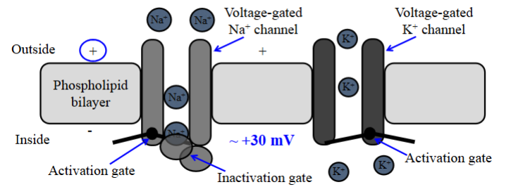
Peak
After reaching +30mV, the Na⁺ channel closes; the inactivation gate slides across the pore and blocks more Na⁺ from entering the cell.
1-1.5 ms.
Open conformation of the voltage-gated Na⁺ channels is only maintained for a few milliseconds.
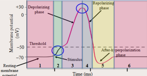
Repolarization
After the inactivation gate blocks off the Na⁺ channel, the activation gate of the K⁺ channel unblocks the channel.
K⁺ rushes outside the cell to make the inside of the cell equal to its equilibrium potential (-90mV).
K⁺ has a positive charge, so it would leave the cell to make the ICF negative.
However, overshoots it a bit.

Hyperpolarization
Membrane potential decreases, the value going closer to the RMP value as K⁺ is leaving the cell.
The permeability of the membrane to K⁺ is greater during hyperpolarization than it is at RMP.
The membrane is more permeable to K⁺.
This is b/c the leak channels are open (where K⁺ can flow) and there are voltage-gated K⁺ that are open.

Absolute Refractory Period (3, 4)
Cannot fire an action potential in response to any stimulus.
Voltage-gated Na⁺ channels are either open or inactivated.
The inactivation gate blocks the voltage-gated Na⁺, closing off the pore.
The channel cannot reopen until the gate leaves the pore and the membrane repolarizes to close it.
After the channels have recovered, another AP can occur.

Relative Refractory Period (5)
Possible to generate a AP, but extremely difficult.
This is because voltage-gated K⁺ channels are still open.
For the cell to reach the threshold (-50 mV) where voltage-gated Na⁺ channels open, Na⁺ permeability must exceed K⁺ permeability, which is already elevated due to open voltage-gated K⁺ channels.

Action Potential Table
Condition | Reason for Change in Potential | Relative Permeability |
1. Rest | K⁺ » Na⁺ | |
2. Depolarizing Input | Sensory of synaptic stimulation | Slight ↑ to Na⁺ |
3. Start of AP (depol) | V-gated Na⁺ channels open | Na⁺ »» K⁺ |
4. Repolarizing Phase | V-gated K⁺ channels open & Na⁺ channels inactivate | K⁺ »» Na⁺ |
5. End of AP | V-gated Na⁺ channels at rest & V-gated K⁺ channels still open | K⁺ »»»» Na⁺ |
6. Rest | V-gated K⁺ channels at rest | K⁺ » Na⁺ |
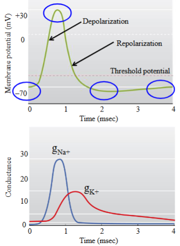
Membrane Conductance
A measure of how easily ions can flow across the membrane, represented by ‘g’.
At the beginning of a AP, there is a increase in Na⁺ conductance.
This tapers off quickly.
At the beginning of a AP for K⁺, there is a slight delay before there is a K⁺ conductance increase (opening of K⁺ voltage channels).
Tapers off.
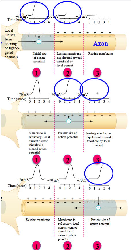
Action Potential: Transmission
Axon at resting membrane potential.
Activation results in opening of voltage-gated Na⁺ channels, local depolarization of membrane.
Local depolarization of membrane causes adjacent voltage-gated Na⁺ channels to activate.
New action potential is generated in adjacent membrane.
Action potential only travels in one direction due to refractory period.
Adjacent Segments
AP begins with Na⁺ flowing into the cell as a response from a stretched sensory receptor.
Once the threshold is reached, the Na⁺ voltage-gated channels open, causing local depolarization in a segment of the axon.
Depolarizes the neighbouring membrane to threshold, activating a AP.
The AP is transmitted from segment to segment in a adjacent, one direction manner.

Electrotonic Conduction
Spread of current inside axon.
AP initiated at one point in membrane. The current spreads electronically to adjacent membrane.
The adjacent membrane depolarizes to threshold and a new AP generated in adjacent membrane.
Proceeds in one direction only.
Time Needed for Electrotonic Conduction
Flow is fast but the AP must be regenerated at every point on the membrane. This requires opening and closing of channels, which takes time!
Moreso if the axon is long, which will take more time for the signal to travel.
The removal of our foot from a nail (as an example) has to be fast. In reality, the removal of our foot is ½ seconds.
Due to myelination.
Myelination
Speeds up AP propagation along the membrane of a neuron.
Virtually all efferent neurons are myelinated.
The production of myelin stem from the glia.
It is wrapped around the neuronal axon.
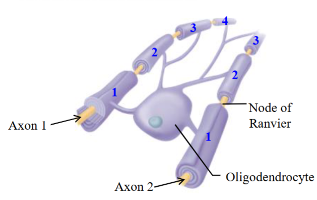
Oligodendrocytes - Myelin
Makes myelin in the Central Nervous System (CNS) only.
i.e. interneuron.
A single oligodendrocyte is able to myelinate several axons and able to myelinate several regions within a given axon.
In the figure, its responsible for myelinating two different neurons.
Potentially able to lay down 7 different myelinated regions in total.
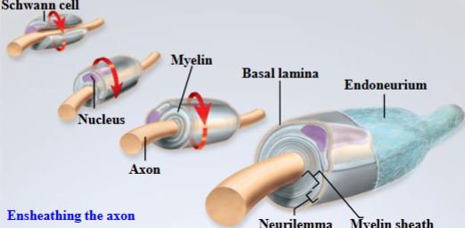
Schwann Cells - Myelin
Makes myelin in the Peripheral Nervous System (PNS) only.
i.e. efferent neuron.
Produced in the nucleus.
Each myelinated segment is generated by a different Schwann cell.
Unsheathing the Axon
Over time, the myelin produced by Schwann cells/oligodendrocytes wraps itself around the axon.
This protein-lipid mix wraps around the axon of a neuron and acts as a insulator.
Myelin Location
Is only found wrapped around the axon.
There is no myelin around the cell body or the dendrites of a neuron.
Myelin stops right before the synaptic terminals.
Begins right after the initial segment of the axon until the axon branches off at the synaptic terminals.
Nodes of Ranvier
Unmyelinated regions of a myelinated axon.
The voltage-gated Na⁺ channels are clustered here.
There are very few voltage-gated Na⁺ channels along the axon in the myelinated region.
The other area where voltage-gated Na⁺ channels are clustered is the axon hillock, which is also unmyelinated.
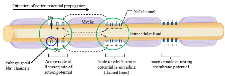
Myelin - AP Propagation
Myelin speeds up AP propagation by acting as an insulator.
It increases conduction speed by improving electrostatic efficiency, so the AP doesn’t need to be regenerated at every point along the membrane.
Regeneration is required only at the nodes of Ranvier.
The AP travels from node to node without diminishing in amplitude.

Myelin - Charged Environment
The protein-lipid substance of the myelin surround and insulates the axon, preventing positively charged ions from entering from either outside of inside the membrane (in the myelinated region).

Myelin - AP Amplification
If an AP is generated at the first node, it travels to the second node without decreasing in size, allowing the neighbouring tissue to depolarize without active depolarization along the entire axon.
Neighbouring tissue doesn’t need to be depolarized at every point along the myelinated region because of myelin.
Depolarization and the opening of voltage-gated Na⁺ channels occur only at the nodes of Ranvier.
This reduces the need for AP regeneration.
Axon Propagation Speed
There are two factors where determine the speed of an axon propagating a action potential.
Size of an axon.
Whether the axon is myelinated or not.
The thicker an axon is, the faster a AP can propagate.
Myelinated axons propagate AP faster than unmyelinated axons.

Multiple Scelerosis
It is thought that the immune system attacks the myelin.
This leads to the speed of saltatory conduction to decrease, as the myelin is not there to increase the speed of transmission.
Loss of myelin = decreased transmission speed.

Classification of Fiber Type
Fiber Type | Group 1 | Group 2 | Group 3 | Group 4 |
Diameter | 12 - 20µm | 6 - 12µm | 1 - 5µm | 0.2 - 1.5µm |
Speed | 80 - 120m/s | 35 - 75m/s | 5 - 30m/s | 0.5 - 2m/s |
Sensory Receptors | Skeletal muscle Proprioceptor | Skin Mechanoreceptor | Pain/temperature | Pain/itch/temperature |
Action potential frequency: Fast →→→ Slow.
Proprioceptor
Sensory receptors which sense the position of our limbs.
Proprioceptors and efferent neurons are included among the thickest diameter neurons that are myelinated.
Conducts AP from 80 - 120m/s.
Mechanoreceptor
Smaller than the larger fibers in the group (group 2).
A type of afferent neuron.
i.e. how you know you’re touching your desk.
One Directional Propagation
AP arrives at axon going from left to right; AP will always travel down the axon to the synaptic terminal.
Absolute refractory period (ARP) lasts 2 milliseconds.
By the time the ARP is over, action potential is between 2 - 20cm down the axon.
Far away enough that depolarization will not occur in neighbouring tissues.
Synaptic Transmission
A physical stimulus causes a sensory neuron to fire an AP.
This generates a receptor potential; the nerve ending depolarizes to threshold, and an AP is produced in the sensory neuron.
The AP travels via electrotonic or saltatory conduction in the CNS.
There's no direct connection between sensory neurons to muscles.
Instead, the presynaptic neuron releases neurotransmitters from vesicles into the synaptic cleft.
These neurotransmitters bind to the postsynaptic neuron, causing an excitatory or inhibitory response that ultimately leads to a physiological effect.
Gap Junction
Allows ions and molecules to pass directly from cell to cell through an actual channel between cells.
Allows for electrical synaptic transmission to occur.
Typically connected at the dendrites.
Dendrite to dendrite, dendrite to cell body, or cell body to cell body.
Found in dendrite/cell body area, where electrical transmission occurs.