Unit 2: The Skull
1/85
There's no tags or description
Looks like no tags are added yet.
Name | Mastery | Learn | Test | Matching | Spaced | Call with Kai |
|---|
No analytics yet
Send a link to your students to track their progress
86 Terms
Functions of bone
support, protection, movement, mineral homeostasis, hemopoiesis, lipid storage
Support
shape, muscle, against gravity. Durable an resistant to a lot of forces
Protection
The role of flat bones, such as the skull and sternum, in protecting our most vital organs.
Movement
The function of long bones in the appendicular skeleton that act as levers for movement.
Mineral homeostasis
The process by which bones build up and break down based on blood calcium levels.
Hemopoiesis
The production of blood cells that occurs in red bone marrow found in various bones.
Lipid storage
The function of yellow bone marrow, which develops from red bone marrow as an individual matures.
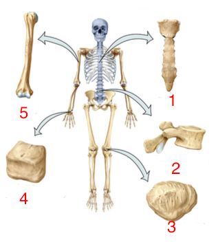
Name these bones
flat bones
irregular bones
sesamoid bone
short bones
long bones
Flat bones
Thin bones primarily found in the skull and sternum, serving mainly for protection.
Irregular bones
Bones like vertebrae that provide attachment points for ligaments and muscles, contributing to support and movement.
Sesamoid bone
A type of bone, such as the patella, that protects ligaments of the knee as we flex and stand so the ligament doesn’t get compressed. Some people have extra ones in their toes.
Short bones
Cubed-shaped bones found in the wrists and ankles that help transfer forces.
Long bones
Bones located in the appendages that are essential for movement.
Osteogenic cell
The stem cell that divides to form osteoblasts. Initial bone cell (baby cell)
Osteoblasts
Bone building active cells that construct the inorganic bone matrix. Juvenile cells
Osteocytes
Mature bone cells that maintain bone tissue after osteoblasts become trapped in the matrix.
Osteoclast
Cells that break down bone tissue, working with osteoblasts to maintain calcium homeostasis.
Compact bone
Dense bone structure containing osteons, which resemble tree rings. Individual rings are called lamellae. Center of osteon is called the central/haversian canal and is where blood vessels pass through.
2 structures made by osteocytes
Lacunae: openings the osteocyte is trapped in, within the lamellae
Canaliculi: canals containing projections of the osteocytes stretching into the lamellae
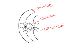
Spongy bone
Bone structure characterized by trabeculae that provide scaffolding for bone marrow. Less compressed canaliculi and osteocytes
Parts of a bone
Diaphysis
epiphysis
articular cartilage
metaphysis
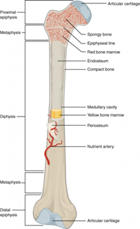
Diaphysis
The shaft of a bone, composed of compact bone on the outside and spongy bone on the inside. Middle of the shaft is the medullary cavity
Epiphysis
The ends of a bone, with proximal and distal regions relative to the body.
Articular cartilage
Hyaline cartilage that protects the ends of bones and can break down during arthritis.
Metaphysis
The region between the diaphysis and epiphysis. Where the epiphyseal line is and where growth is going to occur
Xrays of juvenile vs adult
In juveniles there will be a gap between the diaphysis and epiphysis because the metaphysis is cartilage (metaphysis plate. In adults, it ossifies and turns into bone (metaphysis line)
Intramembranous ossification
The process of bone formation for flat bones, starting before birth and finishing around maturity. General shape is created by multiple layers of fibrous connective tissue
Endochondral ossification
The process responsible for forming all the other bones (not flat), beginning with a cartilage model. Starts before birth in the diaphysis of the developing bone and works its way towards the ends. Shortly after birth ossification begins in the end of the bone
Skull functions
protects the brain and sense organs (taste, smell, sight, hearing)
flat bone
possibly cools the brain when temperatures rise
supports the voicebox and acts as a resonating chamber
vital part of feeding systems of vertebrates
3 parts that make up the skull
chondrocranium, splanchnocranium, dermatocranium
Chondrocranium
Retained in Chondrichthyes to support and protect the brain. Embryonic structure in Osteichthyes. Cartilaginous foundation then it ossifies. Serves as scaffold for developing brain and supports sensory capsules.
Development of chondrocranium
Mesenchyme (embryonic tissue) of the head condenses to form elongate cartilages along the notochord
Sensory capsules, associated with the eyes, nose, and ears develop supporting cartilages laterally (towards the outside of the body
Cartilages fuse into a plate that serves as the bases of the chondrocranium
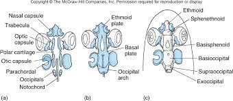
Cartilages (all paired)
anterior trabeculae cartilage
posterior parachordals cartilage
polar cartilage (in some vertebrates) sit in between trabeculae and parachordals
occipital cartilage (more posterior)
Sensory capsules
nasal capsule
otic capsule
optic capsule
Plates
ethmoid plate: front of the skull, fusion of nasal capsule and front portion of the trabeculae
basal plate: in the middle, coming from the parachordals
occipital arch: occipital cartilages expand and grow around the nerve cord/notochord
Splanchnocranium
The oldest part of the skull, associated with filter feeding and later functions in vertebrates such as gill support and attachment for respiratory muscles
Development of splanchnocranium
neural crest cells migrate to the pharynx between the pharyngeal slits to form the pharyngeal/gill/branchial arches. Support structures
Elements that make up each gill arch
pharyngobranchial
epibranchial
ceratobranchial
hypobranchial
basibranchial
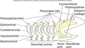
Mandibular arch
The first gill arch that gives rise to the jaw structures in vertebrates and bears teeth. Made up of palatoquadrate (precursor to maxilla) and Meckel’s cartilage (precursor to mandible)
Hyoid arch
second gill arch, follows mandibular arch. Hyomandibula is the prominent element. Gives rise to hyoid bone and the inner ear of tetrapods
types of jaw attachments
Amphistylic
hyostylic
metatostylic
craniostylic
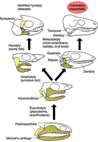
Amphistylic
in chondrichthyans and some actinoptergians. Jaw is attached to the skull by way of ligaments to the palatoquadrate bone and hyomandibula. Very flexible because it’s just ligaments
Hyostylic
Majority of bony fishes. Jaw is attached by the hyomandibula (much closer to the brain case compared to amphistylic). Tighter connection
Metatostylic
Amphibians. Hyomandibula turns into stapes so it doesn’t play a role in jaw attachment. The palatoquadrate turned into the quadrate bone and attaches directly to the skull
Craniostylic
Mammals. Entire maxilla is now apart of the skull itself. Most rigid structure, not flexible
Dermatocranium
Made of dermal bones that forms the sides, roof of the skull, and hard palate.
Series of the dermatocranium
Facial
orbital
temportal
vault
palatal
mandibular
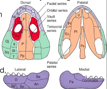
Facial series
encircles the external nares, forming the snout
made up of
maxilla and premaxilla
nasal (sits right next to the nares)
septomaxilla (adjacent to the nasal bones), not always present
Orbital series
encircles the eye, defining the superficial orbit
Made up of:
lacrimal bone (nasal lacrimal duct/tear duct in tetrapods)
prefrontal, postfrontal, and postorbital (fused in tetrapods)
jugal: associated with lower rim of the orbit
scleral ossicles (found in turtles and birds): sitting towards back of the orbit
Temporal series
behind the orbit and completes posterior wall of braincase
made up of:
intertemporal, supratemporal, and tabular (tends to fuse in evolutionary history)
squamosal and quadratojugal: near the cheek area
Vault series
roofing bones that cover the top of the skull
made up of:
frontal: forehead
parietal: major vault bone, primary bone at the top of the head
postparietal
Parietal foreman
Only in amphibians and reptiles. “Third eye” a photosensitive hole through the top of the skull that strikes the pineal gland which regulates sleep/wake cycle. Allows animal to be aware of changes in light intensity around them.
Palatal series
covers much of the roof of the mouth
made up of:
pterygoid: tends to be the largest and most medial (along the midline of the body) of the bones
vomer, palatine, and ectopterygoid: associated with maxillary teeth
parasphenoid: medial, towards the back of the skull. Found in the lower vertebrates (Fishes and some amphibians)
Mandibular series
generally made up of ossified Meckel’s cartilage
Made up of:
lateral side (side facing the outside of the skull (fused in mammals)
medial side
mandibular symphysis: where the 2 halves of the mandibles meet. (fused in mammals)
Lateral side of mandibular series parts
Dentary bone: bares teeth
splenial: 1 or 2 sitting underneath dentary bone
Angular: behind dentary bone
surangular bones: above angular bone
Medial side of mandibular series parts
coronoids: other side of teeth but not holding them in like the dentary bone
prearticular: rest of the mandible
Chondrichthyans skull overview
elaborative cartilage case around the brain (chondrocranium never ossifies), dermatocranium is absent (reflects absense of bone in skeleton)
Bony fishes and tetrapods skull overview
braincase is extensively ossified. Braincase is a bony box with an endoskeletal platform and everything encased by exoskeleton bones
Chondrocranium contribution (embryonic)
provides scaffolding
sphenoid: can be in a series, platform for the brain. Makes up parts of the orbits and the side
occipital: can have up to 4, humans only have 1 at the back of the skull, foramen magnum and occipital condyles
otic capsule: ossifies and surrounds auditory organs and bones
Foramen magnum
where the spinal cord passes, inside the skull the spinal cord is called the medulla oblongata. Basically a giant hole in the skull
Occipital condyles
articulate (make the joint) with the first cervical vertebrae in the column. Allows for rocking back and forth on the vertebra
Splanchnocranium contribution
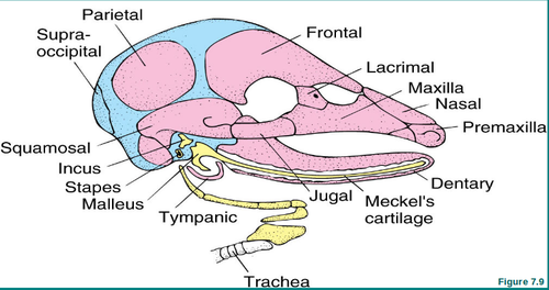
epipterygoid (alisphenoid in mammals): a platform that partly becomes the stapes, incus, and malleus
Dermatocranium contribution
endoskeletal elements, brain, and sensory organs are encased by the exoskeletal elements of it.
Cranial kinesis
Movement between upper jaw and the braincase at the joints between them. The ability of the skull to move, allowing for flexible feeding strategies.
Kinetic skull
majority of vertebrates have this type. Movable skull
Akinetic skull
A skull type that is immovable. Most mammals, crocodiles, and testudines have this type
Advantages of kinetic skull
suction or filter feeding: method of prey capture where the animal gulps water along with the food
Allows tooth bearing (dentary) bones to move rapidly into strategic positions and walk over prey items to push it down the throat
ex) teleost fishes with protrusible jaws and venomous snakes
Unidirectional flow
In animals with gills, excess water exits gill slits at the back of the mouth. Drops pressure in mouth and water flows in. Close mouth to expel water
Bidirectional flow
in animals without gills, excess water must exit back out the mouth. Have a specialized esophagus that stores the water to slowly expel it after they close their mouth without losing food.
Advantages of akinetic skulls
greater jaw strength and crushing
allows offspring to suckle easily (in mammals)
Mastication, more efficiently chew with specialized teeth
Major transitions in the skull: chondrichthyans
Dermatocranium is absent and chondrocranium has expanded
Anterior regions of ethmoid and orbital and posterior oticooccipital merge into an undivided braincase
Hyomandibular and palatoquadrate is loosely attached to the braincase. Makes the jaws very flexible and allow them to jut the mouth out during feeding
Allows sharks to use suction to draw in small prey or to protrude the jaws forward/downward to capture prey
Major transitions in the skull: actinopterygians:
See a proliferation (massive amount) of facial bones
Fishes also have opercula bones (operculum, bony structure that covers the gills) and extrascapulars (series of bones at the back of the skull)
Lots of joints between the different bones, why they have a protrusible jaws. Allows for rapid suction feeding/pipette feeding where the buccal cavity expands to suck in the prey.
Have nares but only do unidirectional flow
Major transitions in the skull: sarcopterygians
In lungfishes, the upper jaw is fused to the braincase and teeth are flattened into plates
Coelacants have strong jaws with large teeth and the braincase is divided into the ethmosphenoid and oticooccipital units. Joint at the top of the skull allow the mouth to open really wide
Nasal capsules contain olfactory epithelium in the form of paired nasal sacs
Nasolacrimal duct connected to the nasal sac which drains excess secretions from the lacrimal gland.
External nare meets with internal nare that goes to the mouth and links up with respiratory system
Major transitions of the skull: Amphibians
Bones of snout reduced, start to fuse
Hyomandibula no longer functions in jaw suspension- now used for hearing as stapes (auditory ossicle/bone that is conserved throughout all tetrapods, middle ear)
Opercular bones and extrascapulars (because we see evolution of the neck) disappear and the cranial kinesis is reduced
Major transitions of the skull: Reptiles (non avian)
Begin to see fenestrae and emarginations
changes in skeletal elements are reflected in degrees of skull mobility
Lizards: ability to rotate parts of the skull and alter the angle of rows of teeth to reduce prey loss
Snakes: have kinetic skull and flexible mandibular symphysis that allow lateral bones to move apart and move independently
Fenestrae
Openings through the cavities of the skull
Emarginations
notches in the skull to accommodate muscles attachment while not fully enclosing them
Anapsids
only opening in the skull is the eye sockets. Thought to have been lost over evolutionary history. ex) turtles
Diapsids
have 2 fenestrae paired on each side in addition to the eye sockets. ex) crocodiles, dinosaurs, squamates
Lingual feeding
chameleons do this. Skull rotation puts pressure on the muscles and squeezes the tongue to make it just out and capture prey
Streptostyly
mandibles move apart and expand
Major transitions in the skull: birds
Braincase is inflated (domed, accommodates for a larger brain theoretically), lost the fenestrae (like anapsids)
Bones are hard to differentiate (total fusion of the skull like mammals)
Jaws are drawn out into a beak that is toothless, though may be serrated to help with feeding
Major transitions in the skull: mammals
single occipital bone with foramen magnum and 2 occipital condyles. Nuchal crest point of attachment for neck muscles on the crown of the skull towards the back
single fused large temporal bone found laterally and moves up a bit to make up part of the eye orbits. Mastoid process attaches to the mandible for mastication
nasal capsule remains largely unossified with the exception of the ethmoid bone (base/back of the nose)
typically perform mastication (chewing food)
exhibit diphyodonty: only 2 sets of teeth erupt. Have specialized teeth
evolution of hard and soft palate so that we can breathe while we eat
Tympanic bulla
bony structure that encloses the middle and inner ear (auditory ossicles)
Splanchnocranium produces 3 ossicles
stapes, incus, and malleus
ethmoid bone
contains 3 sets of scroll like turbinates that increase surface are to condition the air. Cribiform plate is perforated by olfactory nerves, collecting chemical signals and taking the info to the brain. Very important for sense of smell