Unit 3 - Cardiovascular
1/19
There's no tags or description
Looks like no tags are added yet.
Name | Mastery | Learn | Test | Matching | Spaced | Call with Kai |
|---|
No analytics yet
Send a link to your students to track their progress
20 Terms
Describe the general functions of Blood
Transportation
Blood transports oxygen, carbon dioxide, nutrients, waste, and hormones
Regulation
Blood regulates the body temperature by carrying heat across body.
Blood also helps regulate the pH of the body (acidic vs basic) w/ CO2 movement, and regulates fluid balance across the body
Protection
WBC and antibodies in the blood protect against infection.
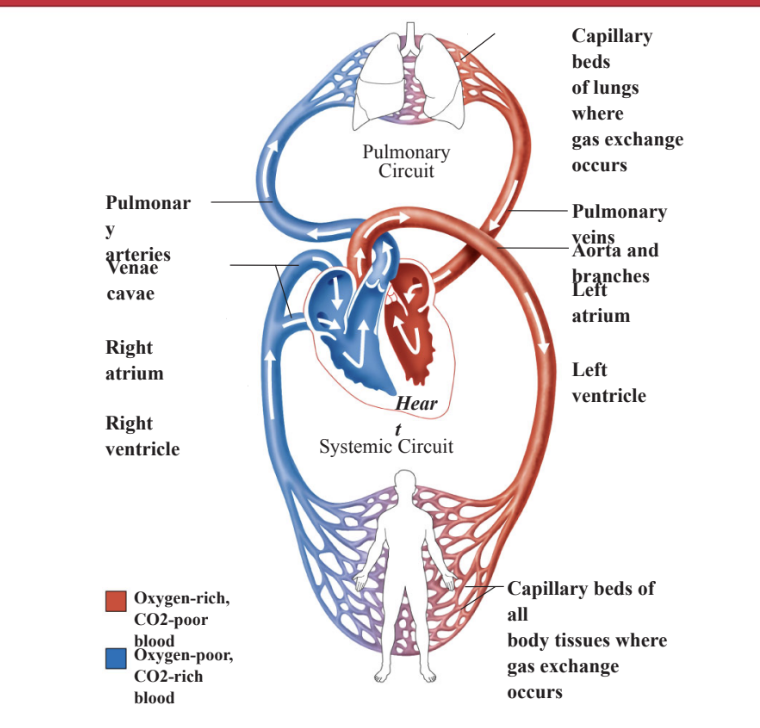
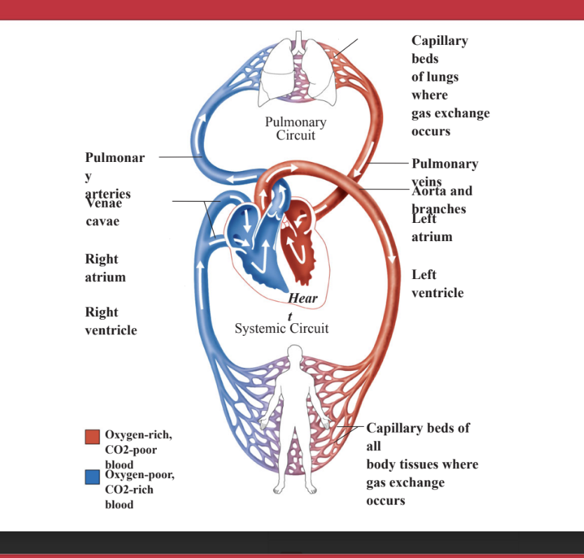
Compare and contrast Pulmonary vs Systemic circulation
Heart: pump @ center moving fluid for 2 circulating system
Pulmonary circulation: delivers blood to the lungs where it gets oxygenated
Systemic circulation: brings blood to all the tissues in the body, thus bringing oxygen to all the cells of the body and removing carbon dioxide
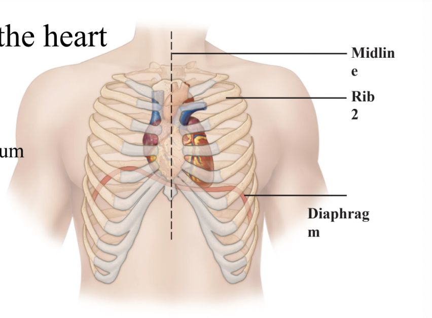
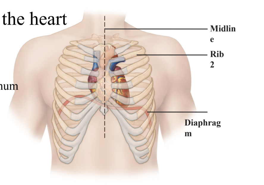
Describe the position of the heart in the thoracic cavity.
Heart is positioned:
in thoracic cavity
left of midline
titled posterior to the sternum for protection of heart
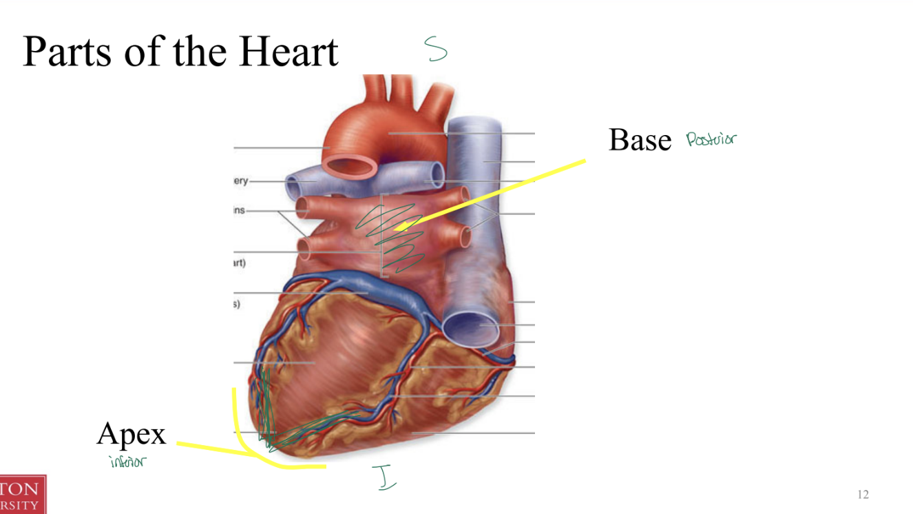
Explain the structural and functional differences between the atria and the ventricles.
The top 2 chambers seen anteriorly are called atria (sing. atrium)
The bottom 2 chambers seen anteriorly are ventricles
The posteriosuperior region is flat and called the base
The inferior, pointed end is the apex
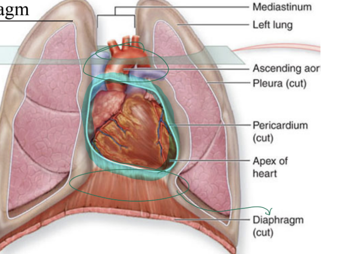
Pericardium
Fibrous sac that contains the heart
Functions:
Restricts heart’s movement within thorax, so it does NOT move from thorax
Prevents overflow (by restricting how expansive the heart can be)
Location:
attached
superior to large vessels leaving heart
inferior to diaphragm
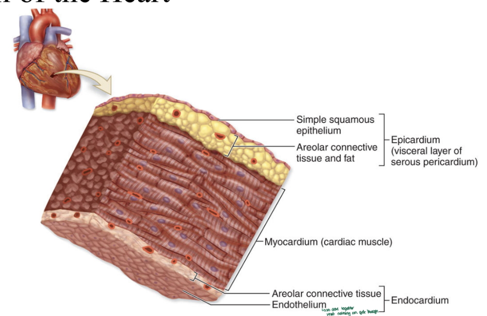
Wall of the heart
Areolar CT and endothelium are so close to each that nothing can get through
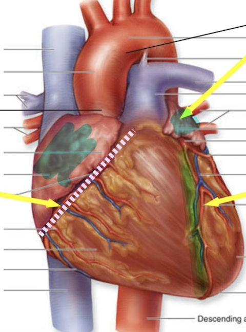
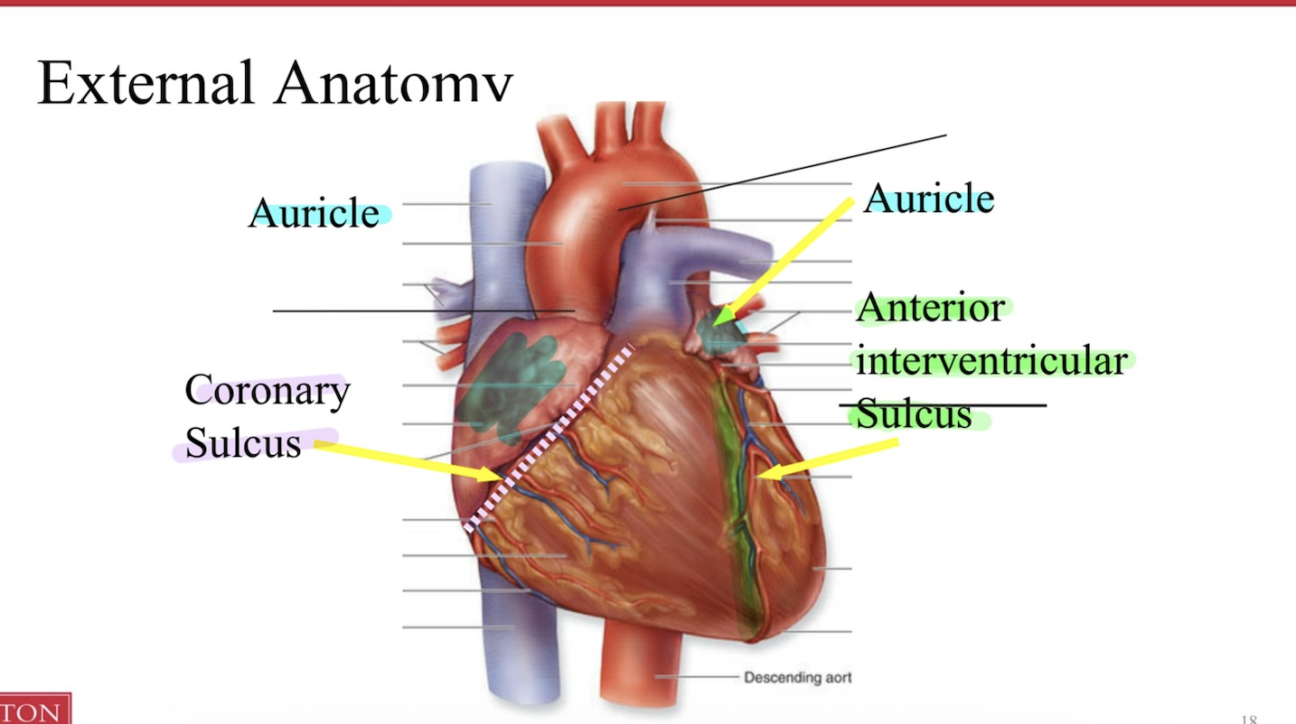
Label the Auricle, Coronary sulcus, Anterior interventricular sulculus
Define them
Auricle: anterior flap-like extension at each atrium that expands the volume
Sulcus: large grooves on external surface of the heart
Coronary vessels: nest in sulci
Coronary sulcus: divides the atria from ventricles
Interventri sulci: divide the ventricles from each other (posterior and anterior)
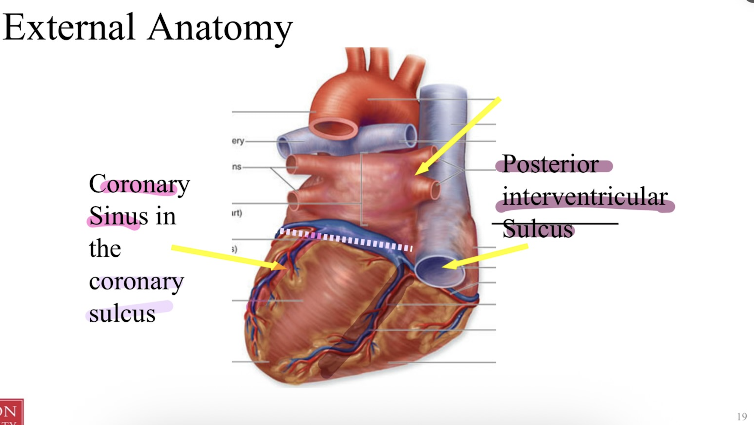
coronary sinus + posterior interventricular sulcus
coronary sinus inside the coronary sulcus
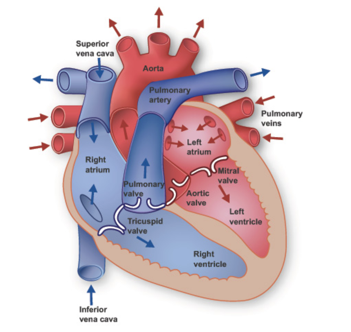
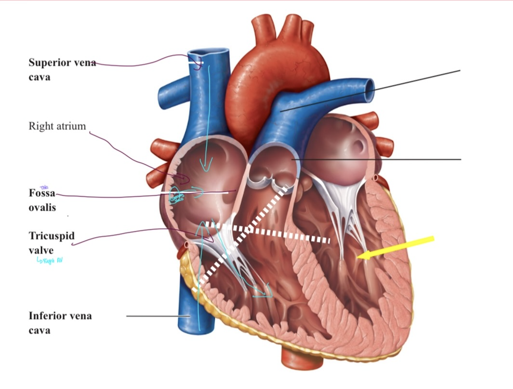
Internal Anatomy - Right atrium
Blood is received from the venous systemic circulation by the right
atrium3 vessels empty into the right atria: superior vena cava, inferior vena cava, and the coronary sinus
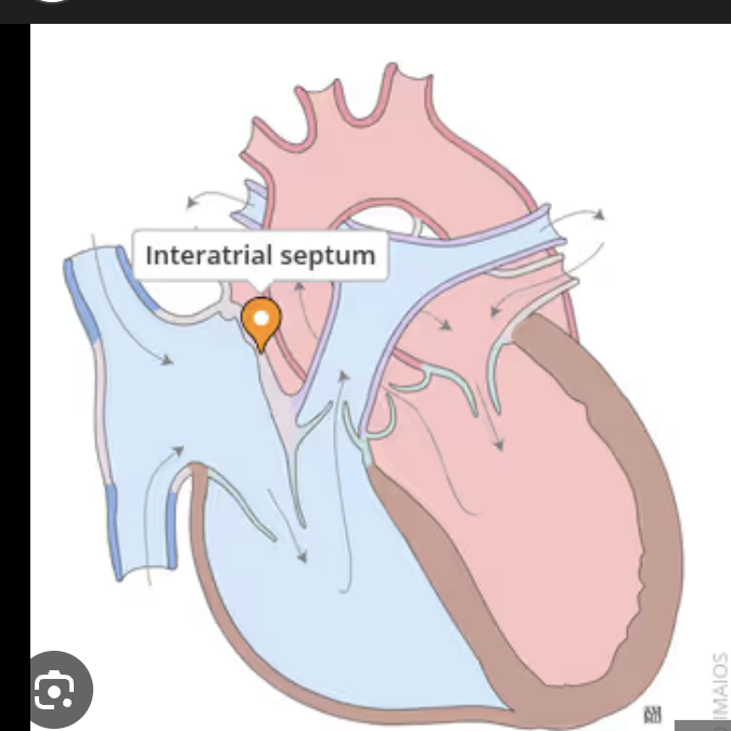
Intratrial septum
is the thin wall between the atria
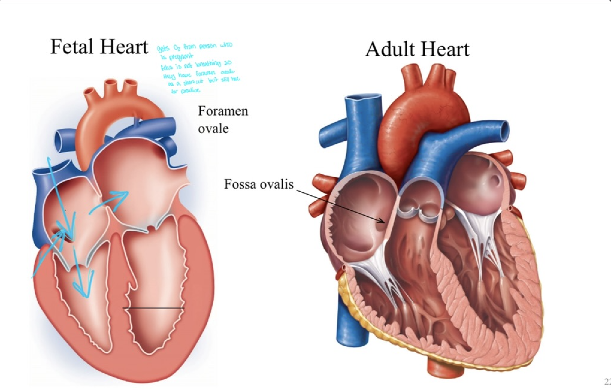
Fossa ovalis
depression in intratrial septum
was a hole from the fetal period that closed.
Blood leaves the R. atria through the atrioventricular (AV) opening
when the AV valve (sometimes called tricuspid valve) opens.
Fetal vs Adult heart
Fetal heart
gets O2 from person who’s pregnant
fetus is NOT breathing so they have foramen ovale as shortcut but still have for practice
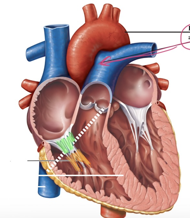
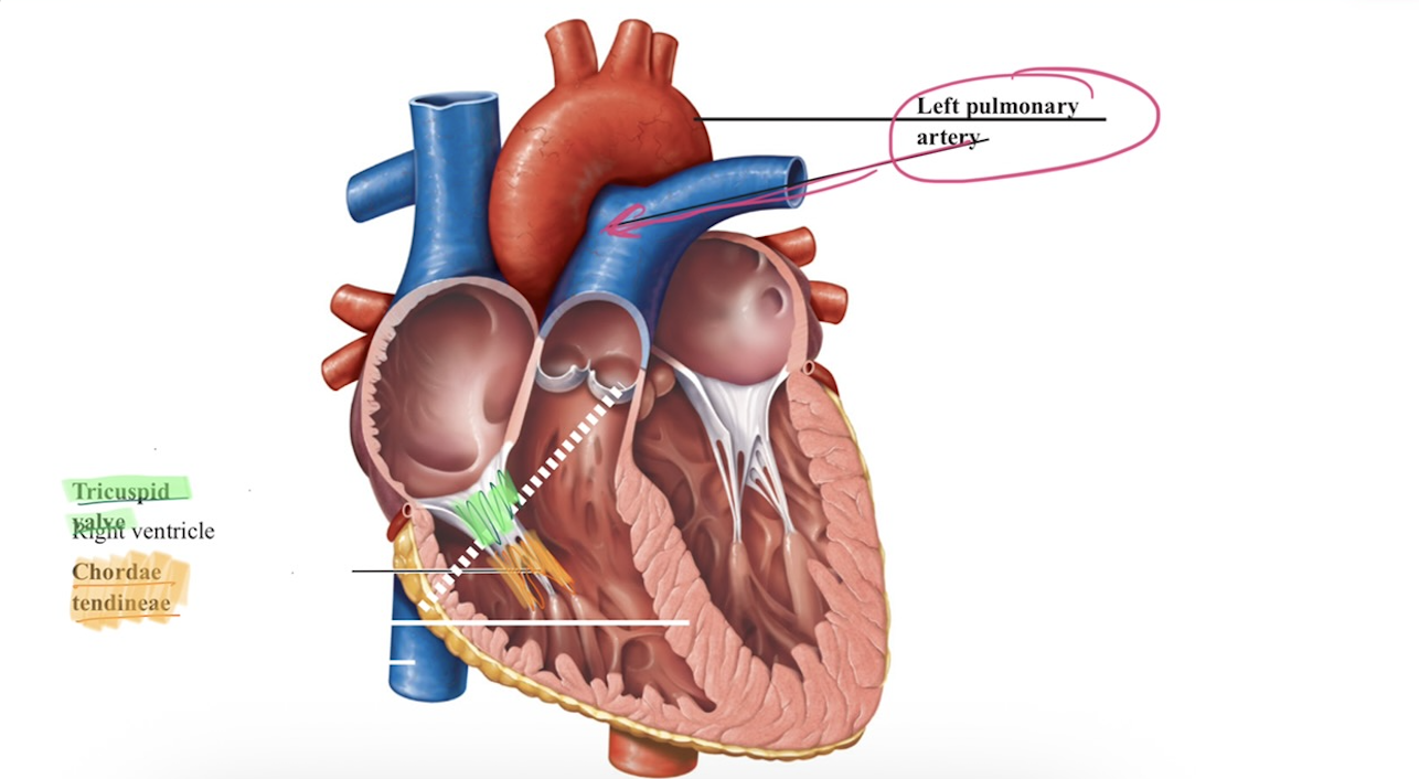
Internal Anatomy - Right Ventricle (Part 1)
Receives blood from Right Atrium
Chordae tendinae:
fibrous cords that anchor AV valve to the ventricle walls
this keep AV valve from flipping backwards when the ventricle contracts —→ one directional flow
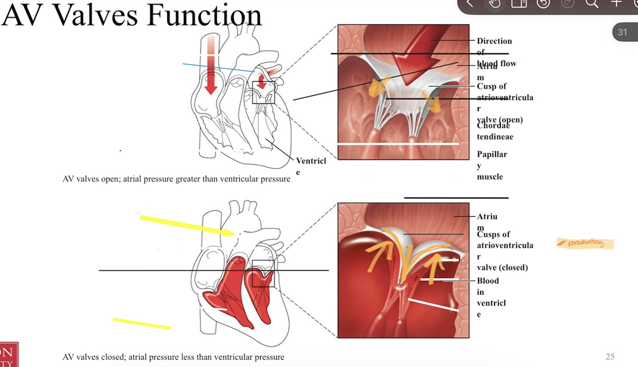
AV valves function
AV valves open when atrial pressure is greater than ventricular pressure
AV valves close when atrial pressure is less than ventricular pressure
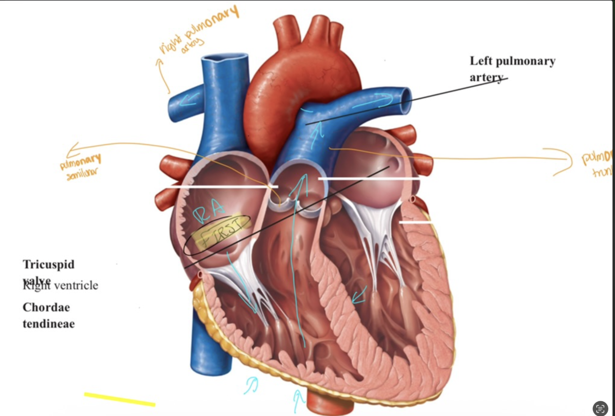
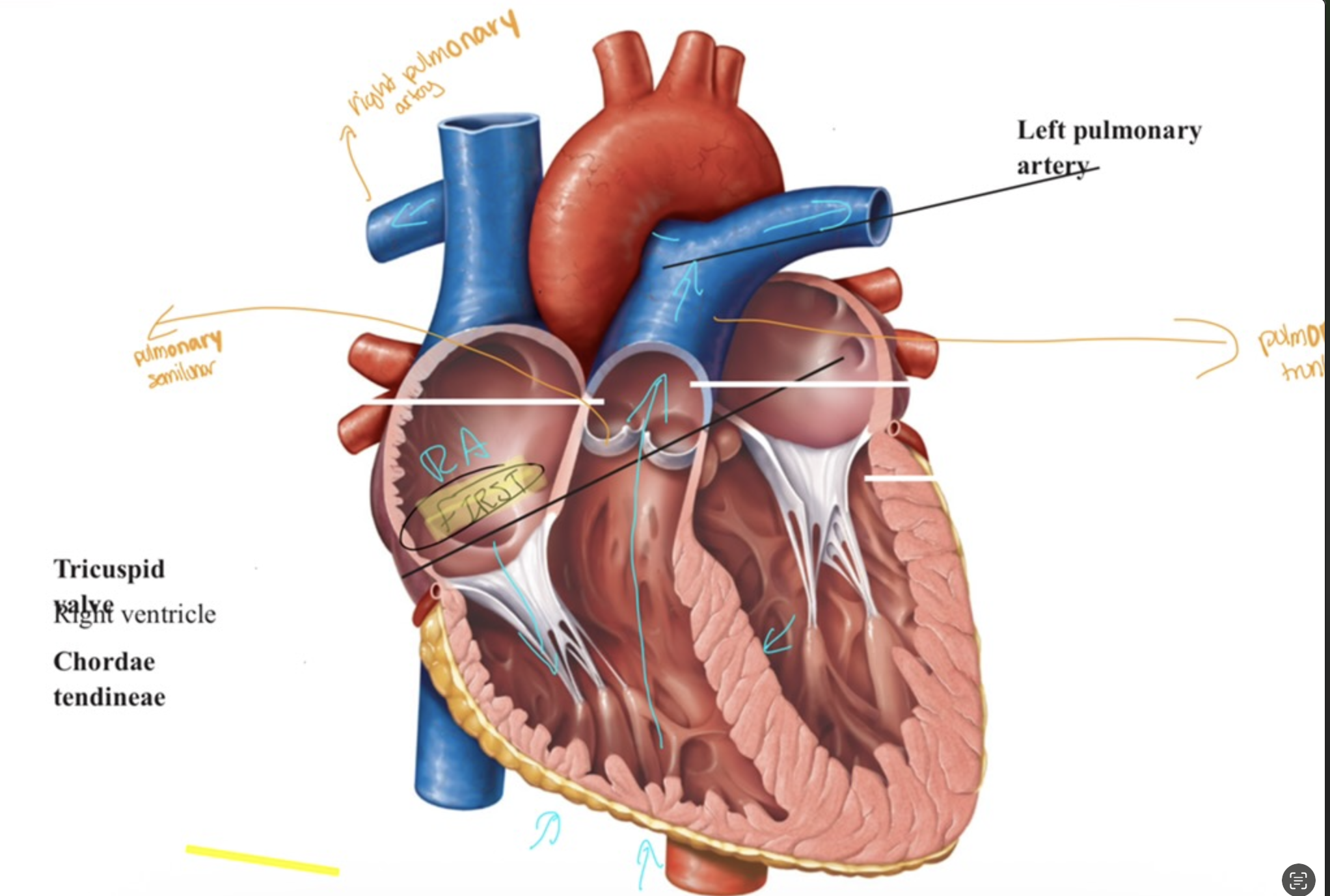
Internal Anatomy - Right Ventricle (Part 2)
Right ventricle contracts,
forces the blood upwards via the pulmonary semilunar valve, into the pulmonary trunk
The pulmonary trunk splits into pulmonary arteries that bring
deoxygenated blood to the lungs for gas exchange.
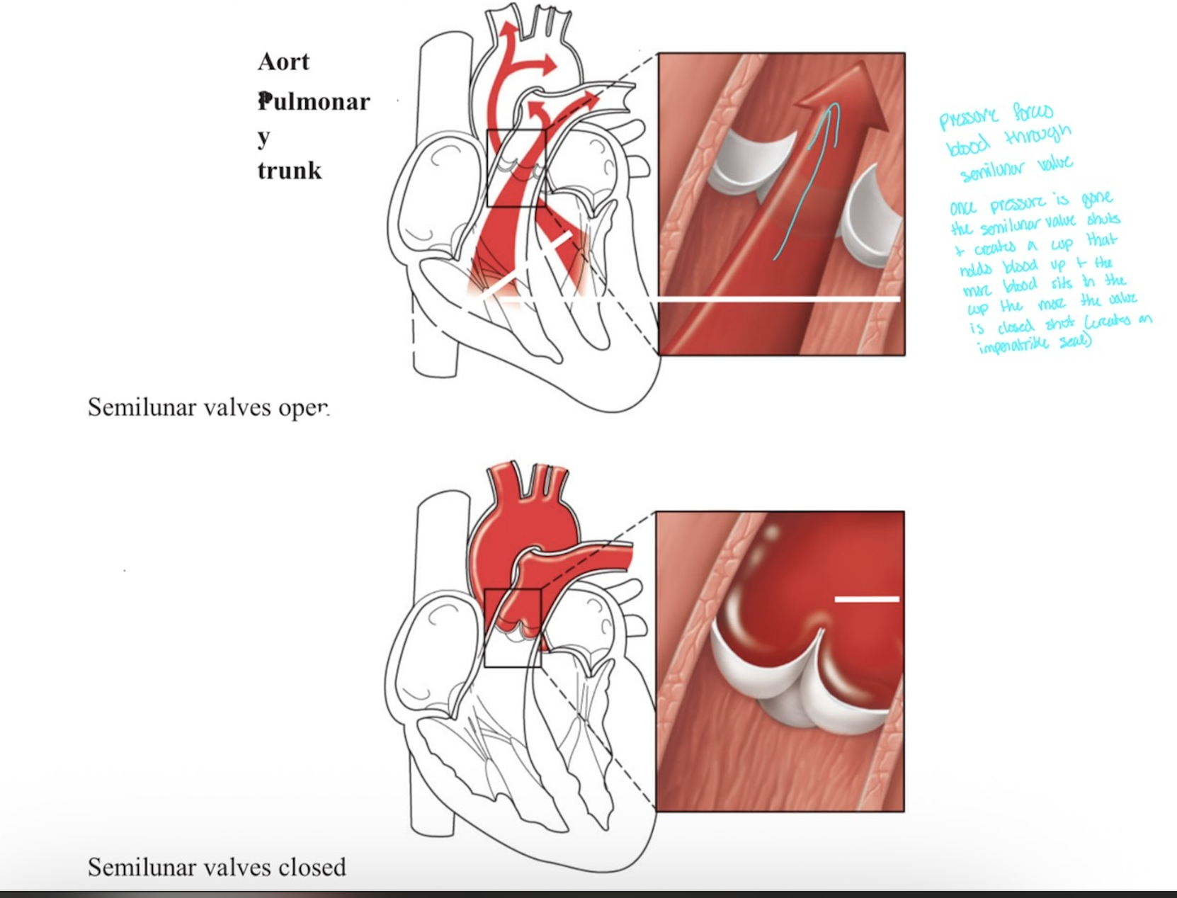
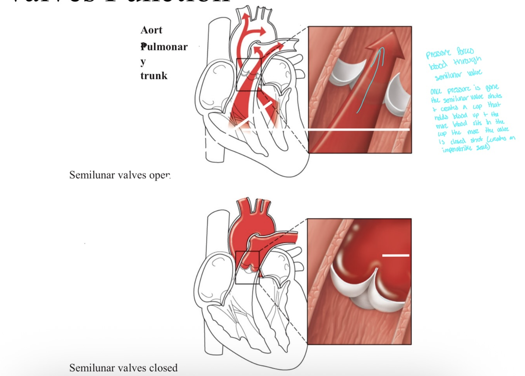
Heart Valves Function
Pressure forces blood via semilunar valves
Pressure goes —> semilunar valves closes,
this makes cup that holds blood up
the more blood sits in cup —> the more the valve is closed shut —> making an impenetrable seal
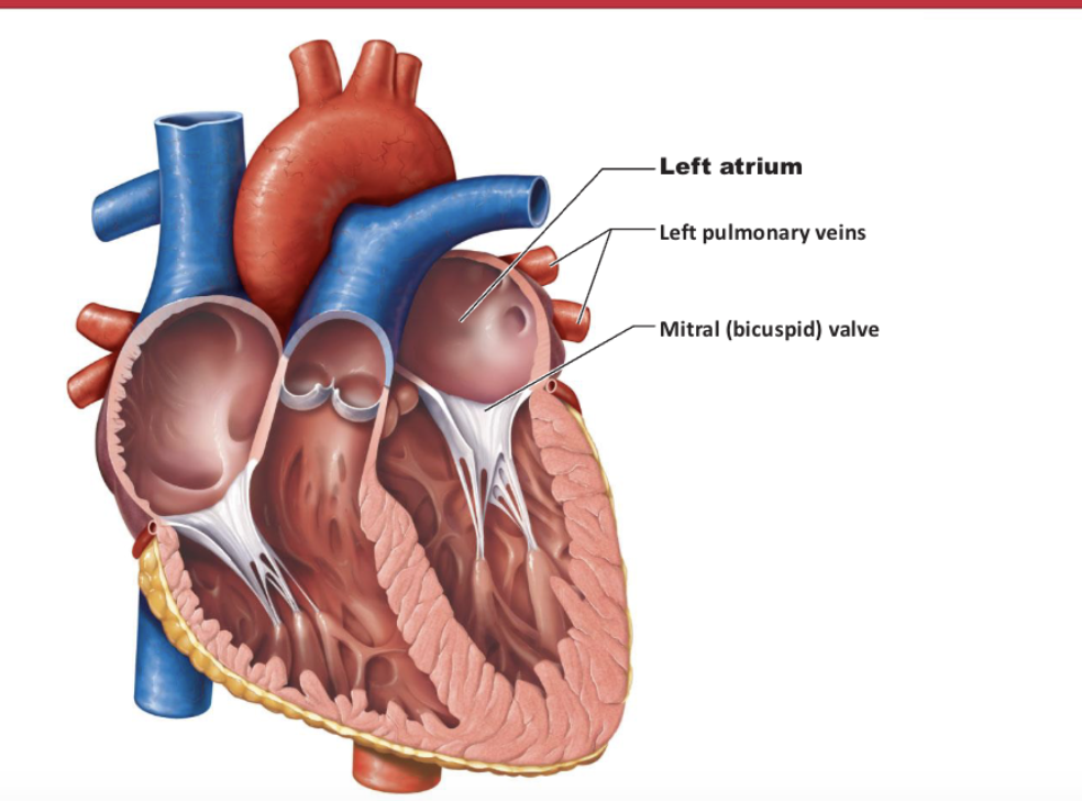
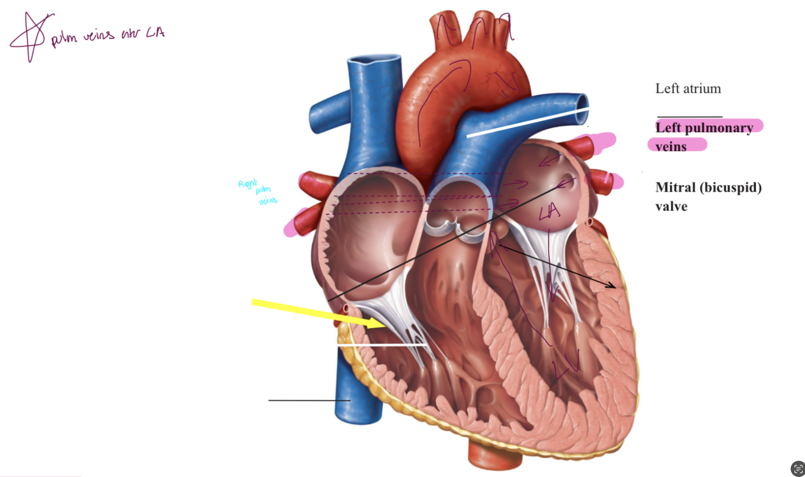
Internal Anatomy - Left Atrium
The pulmonary veins bring the now-oxygenated blood to the left
atriumThe left atrium also has an auricle
The blood flows anterio-inferiorly to the ventricle via the
atrioventricular opening when the AV valve opens
*** left AV valve = bicuspid valve = mitral valve ****
Internal Anatomy - Left Ventricle
left ventricle contracts,
forcing blood through the aortic semilunar valve and into the aorta
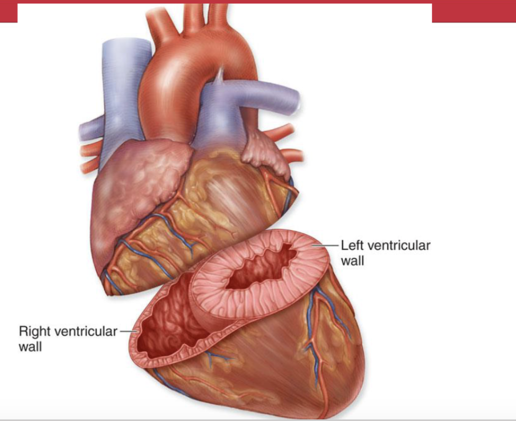
Ventricular walls
Left ventricular wall is thicker than Right ventricular wall
b/c it has to pump for systemic circuit, needing strong pump
Meanwhile right ventricular wall
pumps blood to shorter distance (lungs)
lower pressure, so body is exposed to more oxygen
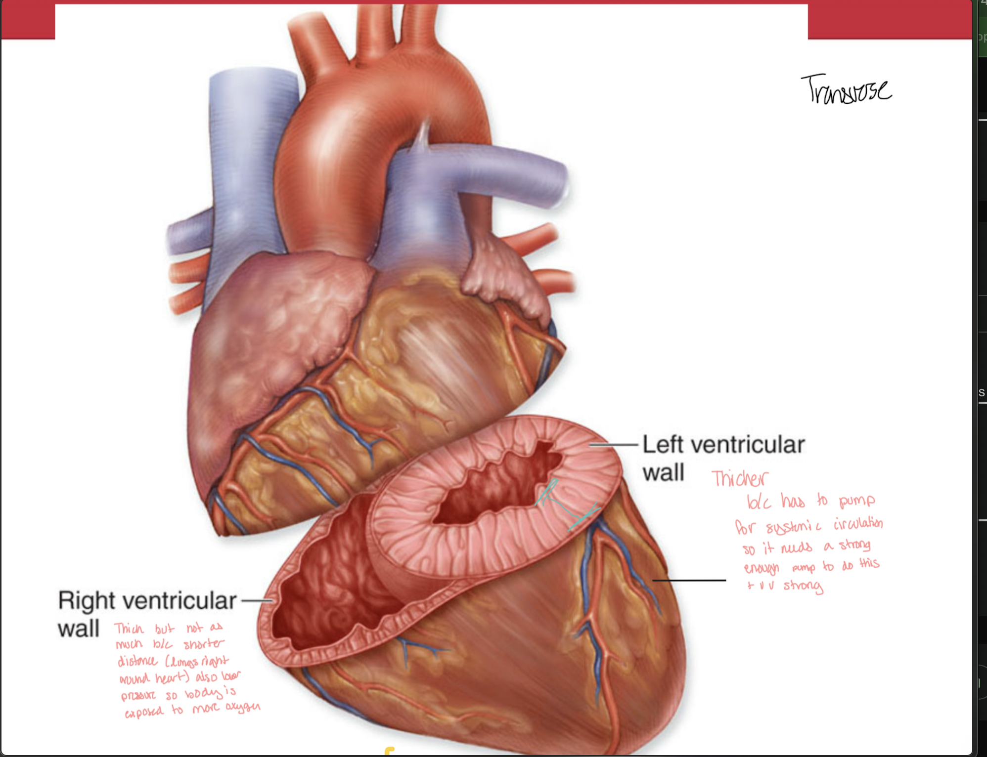
Ventricular Contraction and the Myocardial Vortex
Twist motion
occurs when contracting in order to cause bigger pressure
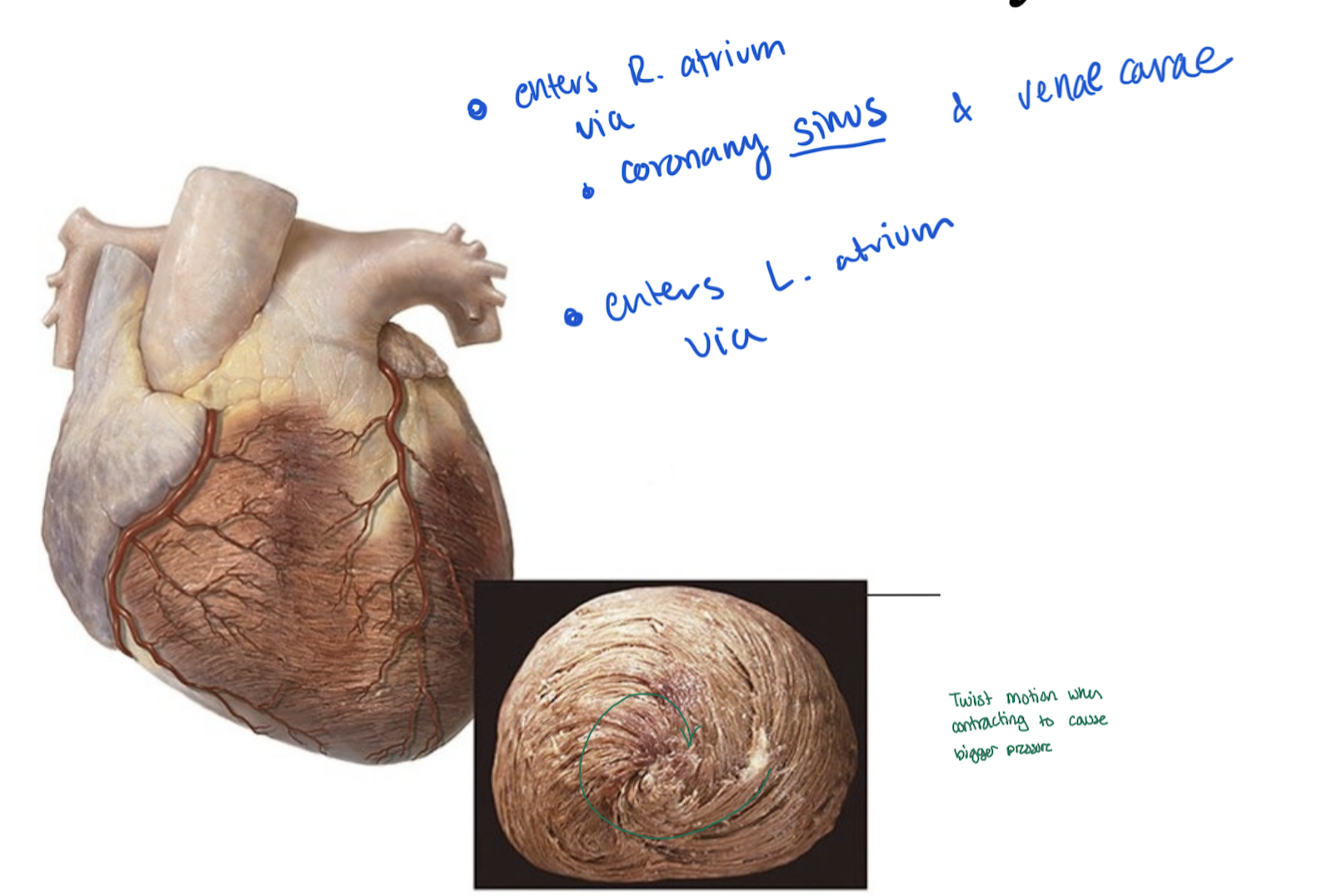
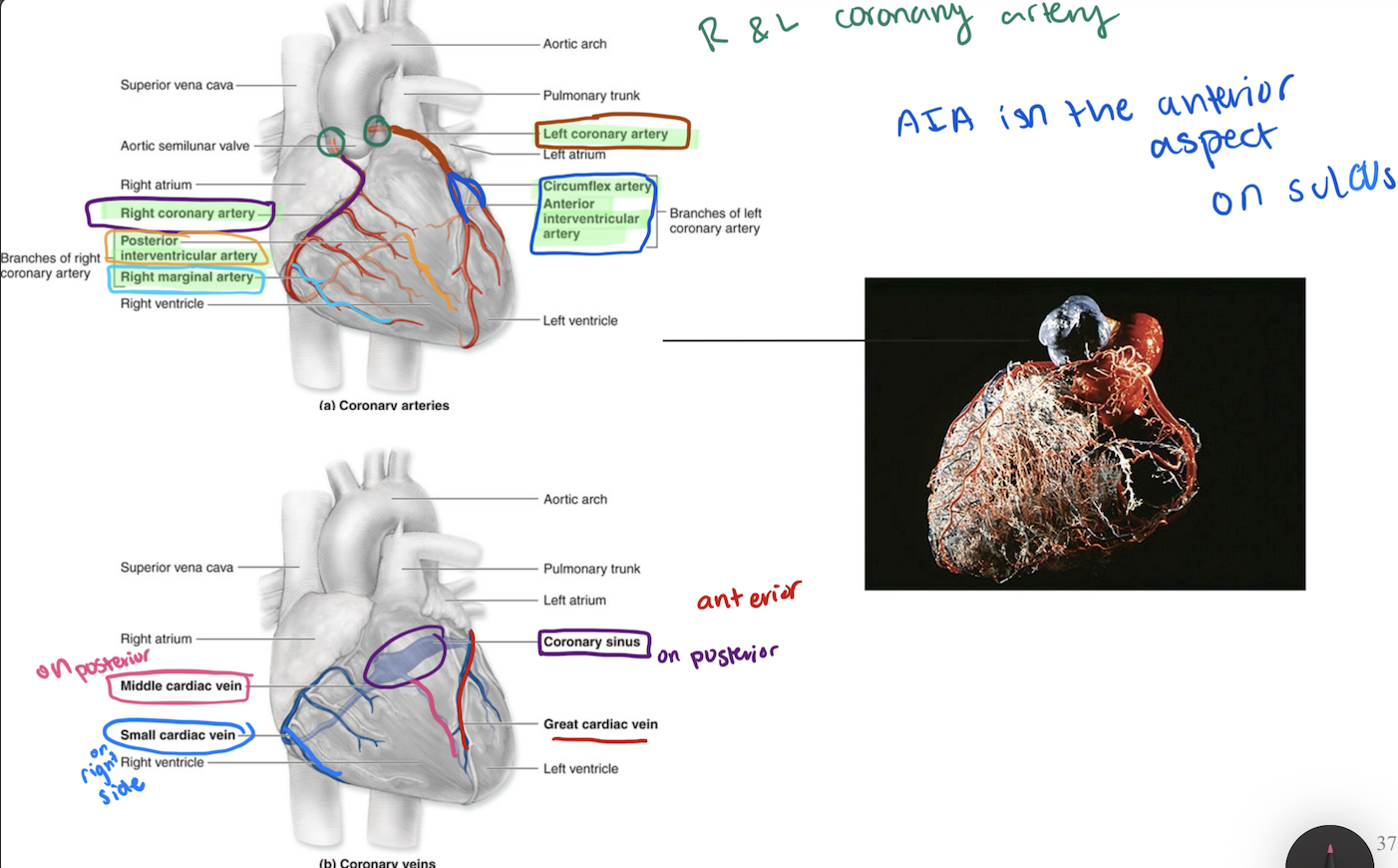
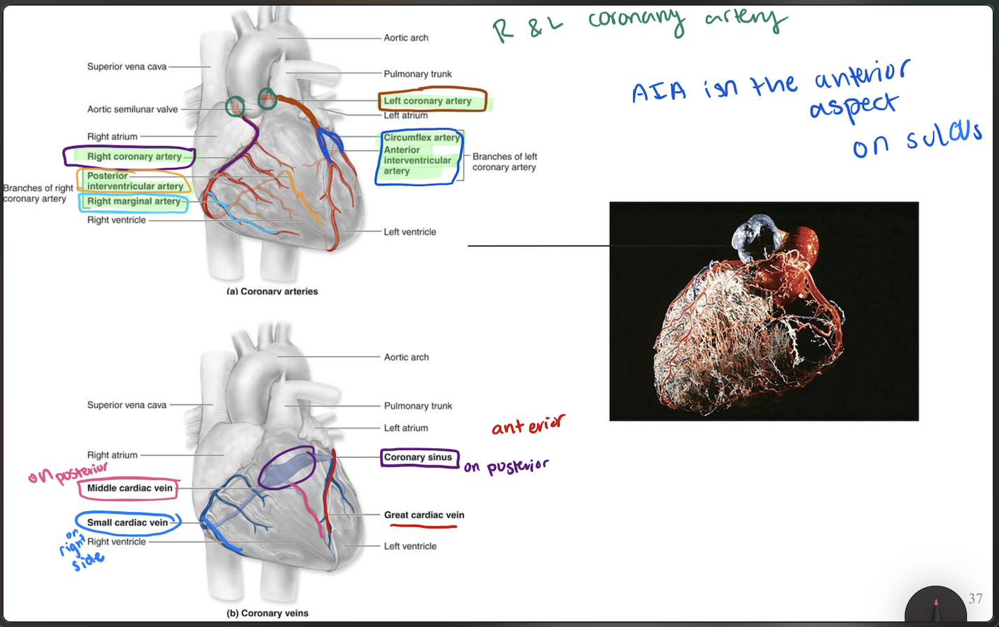
Coronary Circulation
The left and right coronary arteries exit the aorta and feed the cardiac
muscle itself.
They branch into smaller blood vessels
The venous return (deoxygenated blood) occurs through the cardiac
veins (merge into coronary sinus)
Blood flows in the coronary vessels during the periods between heart
contractions.Blood flow is impaired when the heart beats excessively
(tachycardia)
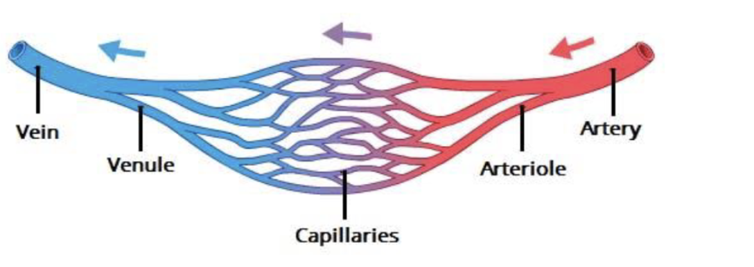
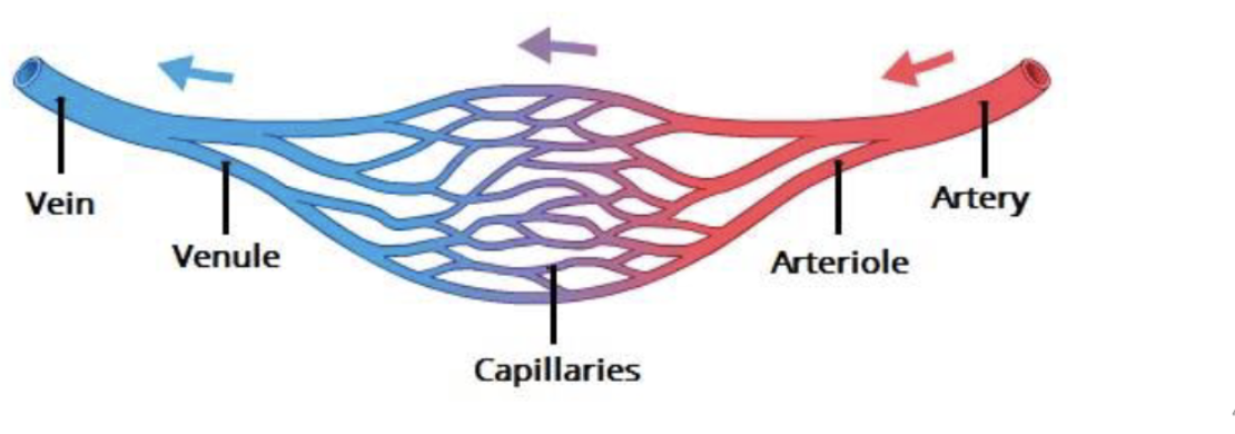
3 Types of Blood vessels
Arteries:
carry blood AWAY from heart
Capillaries
site for O2 diffusion
Veins
carry blood TO from heart