Neuroanatomy
5.0(1)
5.0(1)
Card Sorting
1/90
Earn XP
Description and Tags
Study Analytics
Name | Mastery | Learn | Test | Matching | Spaced | Call with Kai |
|---|
No study sessions yet.
91 Terms
1
New cards
central nervous system
consists of the brain and spinal cord
2
New cards
peripheral nervous system
consists of motor neurons and sensory neurons leading to and from the spinal cord
3
New cards
brain
receives and processes sensory information, initiates responses, stores memories, generates thoughts and emotions
4
New cards
spinal cord
conducts signals to and from the brain, controls reflexes and activities
5
New cards
motor neurons
CNS to muscles to glands (efferent)
6
New cards
sensory neurons
sensory organs to CNS (afferent)
7
New cards
somatic nervous system
division of the peripheral nervous system that controls voluntary movements
8
New cards
autonomic nervous system
division of the peripheral nervous system that controls involuntary responses
9
New cards
sympathetic nervous system
division of the autonomic nervous system that prepares the body for emergencies - fight or fligh
10
New cards
parasympathetic nervous system
division of the autonomic nervous system that promotes normal functioning - rest and digest
11
New cards
functioning of the nervous system
1) Relay Information - sensory system, sensory neurons
2) interpret/make decisions - association system, interneurons
3) carry out some action - motor system, motor neurons
2) interpret/make decisions - association system, interneurons
3) carry out some action - motor system, motor neurons
12
New cards
protection of the CNS
1) bone - skull and vertebral column
2) meninges - 3 layers
3) ventricular system
4) cerebrospinal fluid
5) blood-brain barrier
2) meninges - 3 layers
3) ventricular system
4) cerebrospinal fluid
5) blood-brain barrier
13
New cards
meninges
set of thin membranes that hold the brain and spinal cord in place and act as a protective buffer
14
New cards
dura mater
meninges layer - tough outer layer of fibrous tissue
15
New cards
arachnoid layer
meninges layer - like a spider web; thin sheet of delicate connective tissue (CSF below it)
16
New cards
pia mater
innermost meninge layer - moderately tough inner layer that clings to the brains surface (CSF above it)
17
New cards
ventricular system
responsible for circulating CSF throughout the CNS
18
New cards

1
lateral ventricles
19
New cards

2
interventricular foramen
20
New cards

3
third ventricle
21
New cards

4
cerebral aqueduct
22
New cards

5
fourth ventricle
23
New cards
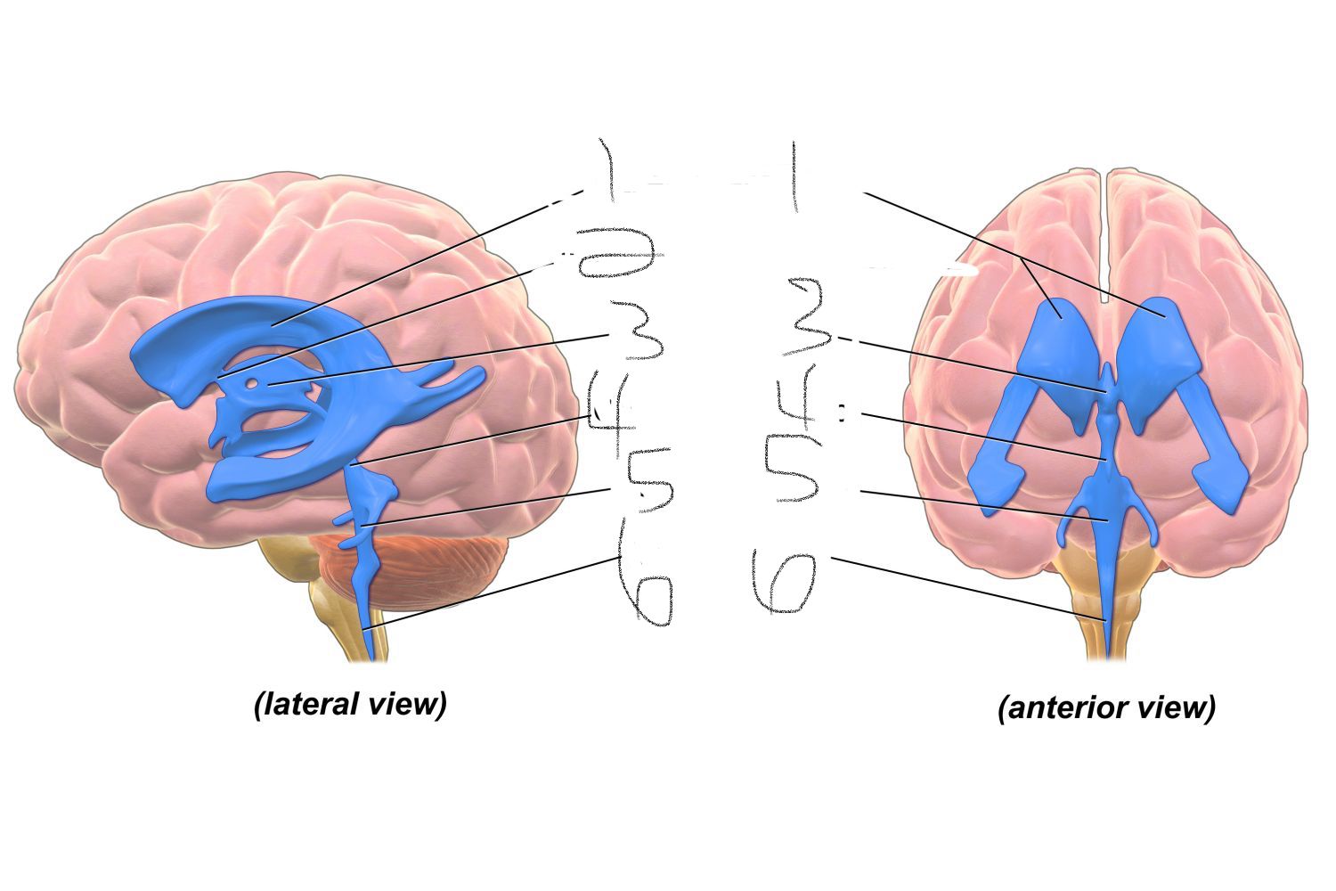
6
central canal
24
New cards
where CSF is found
1) central canal of spinal cord
2) cerebral aqueduct of periaqueductal gray
3) ventricles (lateral, third, fourth)
4) subarachnoid space of meninges
2) cerebral aqueduct of periaqueductal gray
3) ventricles (lateral, third, fourth)
4) subarachnoid space of meninges
25
New cards
roles of CSF
1) provides a medium through which nutrients, hormones, etc, get access to brain cells; dispose of waste products
2) protects brain and spinal cord by acting as a buffer (liquid buffer or cushion to absorb internal as well as external forces)
2) protects brain and spinal cord by acting as a buffer (liquid buffer or cushion to absorb internal as well as external forces)
26
New cards
blood brain barrier
protection of brain from blood born substances
27
New cards
neuraxis
an imaginary line that is drawn through the center of the length of the CNS, from the bottom of the spinal cord to the front of the forebrain
28
New cards
coronal section
cut in the ventricle plane, from the crown of the head down, yielding a frontal view of the brain's internal structures
29
New cards
horizontal section
usually viewed looking down on the brain from above - a dorsal view
30
New cards
sagittal section
cut lengthwise from front to back and viewed from the side - medial view
31
New cards
white matter
nervous tissue of the CNS consisting of neurons and their myelin sheaths
32
New cards
grey matter
nervous tissue containing cell bodies as well as fibers; forms the cerebral cortex consisting of unmyelinated neurons
33
New cards
contralateral
on or relating to the opposite side
34
New cards
ipsilateral
on or relating to the same side
35
New cards
medial
toward the midline
36
New cards
lateral
away from the midline
37
New cards
afferent
towards point of reference (CNS)
38
New cards
efferent
away from point of reference (CNS)
39
New cards
nucleus
collection of cell bodies in the CNS
40
New cards
ganglion
collection of cell bodies in the PNS
41
New cards
tract
collection of axons running together from point A to point B in the CNS
42
New cards
nerve
collection of axons running together from point A to point B in the PNS
43
New cards
frontal lobe
lobe involved in executive functions, thinking, planning, organisation, problem-solving, emotions, behaviour control and personality
44
New cards
parietal lobe
lobe involved in perception, making sense of the world, arithmetic, and spelling
45
New cards
motor cortex
involved in movement, part of the frontal lobe
46
New cards
sensory cortex
involved in sensations, part of the parietal lobe
47
New cards
occipital lobe
lobe involved in vision
48
New cards
temporal lobe
lobe involved in memory, understanding, and language
49
New cards
3 major divisions of the brain
1) forebrain
2) midbrain
3) hindbrain
2) midbrain
3) hindbrain
50
New cards
ventricles in the forebrain
1) Lateral
2) Third
2) Third
51
New cards
subdivisions of the forebrain
1) telencephalon (lateral ventricle)
2) diencephalon (third ventricle)
2) diencephalon (third ventricle)
52
New cards
ventricle in the midbrain
cerebral aqueduct
53
New cards
subdivision of the midbrain
mesencephalon
54
New cards
ventricle in the hindbrain
fourth
55
New cards
subdivisions of the hindbrain
1) metencephalon
2) myelencephalon
2) myelencephalon
56
New cards
principles structures of the forebrain
1) cerebral cortex
2) basal ganglia
3) limbic system
(lateral/telencephalon)
1) thalamus
2) hypothalamus
(third/diencephalon)
2) basal ganglia
3) limbic system
(lateral/telencephalon)
1) thalamus
2) hypothalamus
(third/diencephalon)
57
New cards
principle structures of the midbrain
1) tectum
2) tegmentum
(cerebral aqueduct/mesencephalon)
2) tegmentum
(cerebral aqueduct/mesencephalon)
58
New cards
principle structures of the hindbrain
1) cerebellum
2) pons
(fourth/metencephalon)
1) medulla oblongata
(myelencephalon)
2) pons
(fourth/metencephalon)
1) medulla oblongata
(myelencephalon)
59
New cards
gyri
hills of the cortex
60
New cards
sulci
grooves of the cortex
61
New cards
fissure
especially large, prominent sulcus
62
New cards
central sulcus
divides frontal/parietal lobes
63
New cards
lateral fissure
divides temporal love and parietal/frontal lobes
64
New cards
precentral gyrus
primary motor cortex
65
New cards
postcentral gyrus
primary somatosensory cortex
66
New cards
corpus callosum
large band of axons that connects corresponding part of association of the left and right hemispheres
67
New cards
basal ganglia
-major parts include the caudate nucleus, putamen, and globus pallidus
-consits of a variety of subcortical cell groups engaged primarily in motor control, motor learning, executive functions, behaviour, and emotions
-degredation of neurons in this area causes Parkinsonsl
-consits of a variety of subcortical cell groups engaged primarily in motor control, motor learning, executive functions, behaviour, and emotions
-degredation of neurons in this area causes Parkinsonsl
68
New cards
limbic system
involves many different brain areas and structures that all work together to play a role in out emotions and memory such as the hippocampus and amygdala
69
New cards
hippocampus
small organ wihich regulates emotion, associates w long term memory
70
New cards
amygdala
one of two almond shaped groups of nuclei located deep w/in the lobes of the brain, integrative centre for emotions, emotional behaviour, and motivation
71
New cards
structures of the diencephalon
thalamus + hypothalamus
72
New cards
thalamus
-relays info from sensory receptors to proper brain areas to be processed
-regulation of consciousness, sleep, alertness
-regulation of consciousness, sleep, alertness
73
New cards
hypothalamus
-organizes behaviour related to the survival of the speices
- controls autonomic NS and endocrine system (secretions from the anterior pituitary gland)
-posterior pituitary could be considered an extension of it
- controls autonomic NS and endocrine system (secretions from the anterior pituitary gland)
-posterior pituitary could be considered an extension of it
74
New cards
structures of the mesencephalon
tectum + tegmentum
75
New cards
tectum
Consists of:
-superior colliculi - located at top-role invisual system
-inferior colliculi - located below superior colliculi, role in auditory system
-superior colliculi - located at top-role invisual system
-inferior colliculi - located below superior colliculi, role in auditory system
76
New cards
tegmentum
includes:
-rostal end of the reticular formation (states of consciousness like alertness and sleep)
-several nuclei controlling eye movement
-periaqueductal gray matter (primary control centre for descending pain modulation)
-red nucleus (motor coordination)
-substantia nigra (reward and movement)
-ventral tegmental area (sends dopaminergic projections to both the limbic and cortical areas)
77
New cards
structures of the metencephalon
cerebellum and pons
78
New cards
cerebellum
-receives info from the sensory systems, spinal cord, other parts of the brain and then regulates motor movements
-coordinates voluntary movements such as posture, balance, coordination, speech -- resulting in smmoth + balanced muscular activity
-coordinates voluntary movements such as posture, balance, coordination, speech -- resulting in smmoth + balanced muscular activity
79
New cards
pons
-large bulge in the brain stem, lies between the midbrain and medulla, immediately ventral to the cerebellum
-contains a portion of the reticular formation
-contains a large nucleus that relays information from the cerebral cortex to the cerebellum
-as part of the brain stem it also impacts several automatic functions (breathing/necessary for life)s
-contains a portion of the reticular formation
-contains a large nucleus that relays information from the cerebral cortex to the cerebellum
-as part of the brain stem it also impacts several automatic functions (breathing/necessary for life)s
80
New cards
structures of the myelencephalon
medulla oblongata
81
New cards
medulla oblongata
the most caudal portion of the brain stem; its lower border is the rostal end of the spinal cord
-contains numerous nuclei that act as control centers for the autonomic nervous system (vital functions are controlled -breathing, heat rate, temp - damage usually means rapid deaths
-contains numerous nuclei that act as control centers for the autonomic nervous system (vital functions are controlled -breathing, heat rate, temp - damage usually means rapid deaths
82
New cards
spinal cord
-long, conical structure (about as thick as an adult's little finger)
-principle function is to distribute motor fibres to the effector organs of the body (glands/muscles) and to collect somatosensory info to be passed on to the brain
-has a certain degree of autonomy from the brain; various reflexive control circuits are located there
-principle function is to distribute motor fibres to the effector organs of the body (glands/muscles) and to collect somatosensory info to be passed on to the brain
-has a certain degree of autonomy from the brain; various reflexive control circuits are located there
83
New cards
spinal nerves
31 pairs
- 8 cervical (C1-C8)
- 12 thoracic (T1-T12)
- 5 lumbar (L1-L5)
- 5 sacral (S1-S5)
- 1 coccygeal nerve (Co1)
- 8 cervical (C1-C8)
- 12 thoracic (T1-T12)
- 5 lumbar (L1-L5)
- 5 sacral (S1-S5)
- 1 coccygeal nerve (Co1)
84
New cards
peripheral nervous system
cranial and spinal nervess
85
New cards
somatic nervous system
comprised of
-sensory neurons conveying information form the somatic receptors in head, body wall and limbs and from receptors of special senses
-motor neurons that conduct impulses from CNS to skeletal muscles
-sensory neurons conveying information form the somatic receptors in head, body wall and limbs and from receptors of special senses
-motor neurons that conduct impulses from CNS to skeletal muscles
86
New cards
autonomic nervous system
consists of
-sensory neurons that convey information from the autonomic sensory receptors (located primarily in visceral organs) to CNS
- motor neurons that conduct impulses from CNS to smooth muscle, cardiac muscle and glands
- two main functional divisions (parasympathetic/sympathetic) most organs receive inout from both, but systems are antagonistic (produce opposite effects)
87
New cards
sympathetic nervous system
division of the autonomic nervous system: fight or flight response (emergency functioning)p
88
New cards
parasympathetic nervous system
division of the autonomic nervous system which supports activities involved w increase in body's supply or stored energy (salivation, digestion, increased blood flow to gastrointestinal system) -promotes normal functioning "rest and digest"
89
New cards
autonomic pathway
consists of two neurons that synapse in an autonomic ganglion
- preganglionic neuron from CNS to autonomin ganglion, postganglionic neuron from autonomic ganglion to target tissue
- preganglionic neuron from CNS to autonomin ganglion, postganglionic neuron from autonomic ganglion to target tissue
90
New cards
parasympathetic pathway
preganglionic neuron: soma is usually in the brain stem or sacral (bottom of spinal cord)
-releases acetylcholine
postganglionic neuron: soma is usually in a ganglion near the target organ
-realses NT acetylcholine or nitric oxide
activates: rest and digest
(craniosacral -> long pre-gang axon -> short post gang axon)
-releases acetylcholine
postganglionic neuron: soma is usually in a ganglion near the target organ
-realses NT acetylcholine or nitric oxide
activates: rest and digest
(craniosacral -> long pre-gang axon -> short post gang axon)
91
New cards
sympathetic pathway
preganglionic neuron: soma usually in spine
- NT acetylcholine released
post ganglionic neuron: soma in a sympathetic ganglion located next to the spinal cord
- NT released: norepinephrine
activates: fight or flight
(thoracicolumbar -> short pre gang axon -> long post gang axon)
- NT acetylcholine released
post ganglionic neuron: soma in a sympathetic ganglion located next to the spinal cord
- NT released: norepinephrine
activates: fight or flight
(thoracicolumbar -> short pre gang axon -> long post gang axon)