Muscle Tissue Types and Functions Overview
1/151
There's no tags or description
Looks like no tags are added yet.
Name | Mastery | Learn | Test | Matching | Spaced | Call with Kai |
|---|
No analytics yet
Send a link to your students to track their progress
152 Terms
Skeletal Muscle
Attached to bones via tendons, found throughout the body.
Striated
Skeletal muscle fibers have a banded appearance due to the organized arrangement of actin and myosin filaments.
Multinucleated
Each muscle fiber (cell) contains multiple nuclei, located at the periphery.
Voluntary
Controlled consciously by the somatic nervous system.
Movement
Responsible for voluntary movements like walking, running, and lifting.
Posture
Maintains posture and body position.
Heat generation
Generates heat during contraction (e.g., shivering).
Cardiac Muscle
Found only in the walls of the heart (myocardium).
Single central nucleus
Cardiac muscle cells (cardiomyocytes) typically have one centrally located nucleus, though some may have two.
Intercalated discs
Specialized structures that connect cardiac muscle cells, allowing for synchronized contractions.
Involuntary
Controlled by the autonomic nervous system and hormones, without conscious effort.
Pump blood
Cardiac muscle contracts rhythmically to pump blood throughout the body.
Self-exciting
Has autorhythmicity, meaning the heart can initiate its own contractions due to pacemaker cells.
Smooth Muscle
Found in the walls of hollow organs (e.g., stomach, intestines, blood vessels, bladder) and other structures like the respiratory and reproductive tracts.
Non-striated
Smooth muscle fibers do not have visible striations because actin and myosin are arranged differently than in skeletal and cardiac muscle.
Spindle-shaped cells
Smooth muscle fibers are long, thin, and spindle-shaped.
Movement of substances
Smooth muscle controls movements like the contraction of blood vessels, peristalsis in the digestive tract, and regulation of airflow in the respiratory system.
Regulation of organ volume
Helps in controlling the diameter of blood vessels and hollow organs.
Sliding Filament Theory
Explains how muscles contract to produce movement, describing the interaction between actin and myosin within the sarcomere.
Sarcomere
The functional unit of muscle contraction, located between two Z-discs.
Myosin
Thick filaments with 'heads' that bind to actin during contraction.
Actin
Thin filaments that slide past myosin during contraction.
Tropomyosin
A protein that blocks the binding sites on actin.
Troponin
A protein that binds calcium and helps expose the active sites on actin by moving tropomyosin.
Power Stroke
The myosin head pivots, pulling the actin filament toward the center of the sarcomere.
Detachment
After the power stroke, ATP binds to the myosin head, causing it to detach from actin.
Re-cocking the Myosin Head
ATP is hydrolyzed into ADP and Pi (inorganic phosphate), re-cocking the myosin head into its original position.
Atrophy
Muscle shrinking.
Hypertrophy
Muscle growing.
Type I (Slow-Twitch Fibers)
Contraction speed: Slow; Fatigue resistance: High (good for endurance); Energy source: Aerobic (uses oxygen).
Type IIa (Fast-Twitch, Intermediate Fibers)
Contraction speed: Fast.
Type IIb (Fast-Twitch, Glycolytic Fibers, white fibers)
Contraction speed: Very fast. Fatigue resistance: Low (for short bursts of power). Energy source: Anaerobic (without oxygen). Example: Used in explosive activities like sprinting or weightlifting.
Agonist (Prime Mover)
The main muscle responsible for producing a specific movement. Example: The biceps brachii is the agonist for elbow flexion.
Antagonist
A muscle that opposes the action of the agonist, relaxing or lengthening during the movement. Example: The triceps brachii is the antagonist to the biceps during elbow flexion.
Synergist
Muscles that assist the agonist by stabilizing joints or adding extra force to the movement. Example: The brachialis assists the biceps in elbow flexion.
Orbicularis oris
Compresses lips, purses lips 'pout'.
Buccinator
Molar region of maxilla and mandible; compresses cheeks.
Mentalis
Mandible below incisors; elevates and protrudes lower lip.
Risorius
Lateral Fascia associated with masseter muscle; draws corner of mouth to the side.
Zygomaticus
Zygomatic bone; raises lateral corners of mouth upward (smiling muscle).
Depressor labii inferioris
Body of mandible lateral to its midline; draws lower lip down (pouting muscle).
Corrugator supercilii
Arch of frontal bone; pulls skin inferiorly and anteriorly; wrinkles brow.
Levator labii superioris
Zygomatic bone and infraorbital margin of maxilla; opens lips; raises and furrows upper lip.
Orbicularis oculi
Frontal and maxillary bones and ligaments around orbit; closes eye.
Occipitofrontalis
Epicranial aponeurosis; raises eyebrows, wrinkles forehead.
Platysma
Mandible & skin of cheek; tenses skin of neck, depresses mandible.
Masseter
Zygomatic arch; elevates mandible & closes jaw.
Temporalis
Temporalis fossa; elevates mandible & closes jaw.
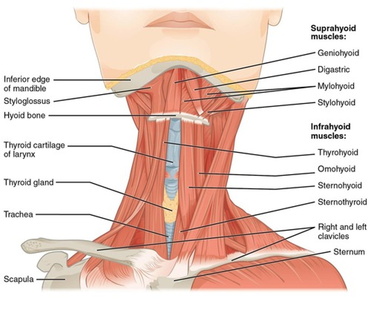
Digastric
Inferior surface of mandible at chin; depresses mandible, opening mouth, and/or elevates larynx.
Sternocleidomastoid
Clavicular head; together they flex the neck; alone one side bends neck toward shoulder & turns face to opposite side.
Splenius
Ligamentum nuchae, spinous processes of C7-T6; extends or hyperextends head.
Erector Spinae
Spinalis group (medial); extends vertebral column.
Quadratus lumborum
Iliac crest & iliolumbar ligament; depresses ribs, lateral flexion of vertebral column.
Oblique and Rectus Muscles
The muscle of the oblique and rectus groups lie between the vertebral column and the ventral midline.
Origin
The point where a muscle attaches to a stationary bone.
Insertion
The point where a muscle attaches to a movable bone.
Action
The specific movement produced by a muscle when it contracts.
Scalenes
Muscles in the cervical region that elevate ribs and flex the neck.
External intercostals
Muscles that elevate ribs.
Internal intercostals
Muscles that depress ribs.
External oblique
Muscle in the abdominal region that compresses the abdomen and rotates the vertebral column.
Internal oblique
Muscle that compresses the abdomen and rotates the vertebral column to the same side.
Transversus abdominis
Muscle that compresses the abdomen.
Rectus abdominis
Muscle that depresses ribs, flexes the vertebral column, and compresses the abdomen.
Pectoralis Minor
Muscle that draws the scapula forward and downward.
Serratus anterior
Muscle that stabilizes and rotates the scapula.
Subclavius
Muscle that helps stabilize and depress the pectoral girdle.
Trapezius
Muscle that stabilizes, raises, retracts, depresses, and rotates the scapula.
Levator scapulae
Muscle that elevates and adducts the scapula.
Rhomboid major and minor
Muscles that stabilize the scapula.
Bulbospongiosus
Muscle that compresses the base of the penis/clitoris and ejects urine or semen.
Ischiocavernosus
Muscle that compresses and stiffens the penis or clitoris.
Deep transverse perineal
Muscle that supports the pelvic floor.
External urethral sphincter
Muscle that closes the urethra and compresses glands.
Pelvic diaphragm
Muscle group that supports the organs of the pelvic cavity and flexes the joints of the sacrum and coccyx.
Appendicular muscles
Account for roughly 40 percent of the skeletal muscles in the body.
Muscles of the pectoral girdle and upper limbs
One of the two major groups of appendicular muscles.
Muscles of the pelvic girdle and lower limbs
One of the two major groups of appendicular muscles.
Pectoralis major
Prime movers of arm flexion, rotates medial, adducts; originates from sternal and inferior portion of clavicle, body of sternum, cartilage of ribs 1-6, and aponeurosis of external oblique muscle.
Latissimus dorsi
Prime mover of arm flexion, rotates medially, adducts; originates from spinous process of inferior thoracic and all lumbar vertebrae, ribs 8-12, and thoracolumbar fascia.
Deltoid
Prime mover of arm abduction; originates from clavicle and scapula.
Teres major
Extends, medially rotates, adducts humerus; originates from inferior angle of scapula.
Coracobrachialis
Flexion and adduction of humerus; originates from coracoid process.
Subscapularis
Chief medial rotator of humerus; originates from subscapular fossa of scapula.
Supraspinatus
Initiates abduction of shoulder; originates from supraspinous fossa of scapula.
Infraspinatus
Rotates humerus laterally; originates from infraspinous fossa of scapula.
Teres minor
Rotates humerus laterally; originates from lateral border of dorsal scapular surface.
Triceps brachii
Powerful forearm extensor; long head originates from infraglenoid tubercle of scapula.
Anconeus
Abducts ulna during pronation; originates from lateral epicondyle of humerus.
Biceps brachii
Flexion at elbow and shoulder, supinates forearm; short head originates from coracoid process.
Brachialis
Major forearm flexor; originates from anterior, distal surface of humerus.
Brachioradialis
Synergist in forearm flexion; originates from lateral supracondylar ridge at distal end of humerus.
Pronator teres
Pronates forearm; originates from medial epicondyle of humerus and coronoid process.
Flexor carpi radialis
Powerful wrist flexor, abducts hand; originates from medial epicondyle of humerus.
Palmaris longus
Tenses skin and fascia of palm, hand movements; originates from medial epicondyle of humerus.
Flexor digitorum superficialis
Flexion at middle phalanges (2-5) + wrist; has four tendons into middle phalanges of fingers 2-5.
Flexor pollicis longus
Flexes distal phalanx of thumb; originates from anterior surface of radius and interosseous membrane.
Flexor digitorum profundus
Flexes distal interphalangeal joints; has four tendons into distal phalanges 2-5.
Pronator quadratus
Prime mover of forearm pronation; originates from distal portion of anterior ulnar shaft.
Extensor carpi radialis longus
Extends and abducts wrist; originates from lateral supracondylar ridge of humerus.