Clinical Skills 3: Exam One (Wagner College)
1/78
There's no tags or description
Looks like no tags are added yet.
Name | Mastery | Learn | Test | Matching | Spaced | Call with Kai |
|---|
No analytics yet
Send a link to your students to track their progress
79 Terms
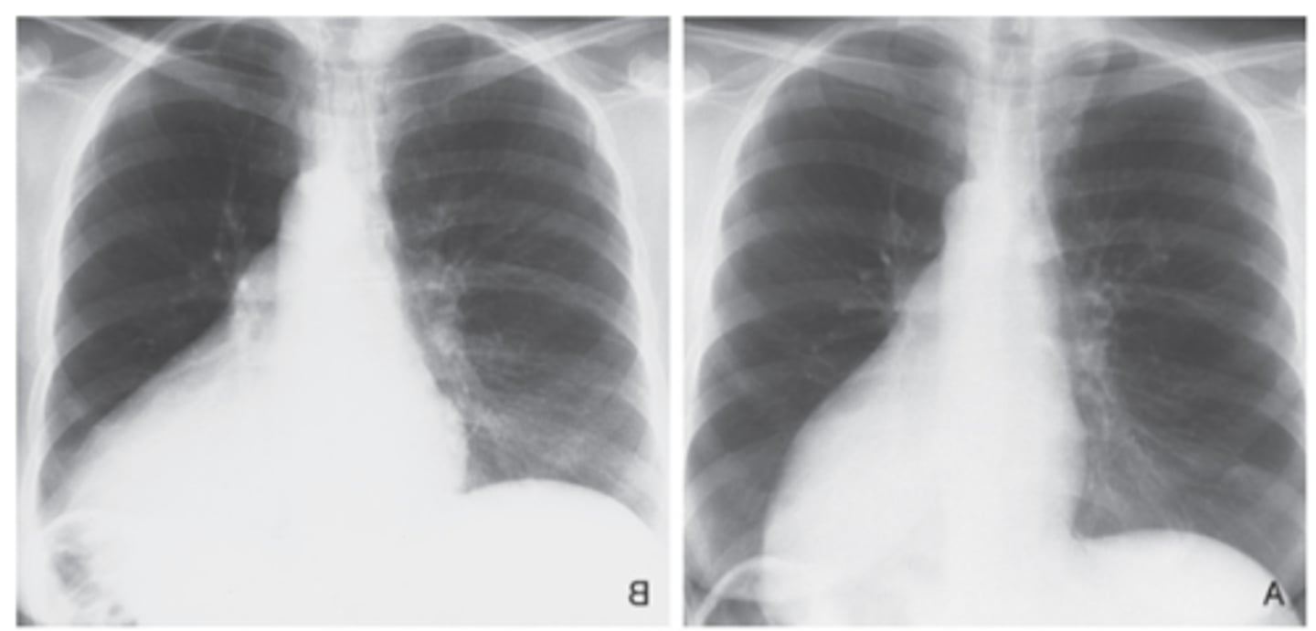
What does a Pericardial effusion look like on a chest X-ray?
-acute and marked enlargement of cardiac silhouette
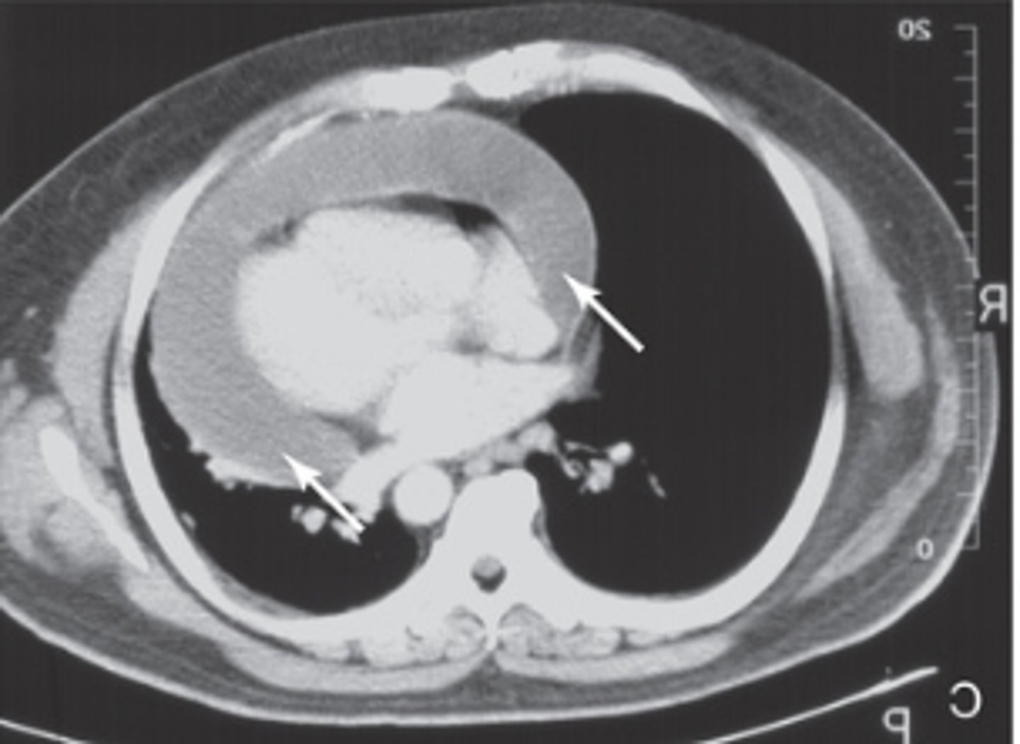
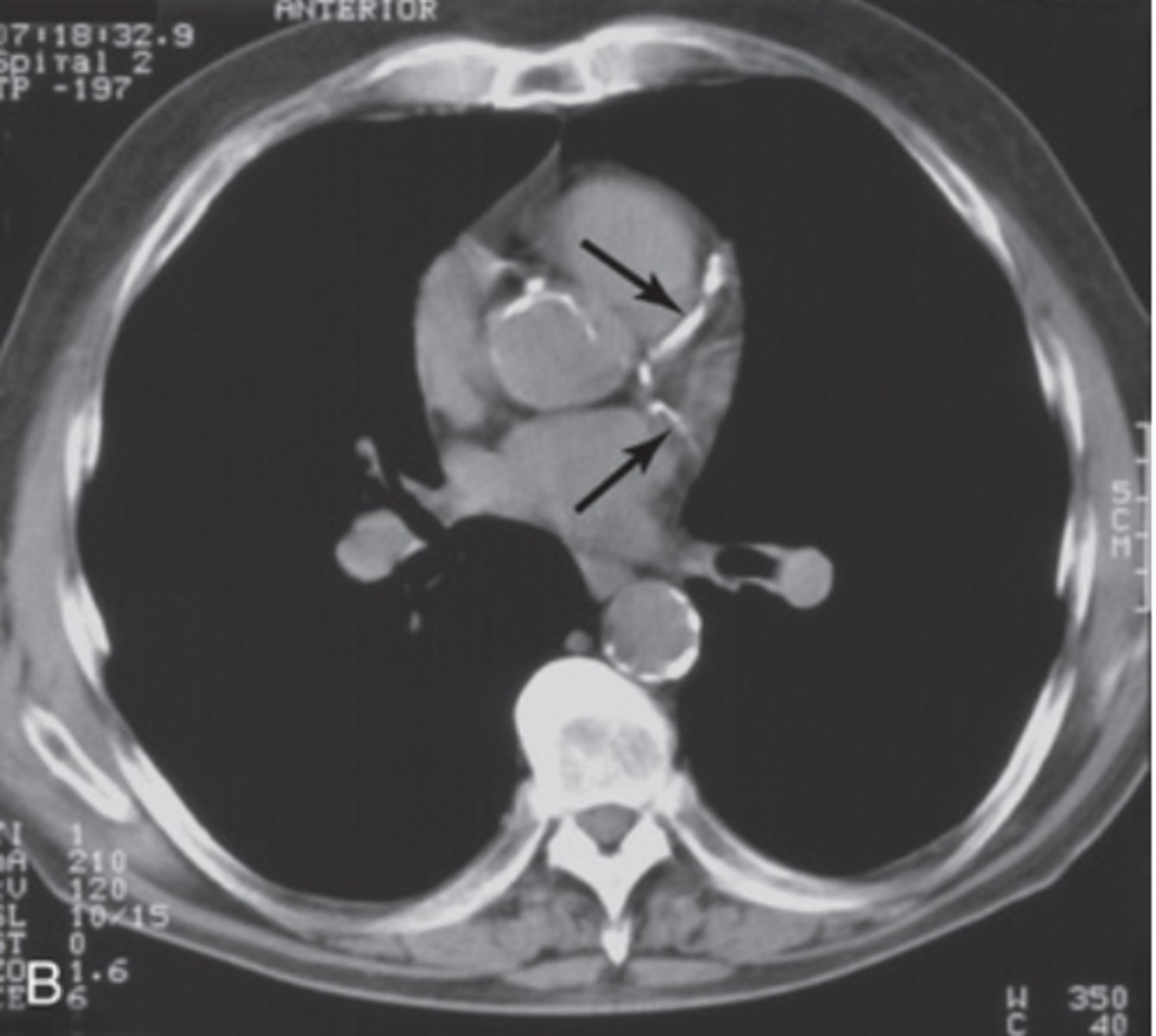
What does a Pericardial effusion look like on a chest CT?
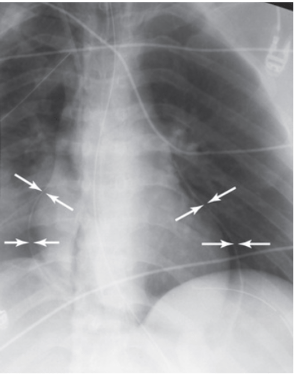
What does air in the pericardial sac look like on chest X-ray?
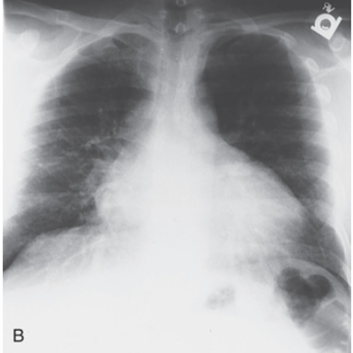
What does the heart look like on chest x-ray in patients on Doxorubin?
Multi-chamber dilation
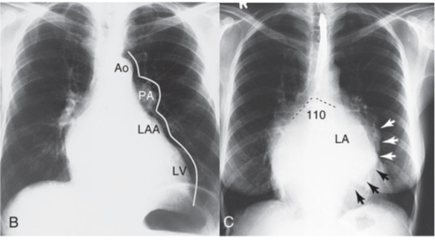
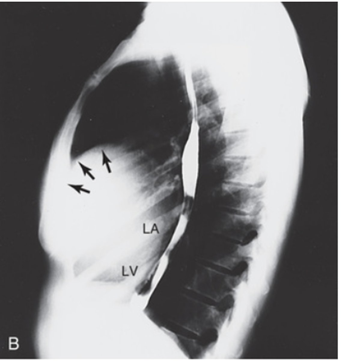
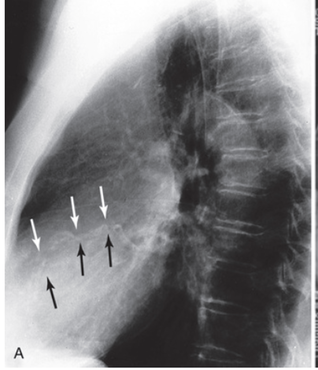
What does left atrial enlargement look like on a lateral view?
Displaced esophagus

What does left atrial enlargement look like on a PA x-ray?
- Ski mogul heart
- Carnia widens over 75 degrees
Mitral heart on PA chest xray also called
ski mogul heart
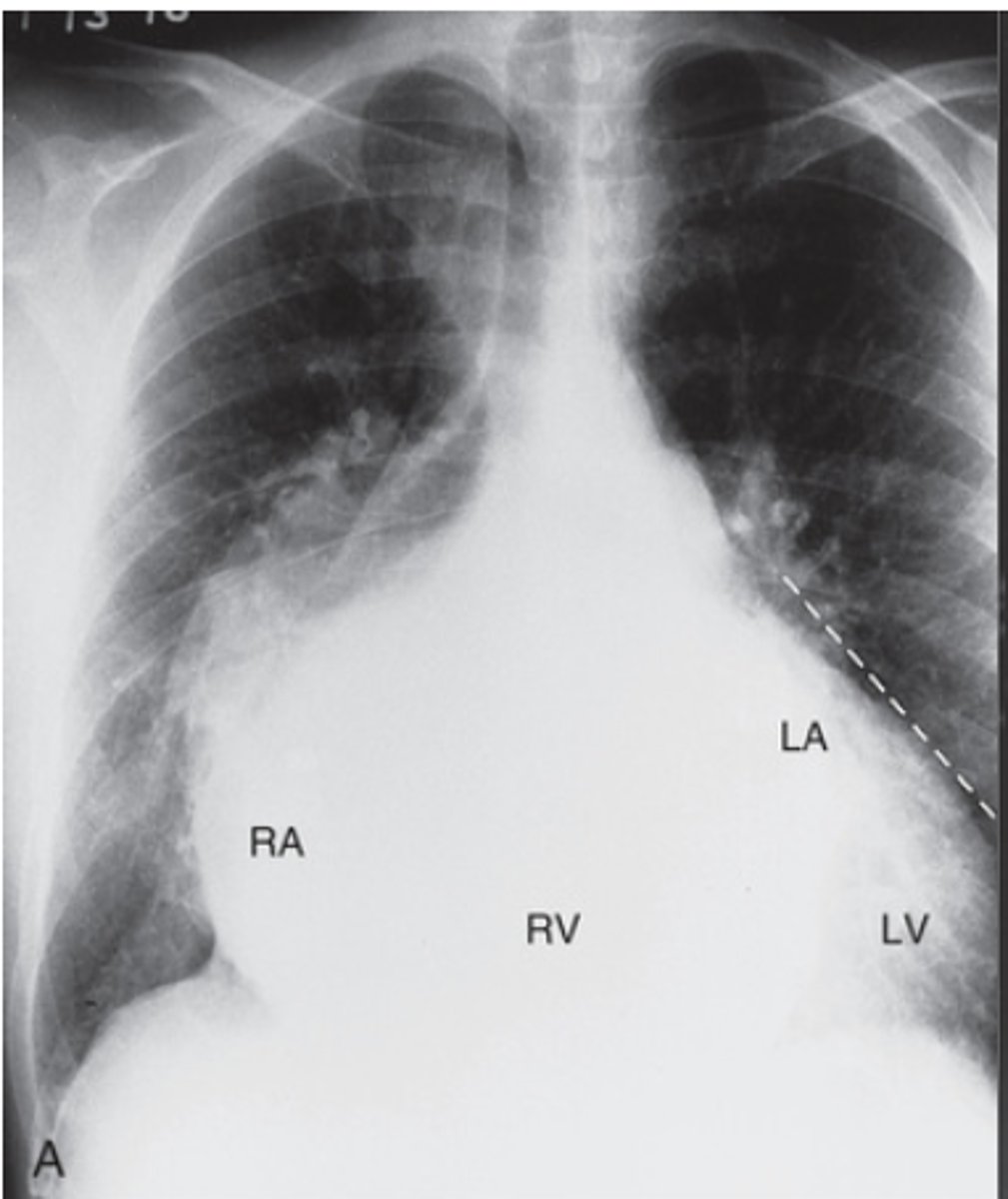
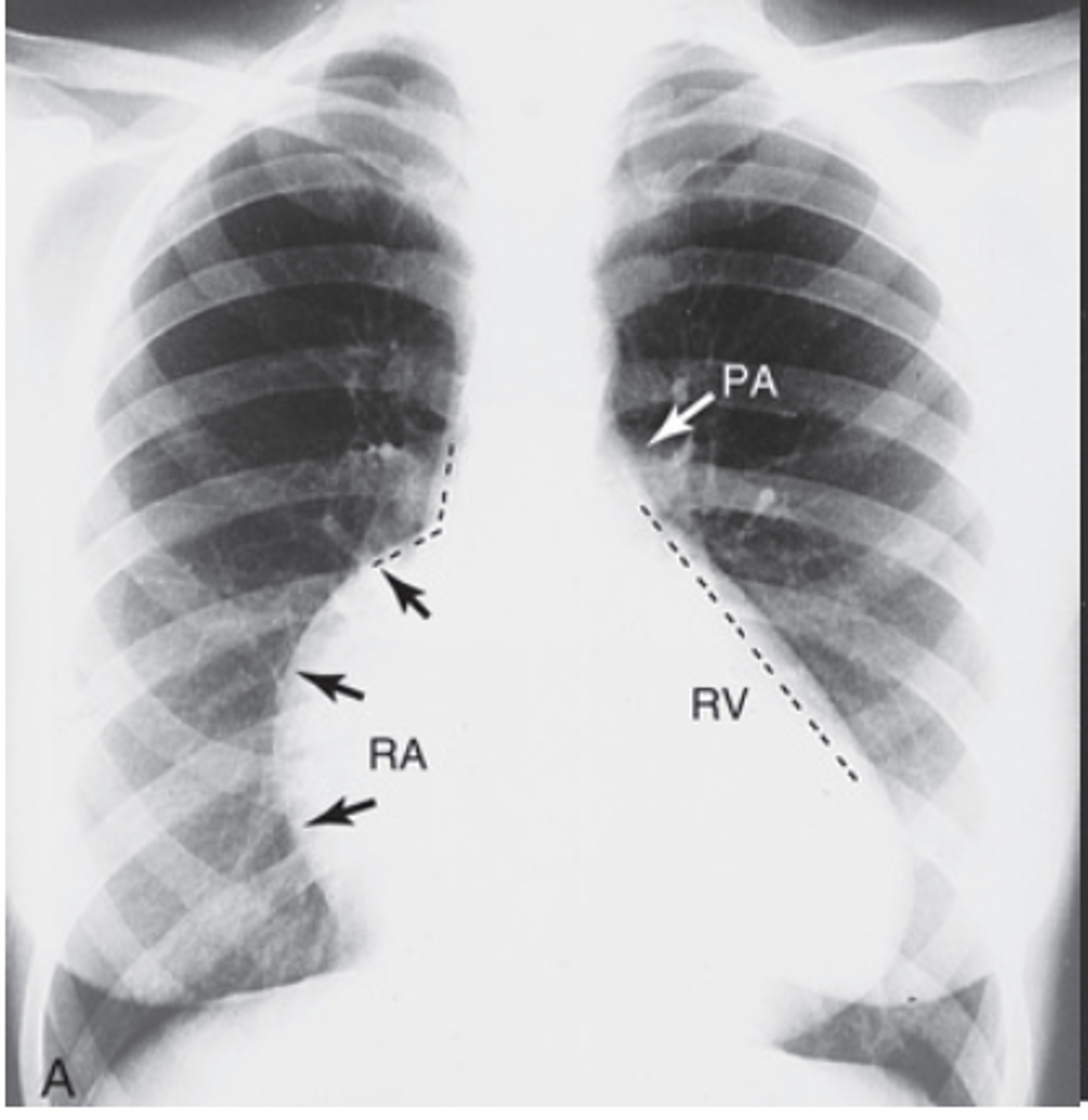
What does mitral and tricuspid disease look like on a PA x-ray?
- Complete enlargement of the right side
- Complete straightening of left wall
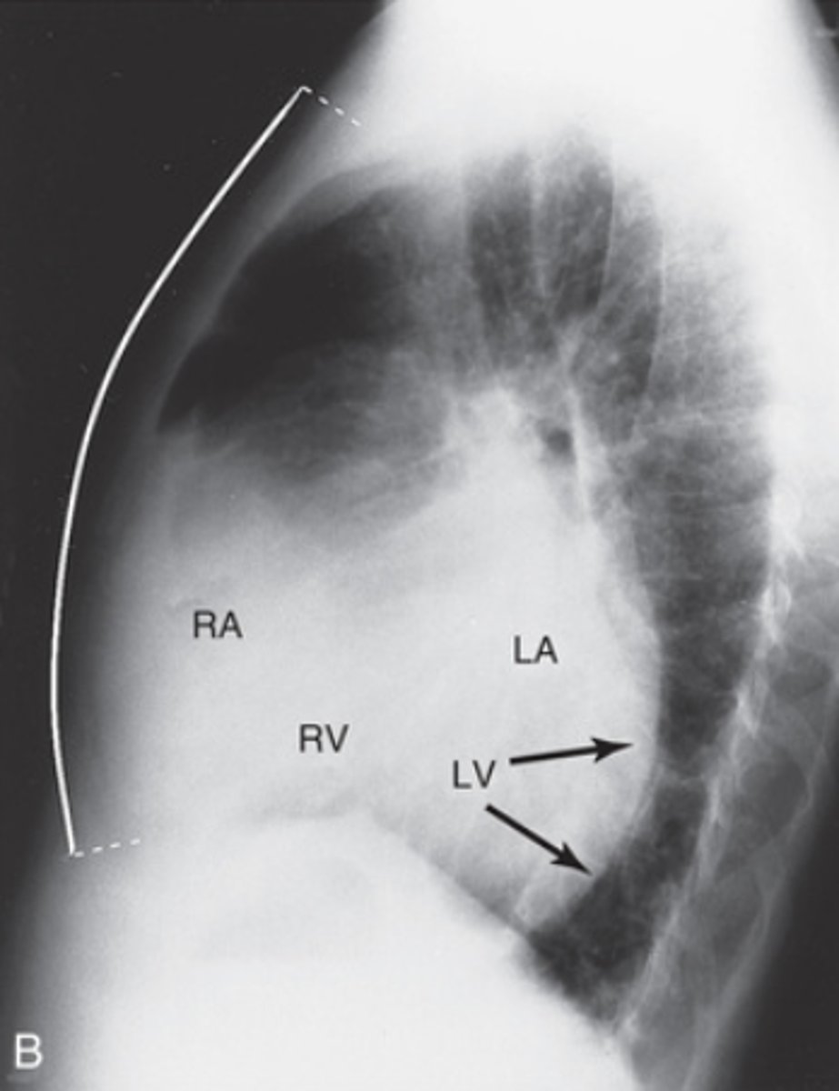
What does mitral and tricuspid disease look like on a lateral x-ray?
- The right side of the heart is filling retrosternal space
- Image of heart is imposing on the image of spine
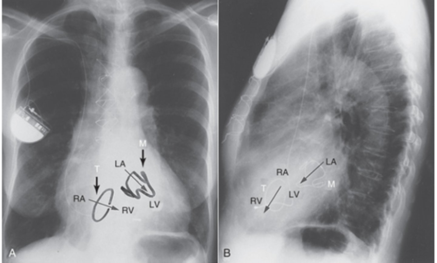
What do prosthetic mitral and tricuspid valves look like on x-ray?
- The mitral valve is posteriorly and to the left side
- The tricuspid valve is anterior and toward the right side
What does Aortic insufficiency look like on PA X-ray?
- rounding of the cardiac apex
- Apex is pointing down and projects below left hemidaphigram
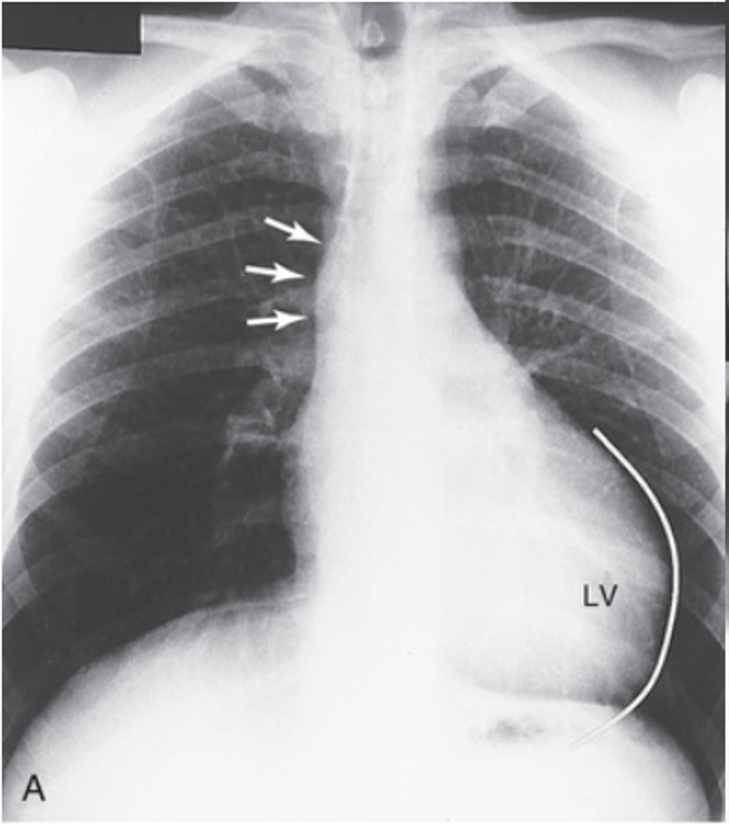
What does Aortic insufficiency look like on lateral x-ray?
- Convex left ventricle
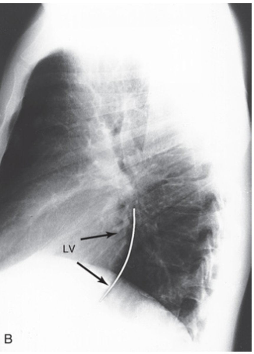
What does prosthetic aortic valve look like?
- located at the root of the ascending aorta
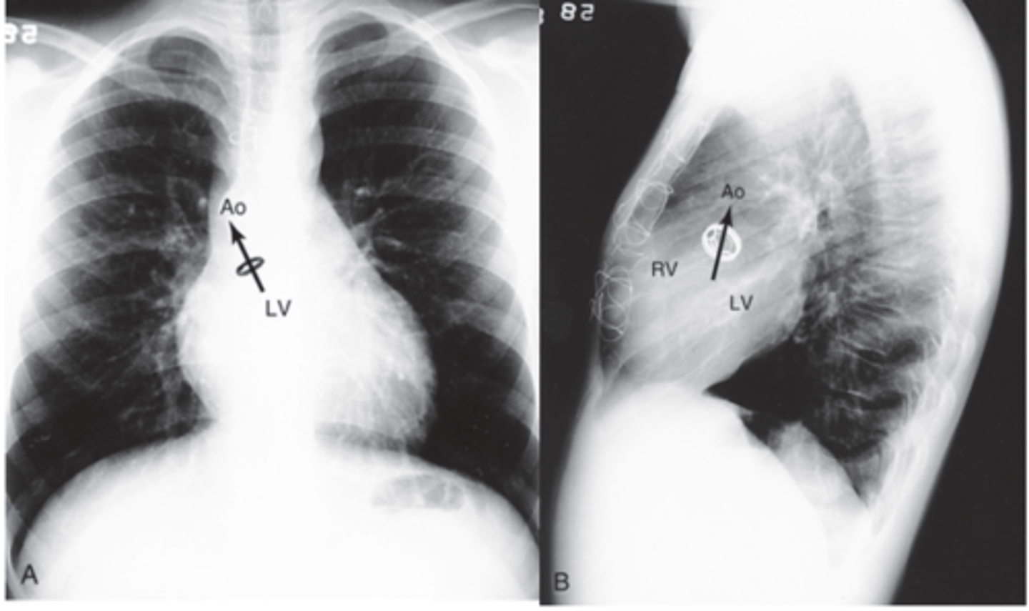
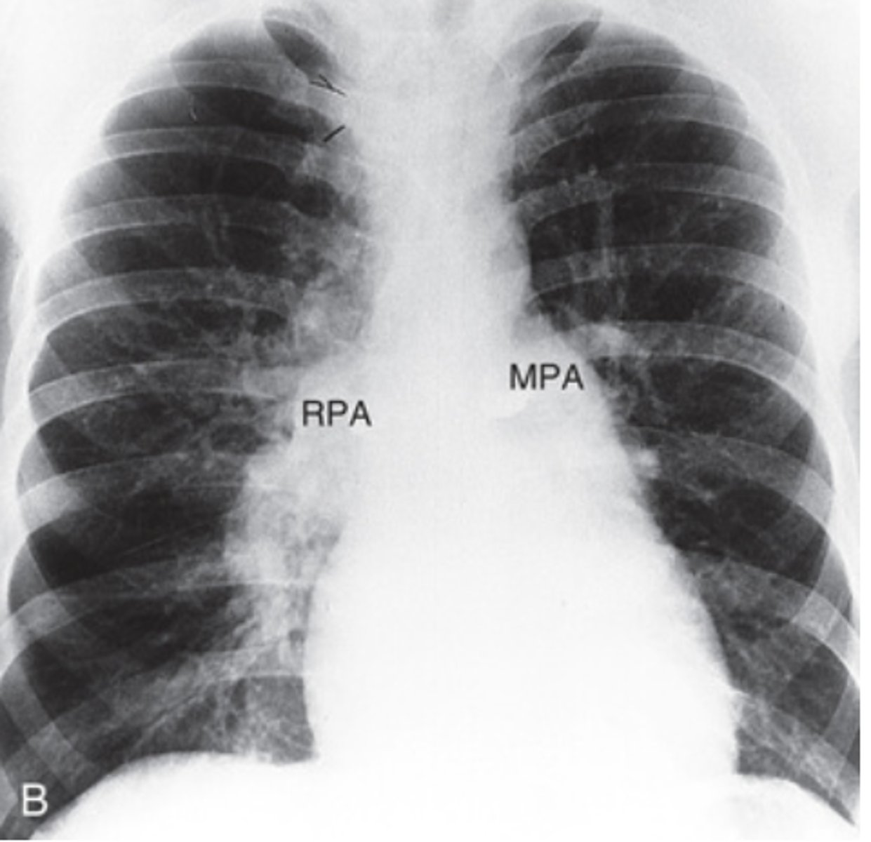
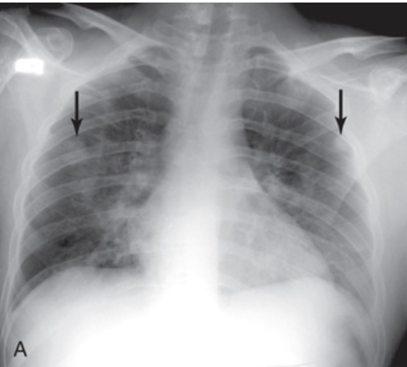
What does pulmonary hypertension look like on a PA x-ray?
- Enlargement of both the left and right main pulmonary arteries is present
What does the Tetralogy of Fallot look like on a PA x-ray?
-Pulmonic stenosis
-VSD
-Overriding aorta
-Right ventricular Hypertrophy
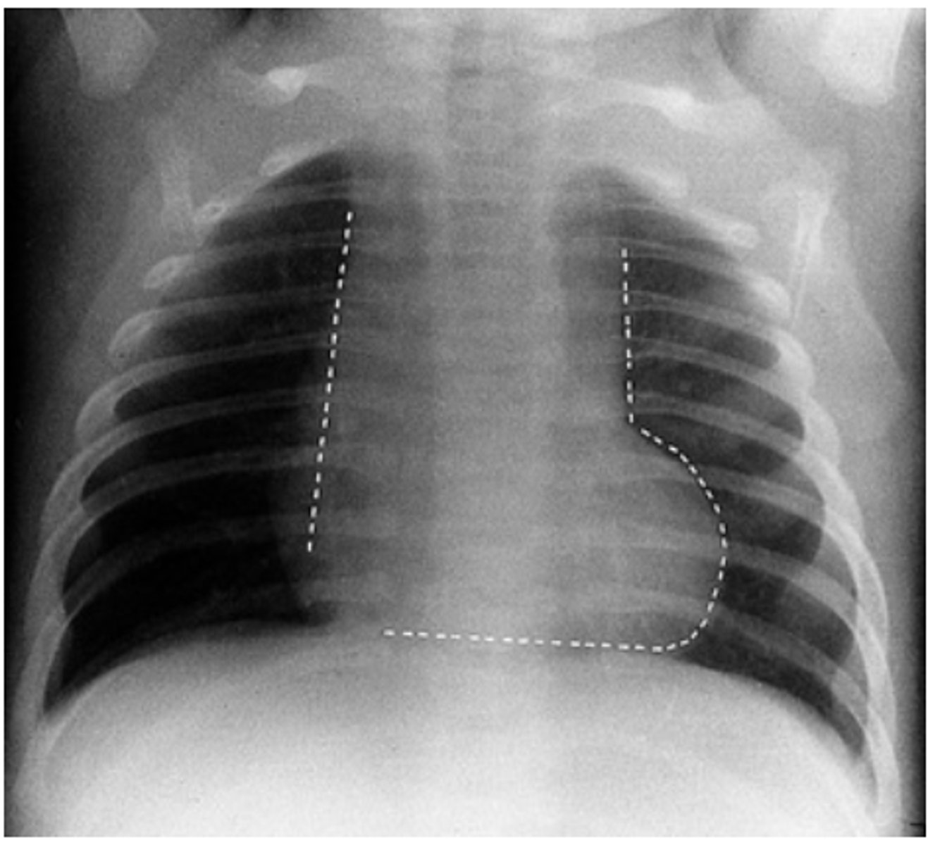
What does Ebstein malformation look like on PA x-ray?
- giant right atrium appears
- shoulder along the right side
- very small pulmonary artery
What does Ebstein malformation look like on lateral x-ray?
Filling retrosternal space
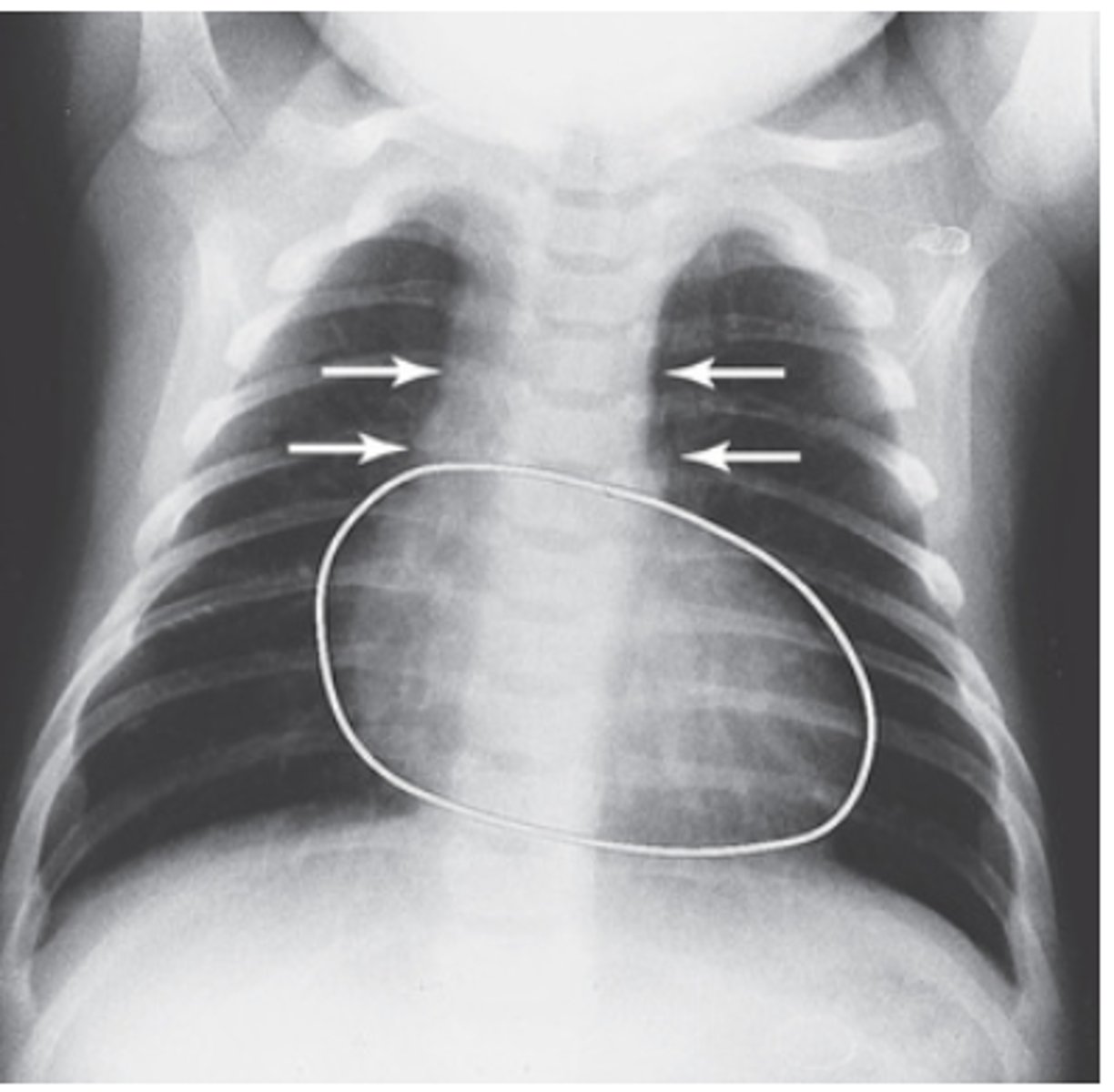
What does transposition of the great vessels look like on PA x-ray?
- egg on side shape
What does ASD look like on PA X-rays?
- Enlargement of pulmonary artery
- increase in the size of the right atrium and right ventricle
What does ASD look like on lateral X-rays?
- Right ventricle enlargement
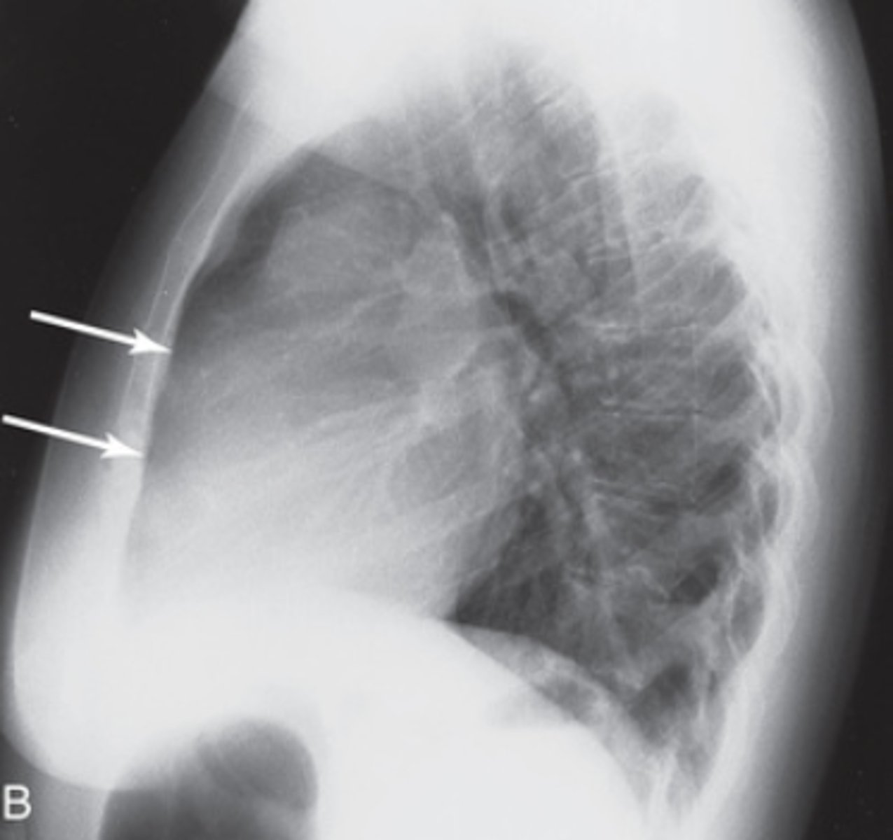
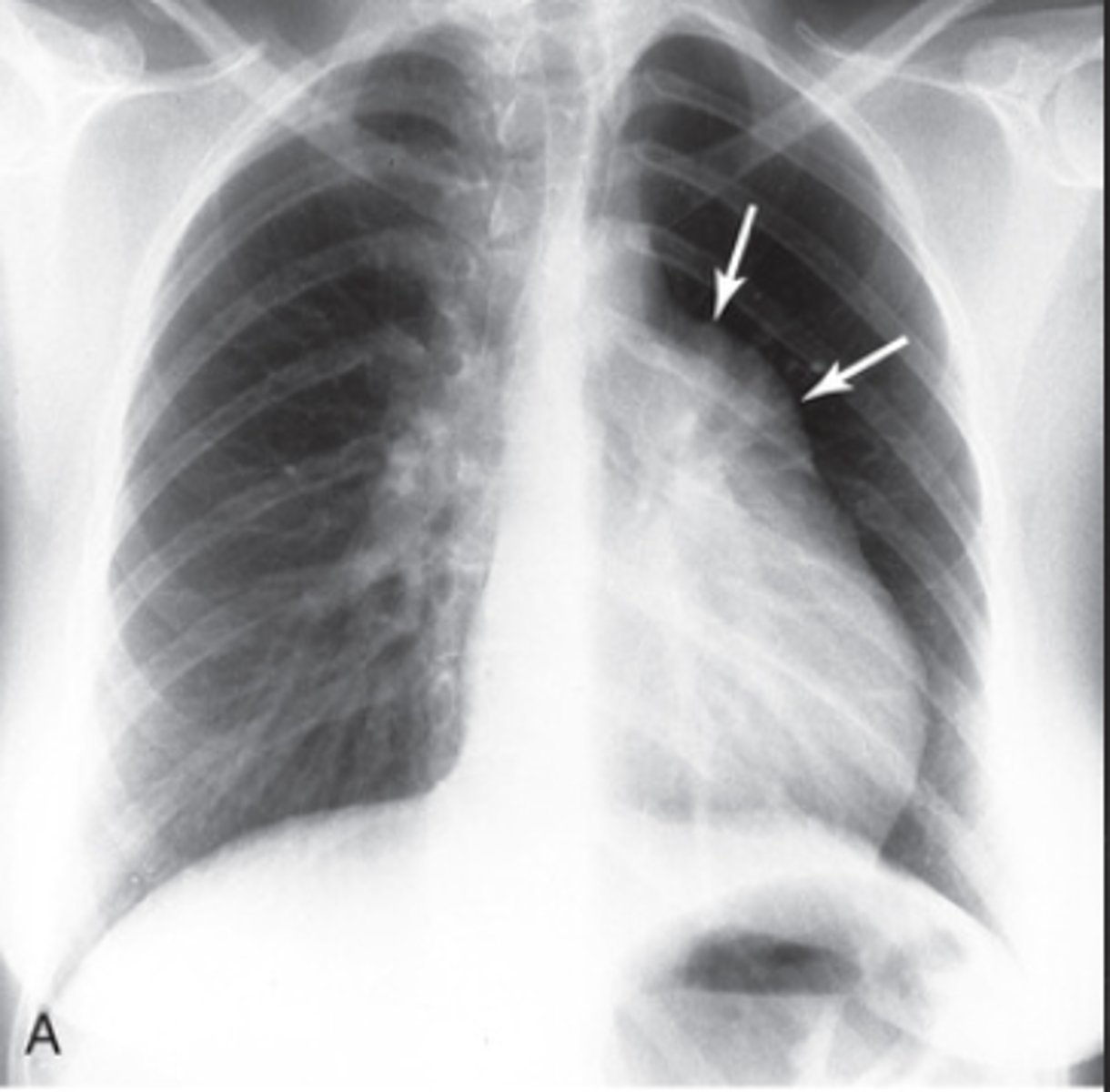
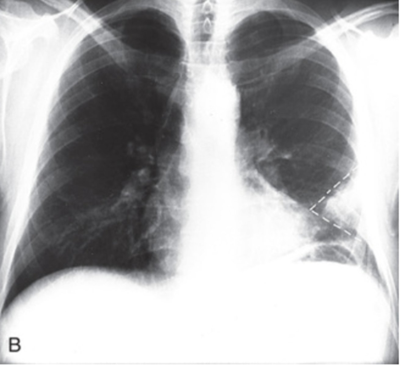
What does a pulmonary embolism look like on PA x-ray?
- Wedge-shaped infiltrate
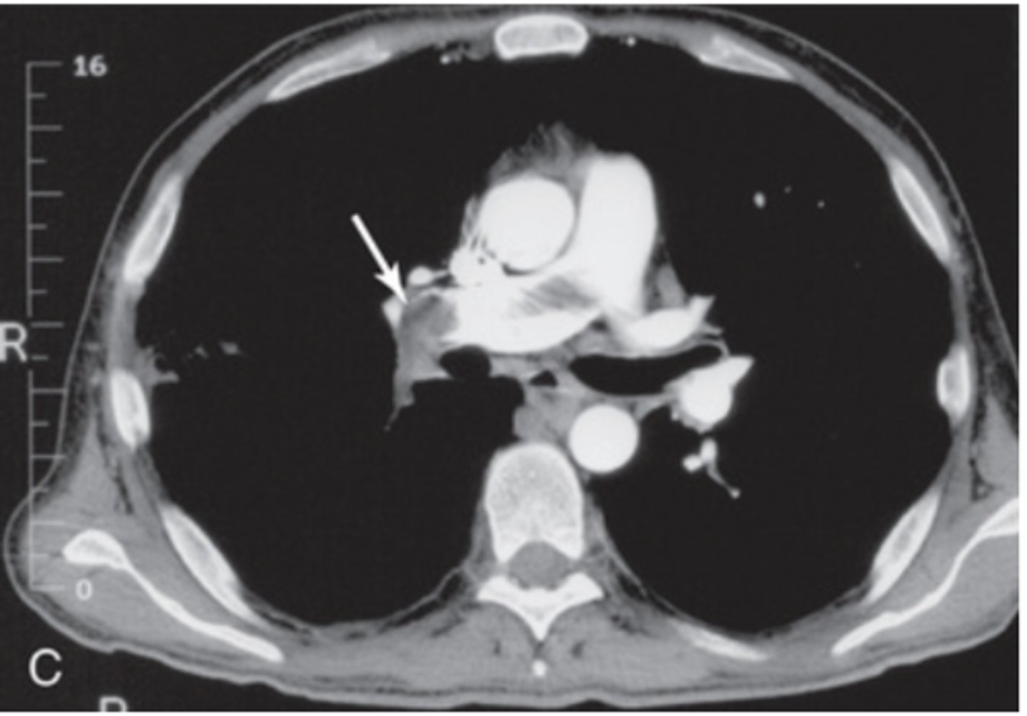
What does a pulmonary embolism look like on a CT of the chest?
What does a septic PE look like on PA chest x-ray?
ill-defined pulmonary nodules that can cavitate
What does coronary artery calcification look like on a lateral x-ray?
What does coronary artery calcification look like on a chest CT?
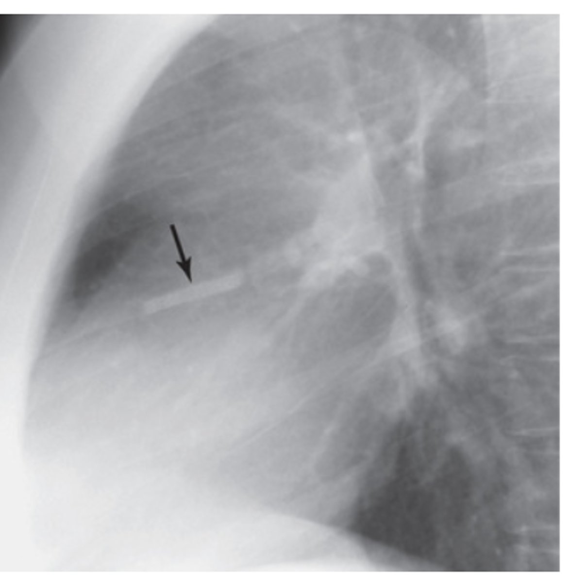
What does a wire mesh stent look like on lateral chest x-rays?
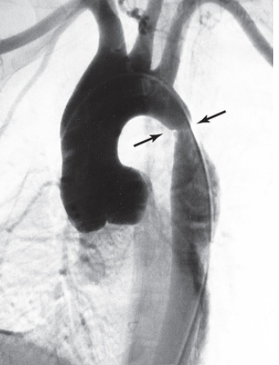
What does Coarctation of the Aorta look like on PA chest x-rays?
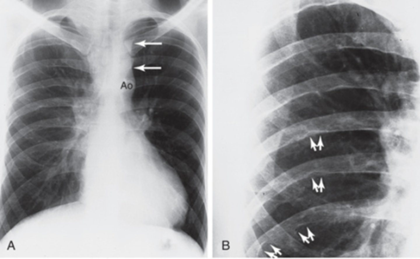
What does the Coarctation of the Aorta look like on CT/MR angiography?
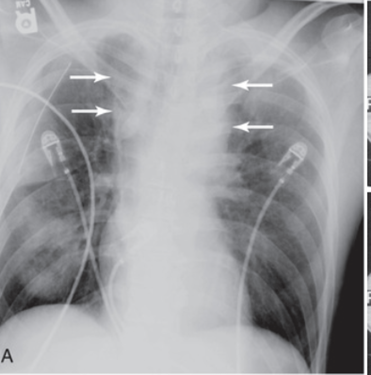
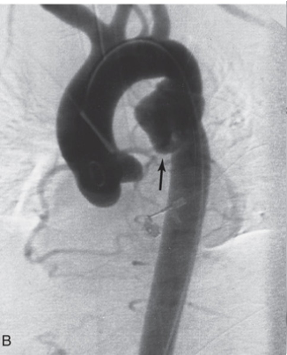
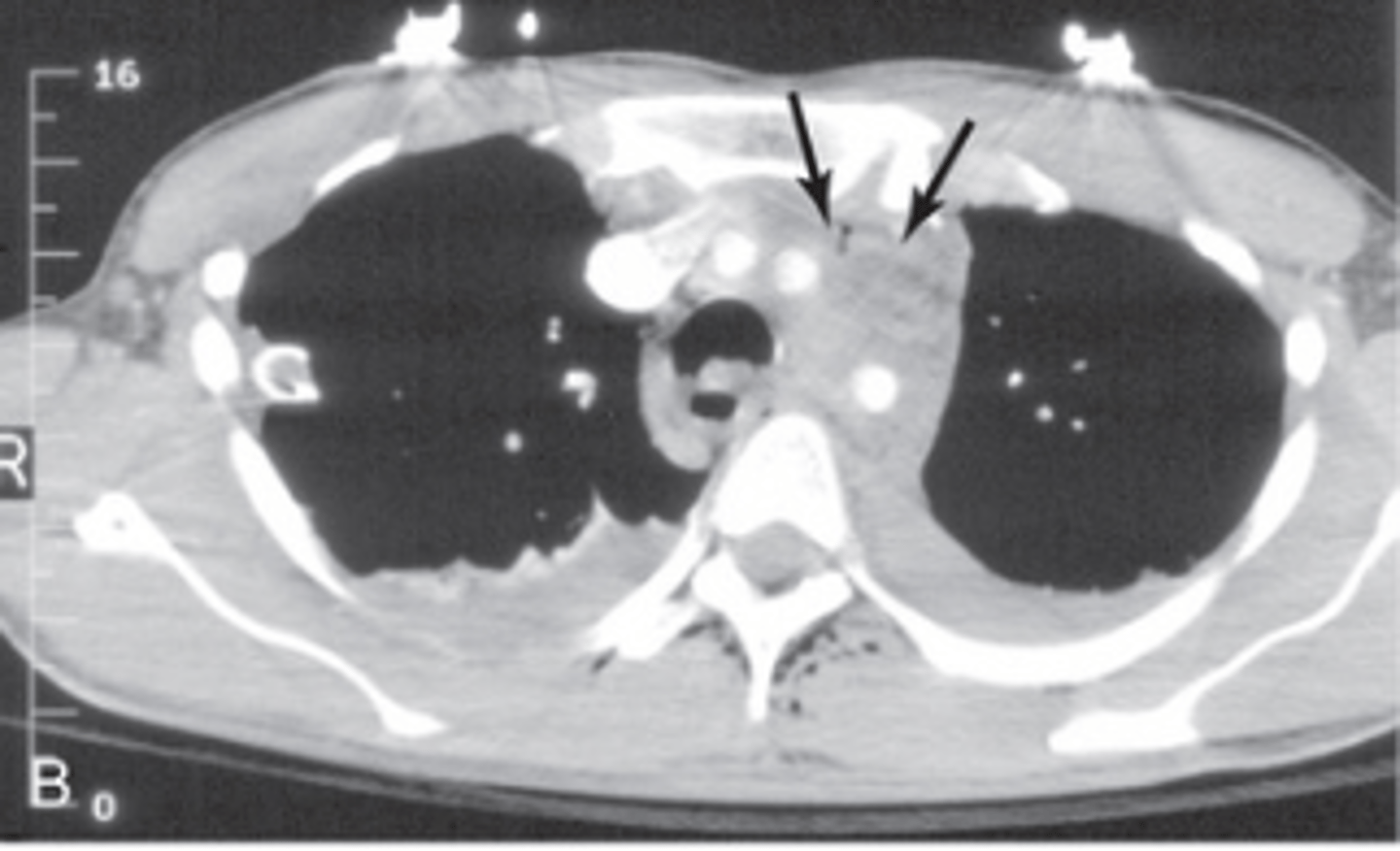
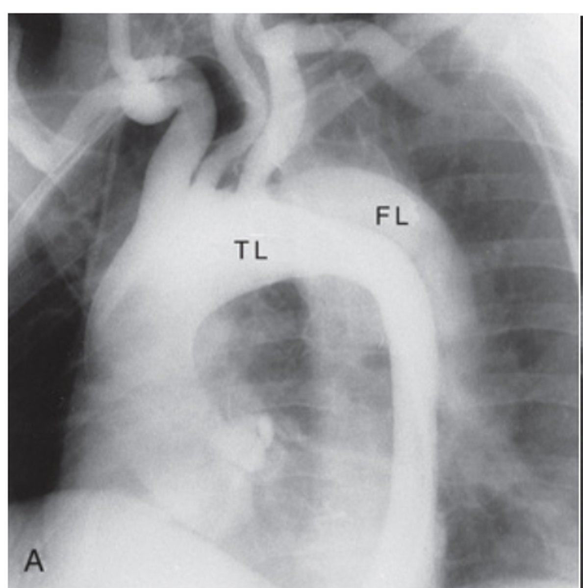
What does an aortic tear look like on a PA chest x-ray?
What does an aortic tear look like on a CT/MR angiogram?
What does an aortic tear look like on a chest CT?
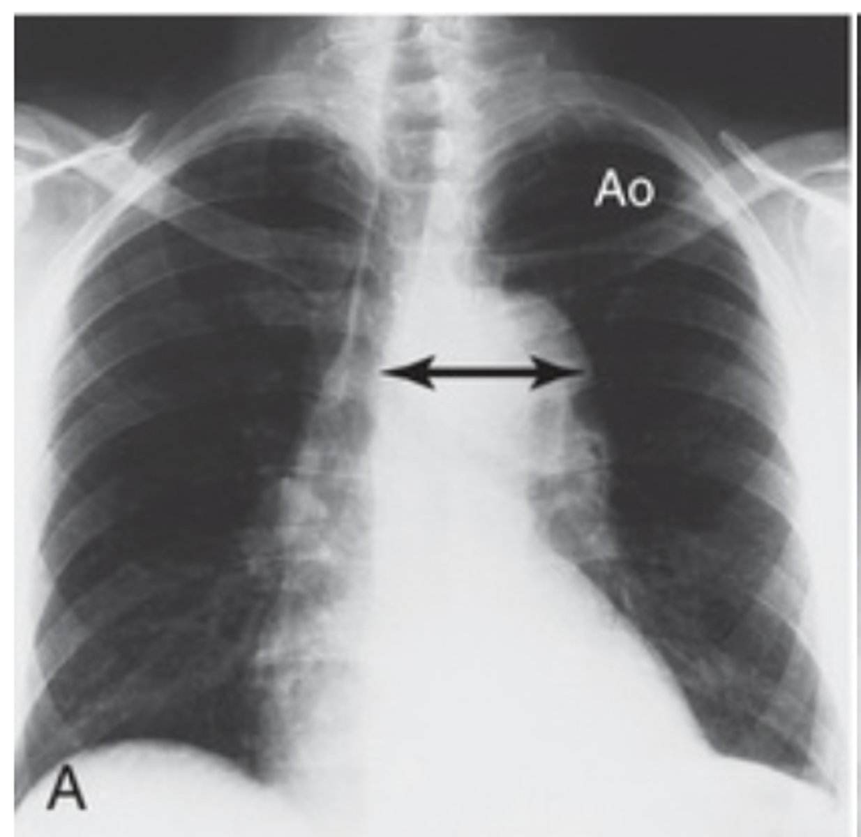
What does a Thoracic Aortic Aneurysm look like on PA chest x-ray?
- widening of the ascending aorta or aortic arch
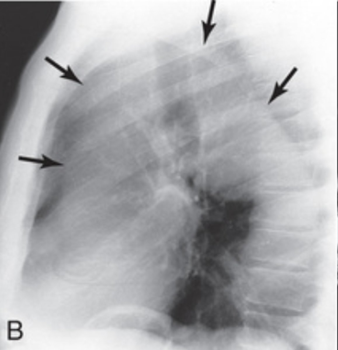
What does a Thoracic Aortic Aneurysm look like on lateral chest x-ray?
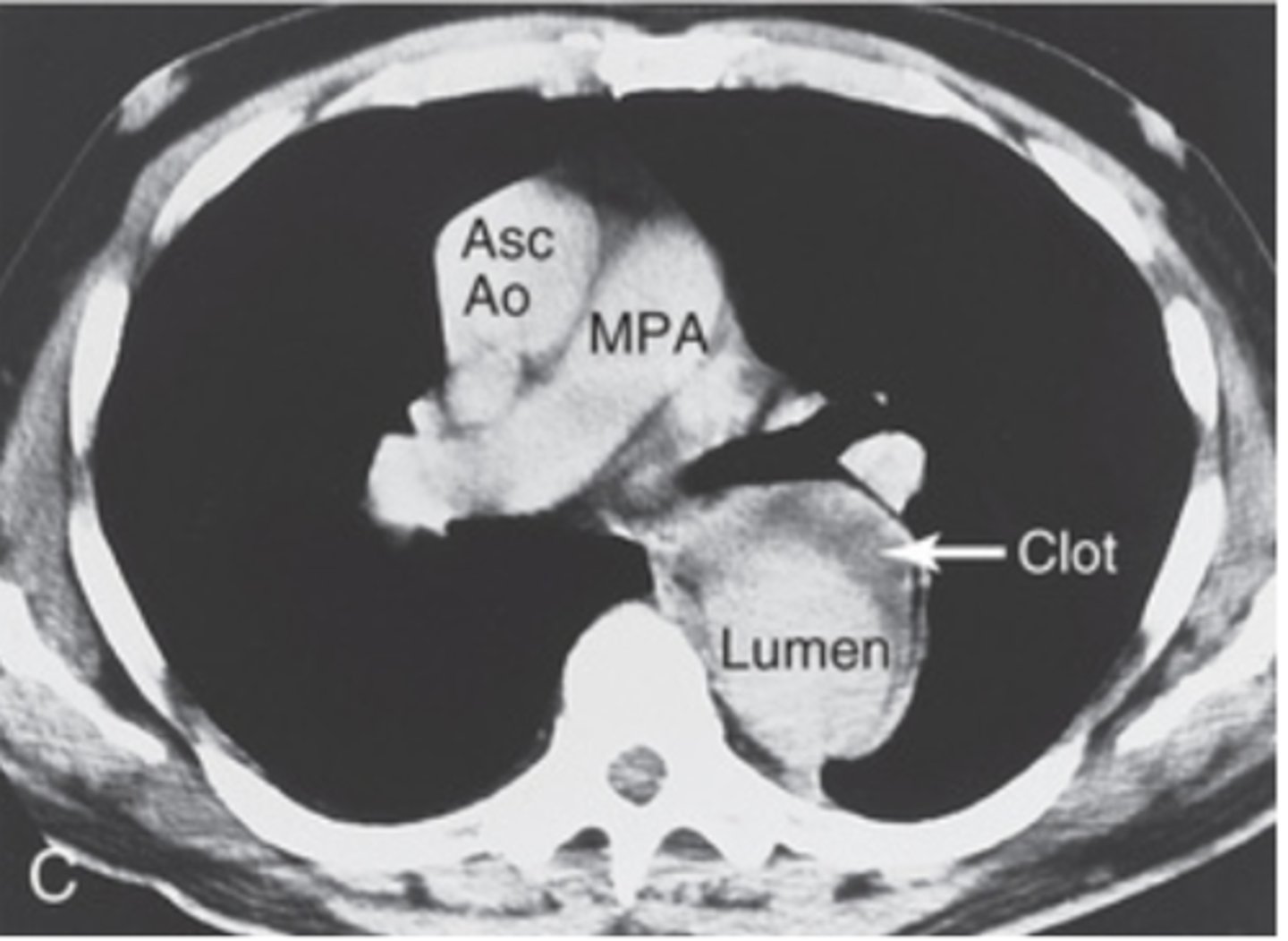
What does a Thoracic Aortic Aneurysm look like on chest CT?
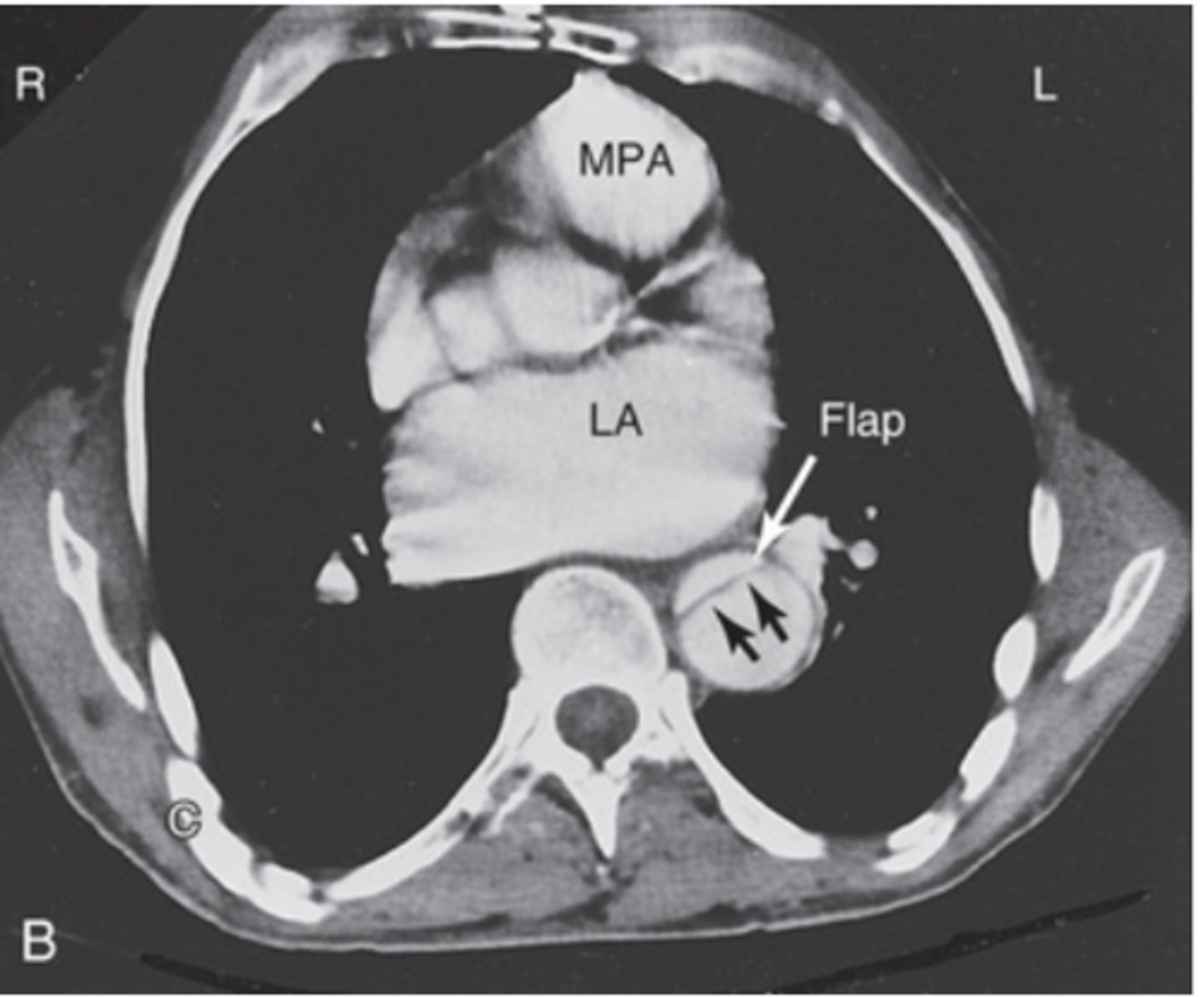
What does an aortic dissection look like on a CT angiogram?
What does an aortic dissection look like on a chest CT?
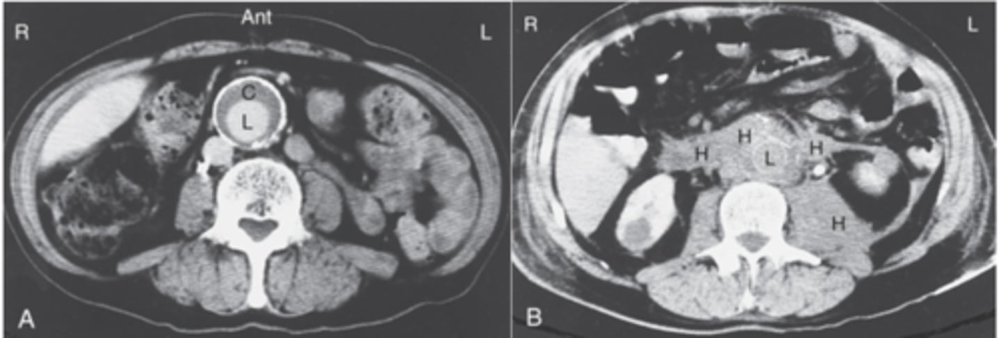
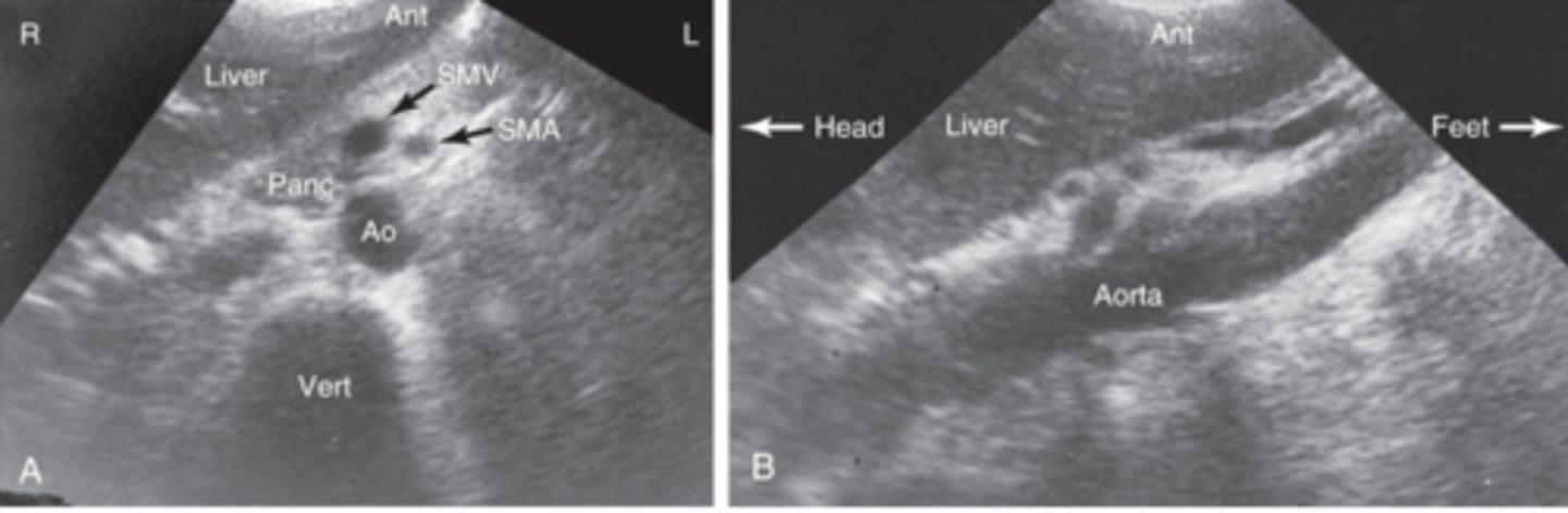
What does an abdominal aneurysm look like on CT of abdomen?
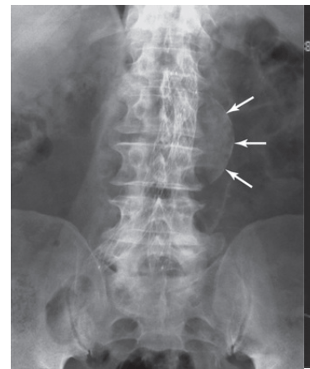
What does an abdominal aneurysm look like on PA x-ray of abdomen?
What does an abdominal aneurysm look like on ultrasound of abdomen?
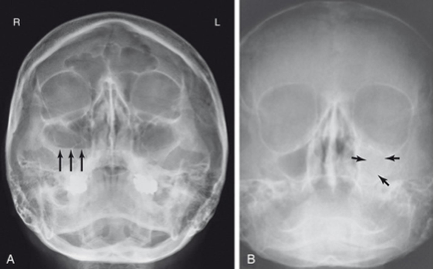
What does Acute sinusitis look like on x-ray?
- air-fluid level in the sinus
- complete opacification is found
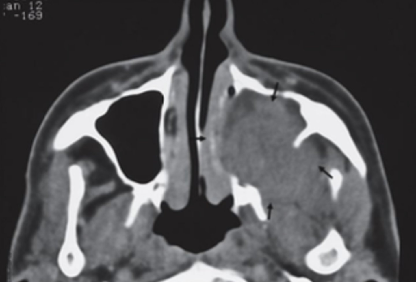
What does nasopharyngeal carcinoma look like on CT of the head
bony margins
What does Thyroid eye disease look like on CT of the head?
enlargement of the EOM frequently bilateral
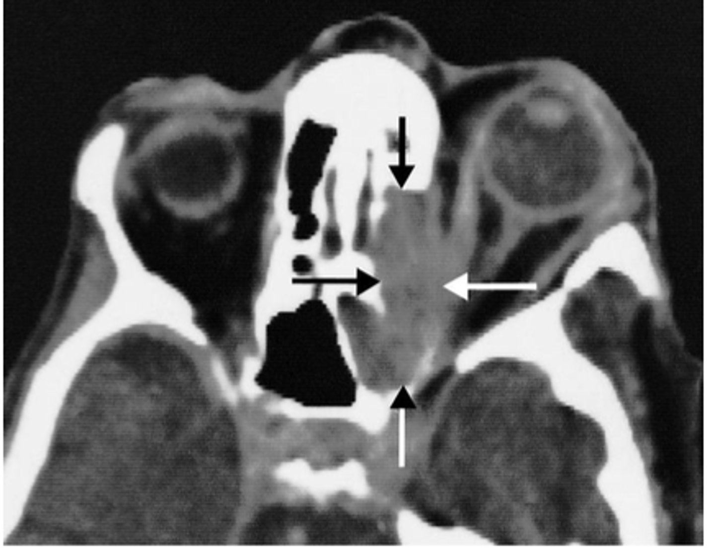
What does severe Thyroid eye disease look like on CT of the head?
compression of the optic nerve in the apex
of the orbit

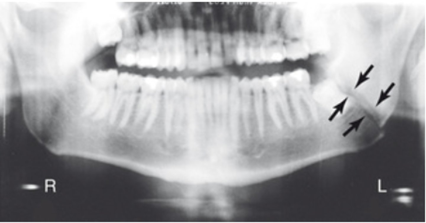
What does a Mandibular fracture look like?
What does epiglottis look like on lateral soft tissue view of the neck?
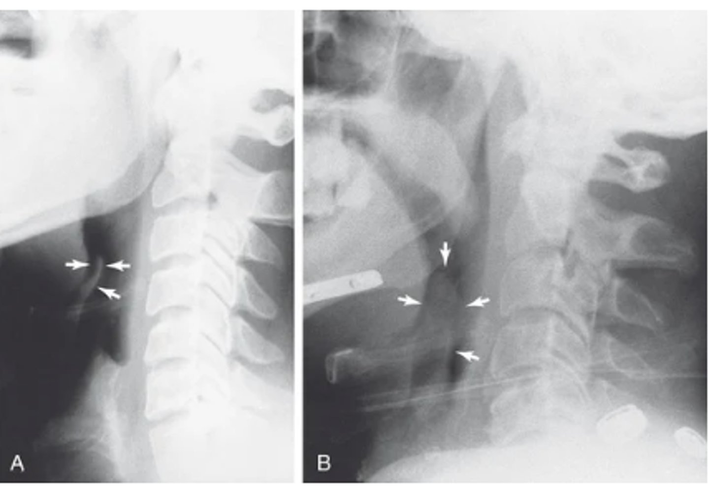
What does a retropharyngeal abscess look like on a lateral soft tissue view of the neck?
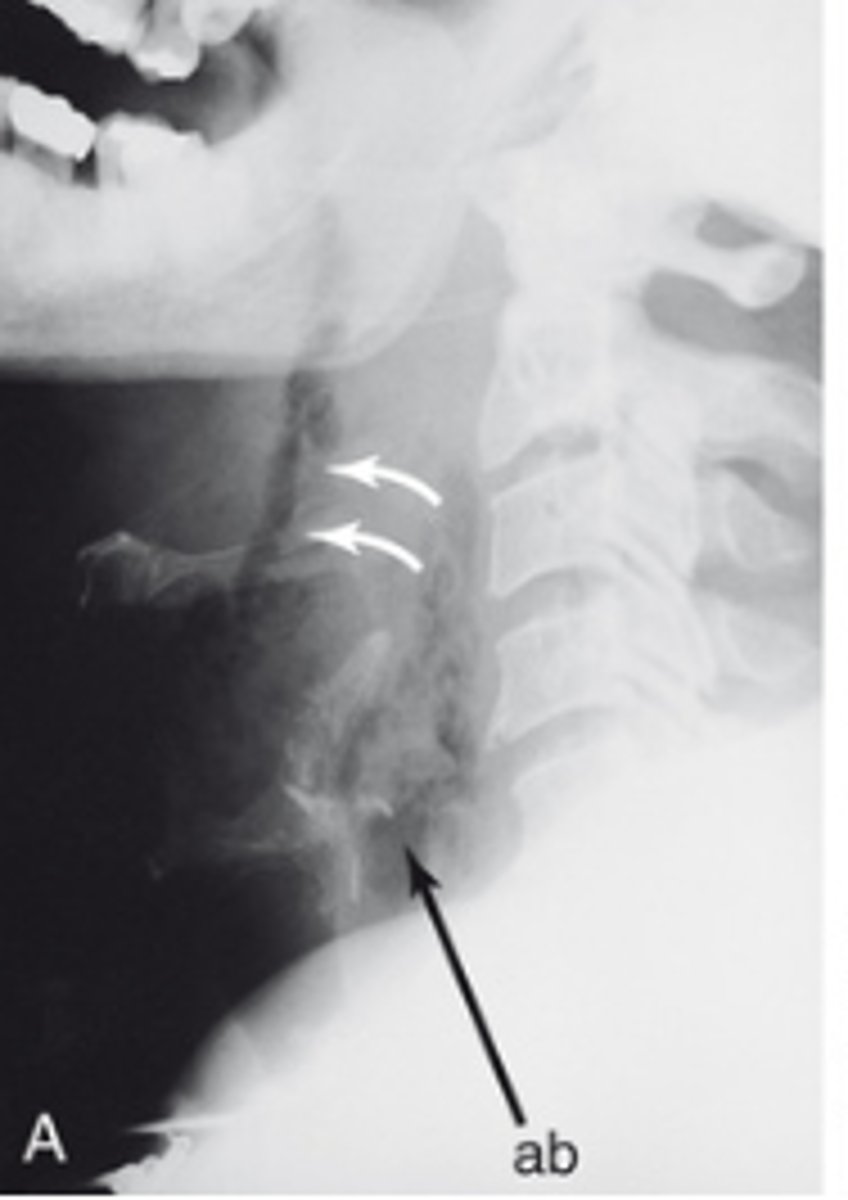
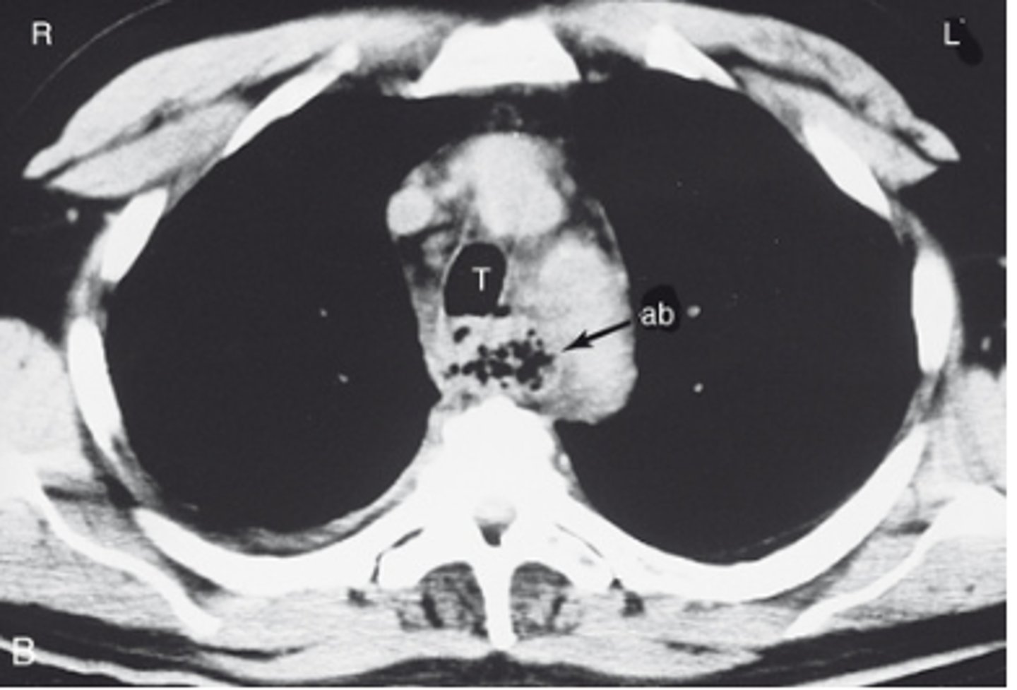
What does a retropharyngeal abscess look like on a CT of the neck?
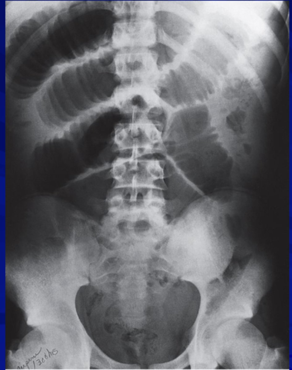
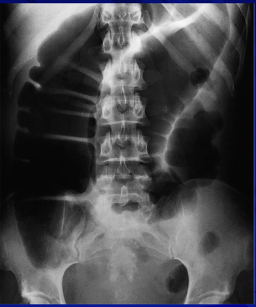
What does dilated small bowel look like?
A stacked coin appearance
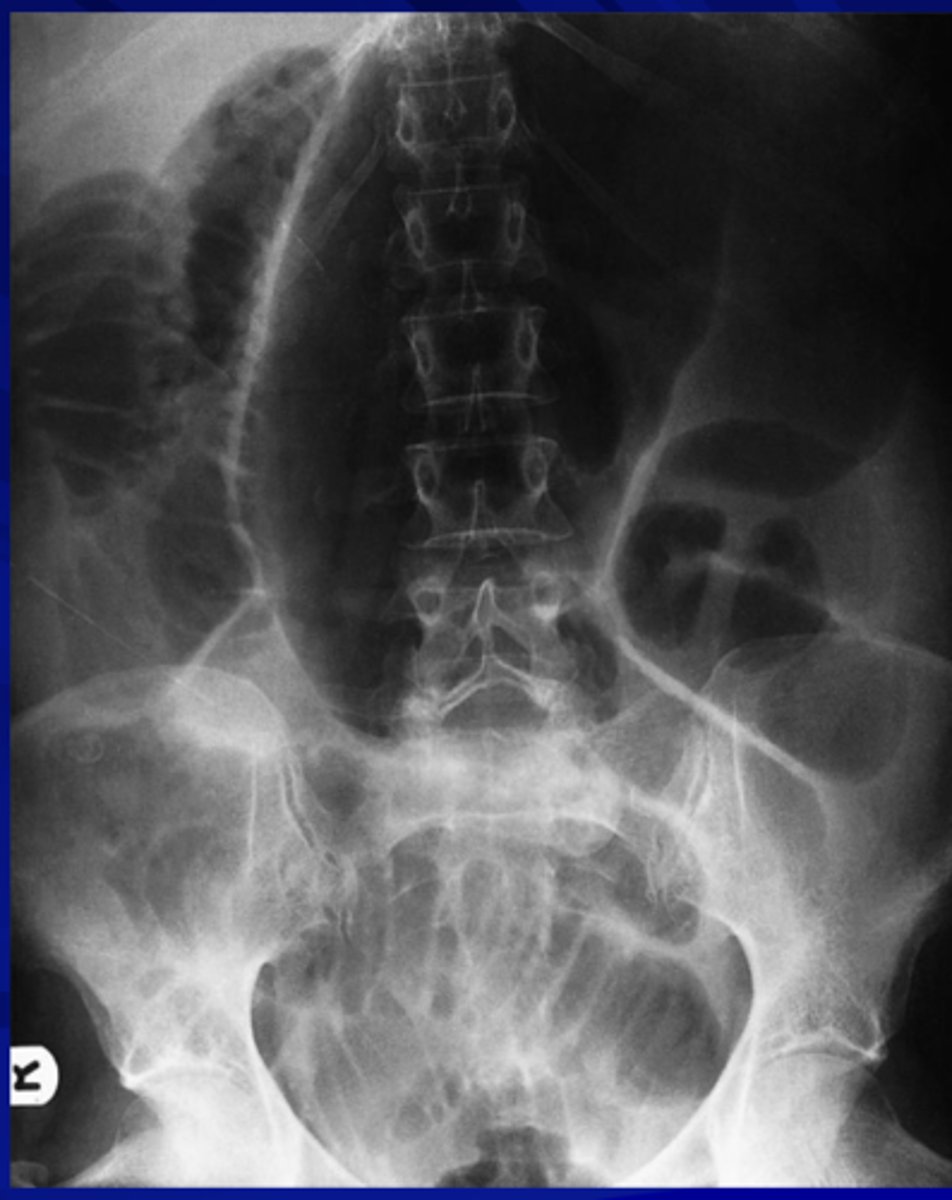
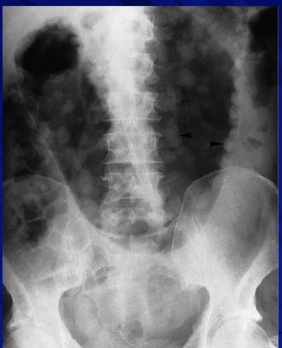
What does a large bowel obstruction look like?
What does a cecum volvulus look like on x-ray?
What does ulcerative colitis look like on x-ray?
Look for presence of pseudo-polyps
What does a perforated peptic ulcer cause on an x-ray?
pneumoperitoneum
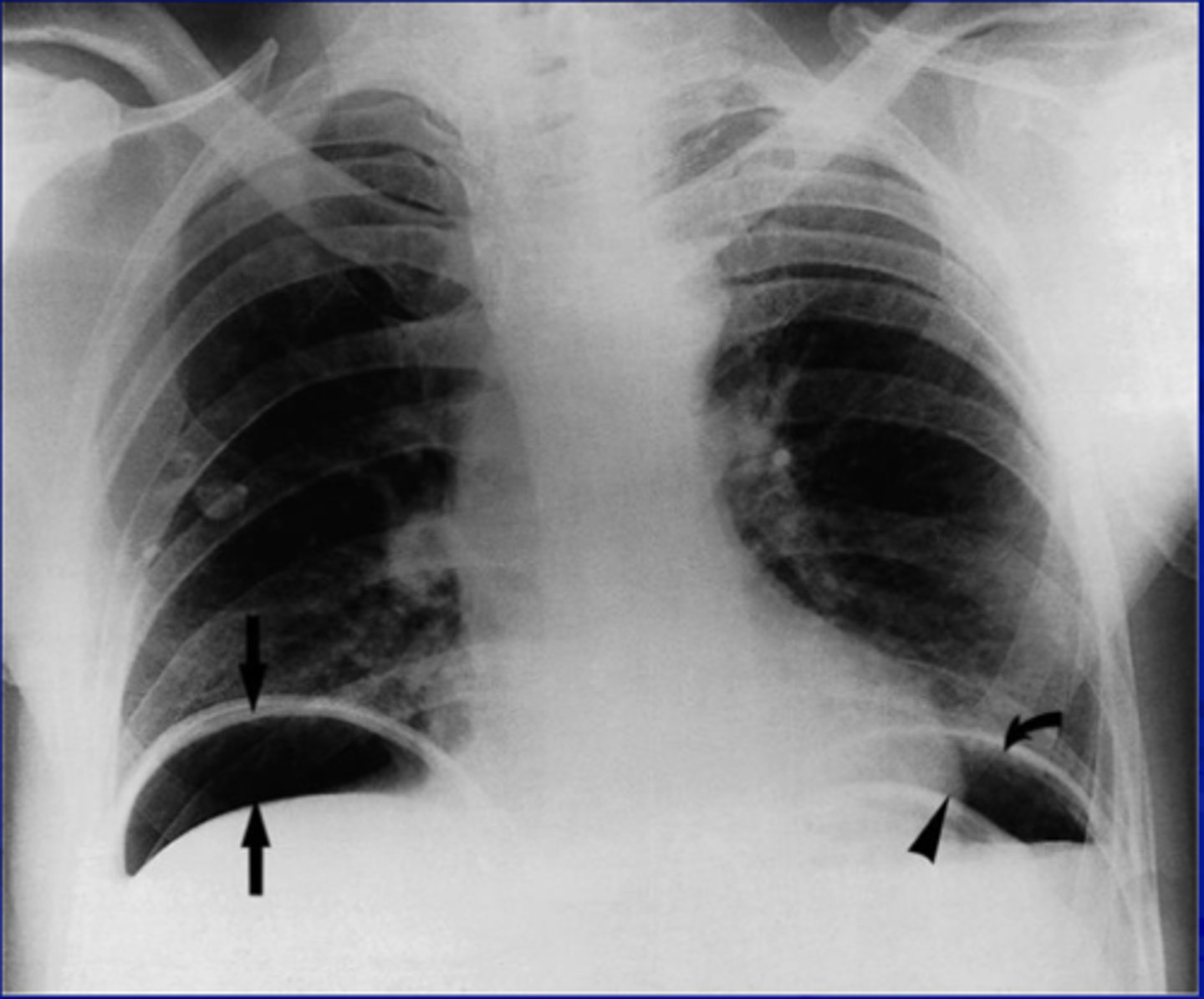
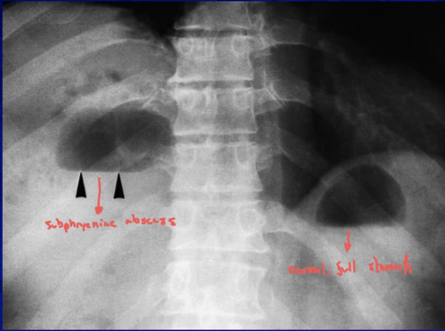
What does a subphrenic abscess look like on an x-ray? And what does it signify?
pleural effusion or pulmonary
collapse/consolidation
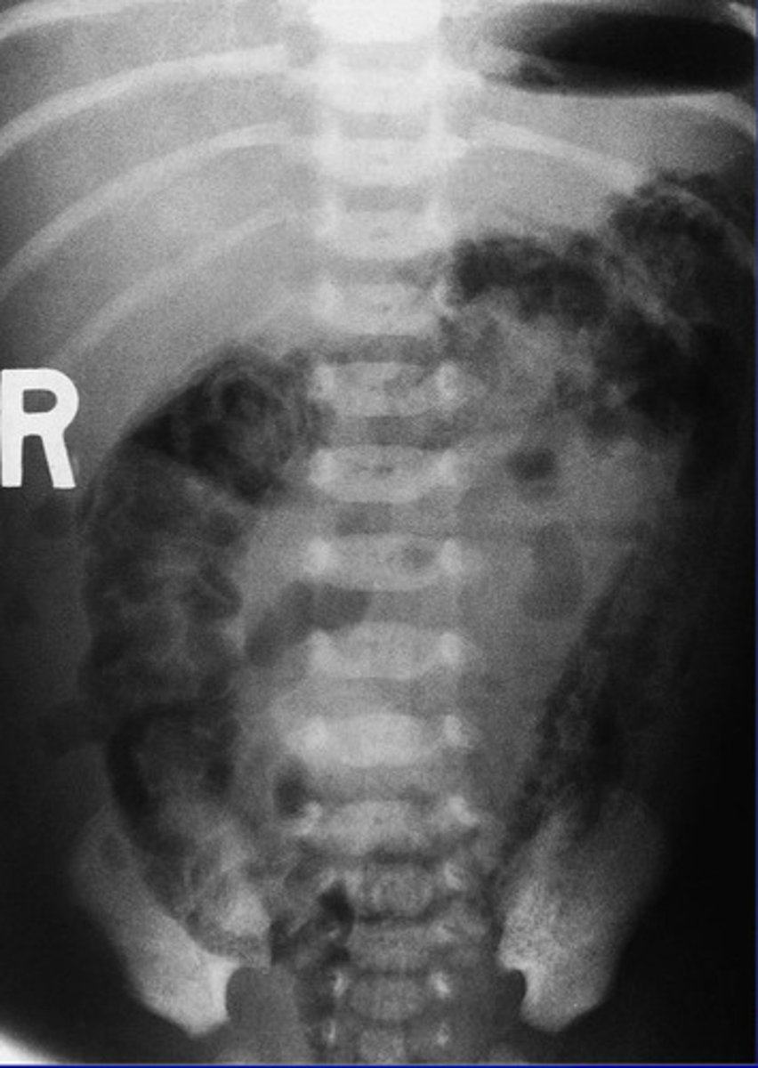
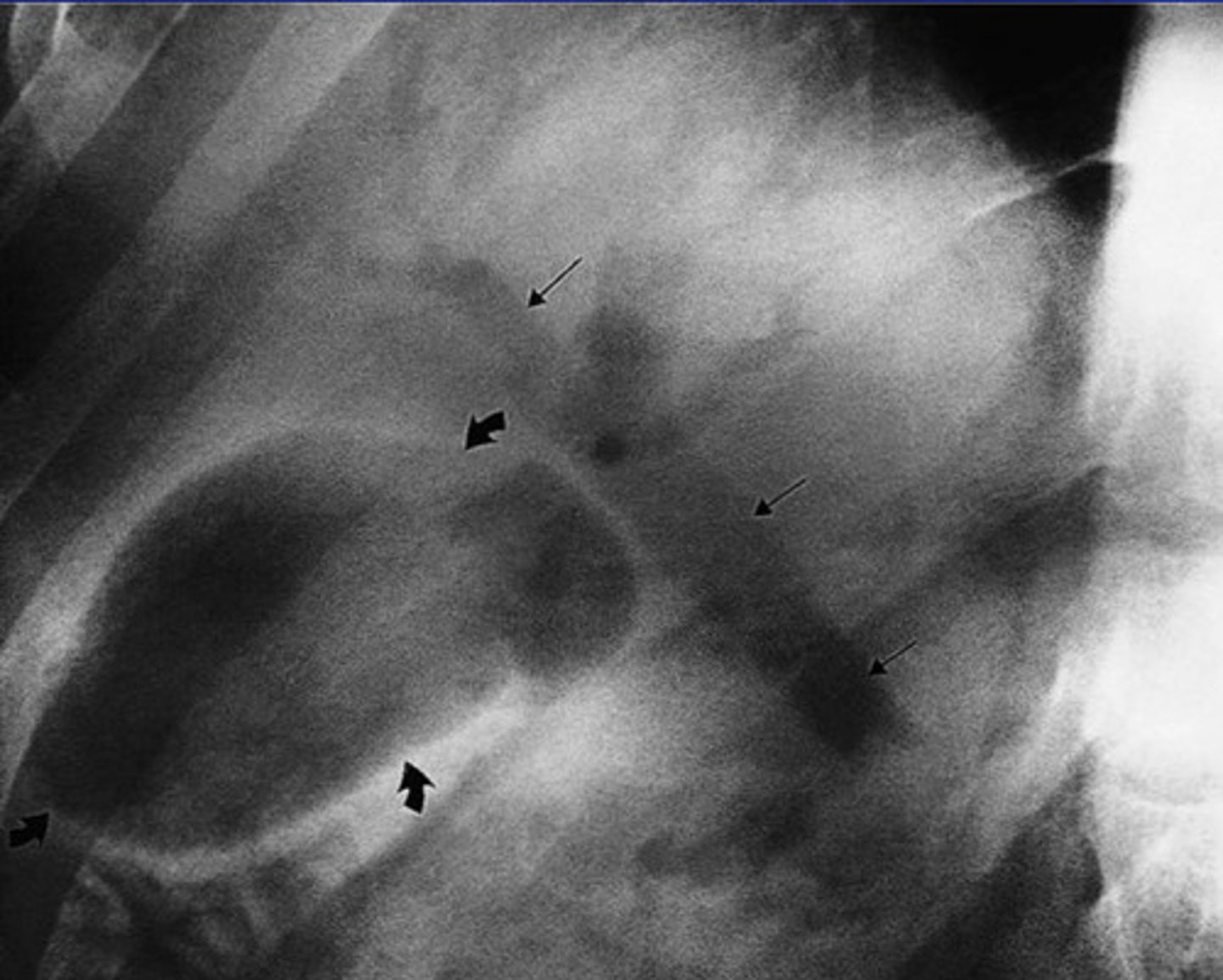
What does necrotizing enterocolitis look like on an x-ray?
What does gas in the biliary system look like on an X-ray?
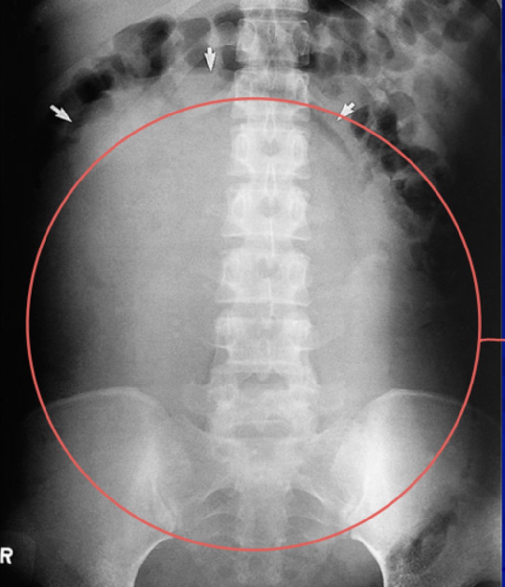
What does ascites look like on an x-ray?
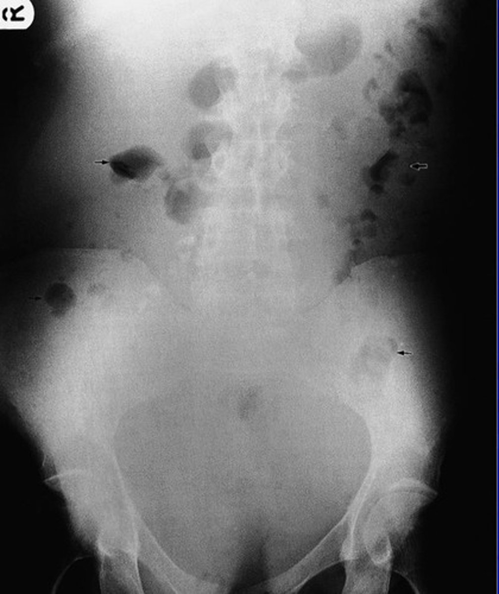
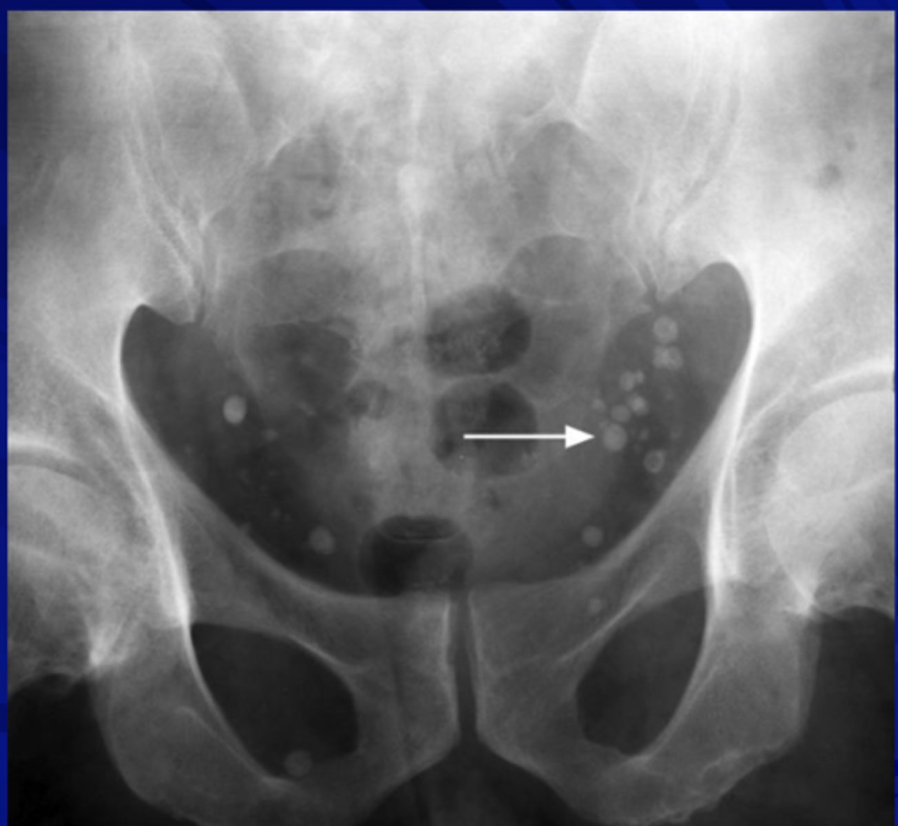
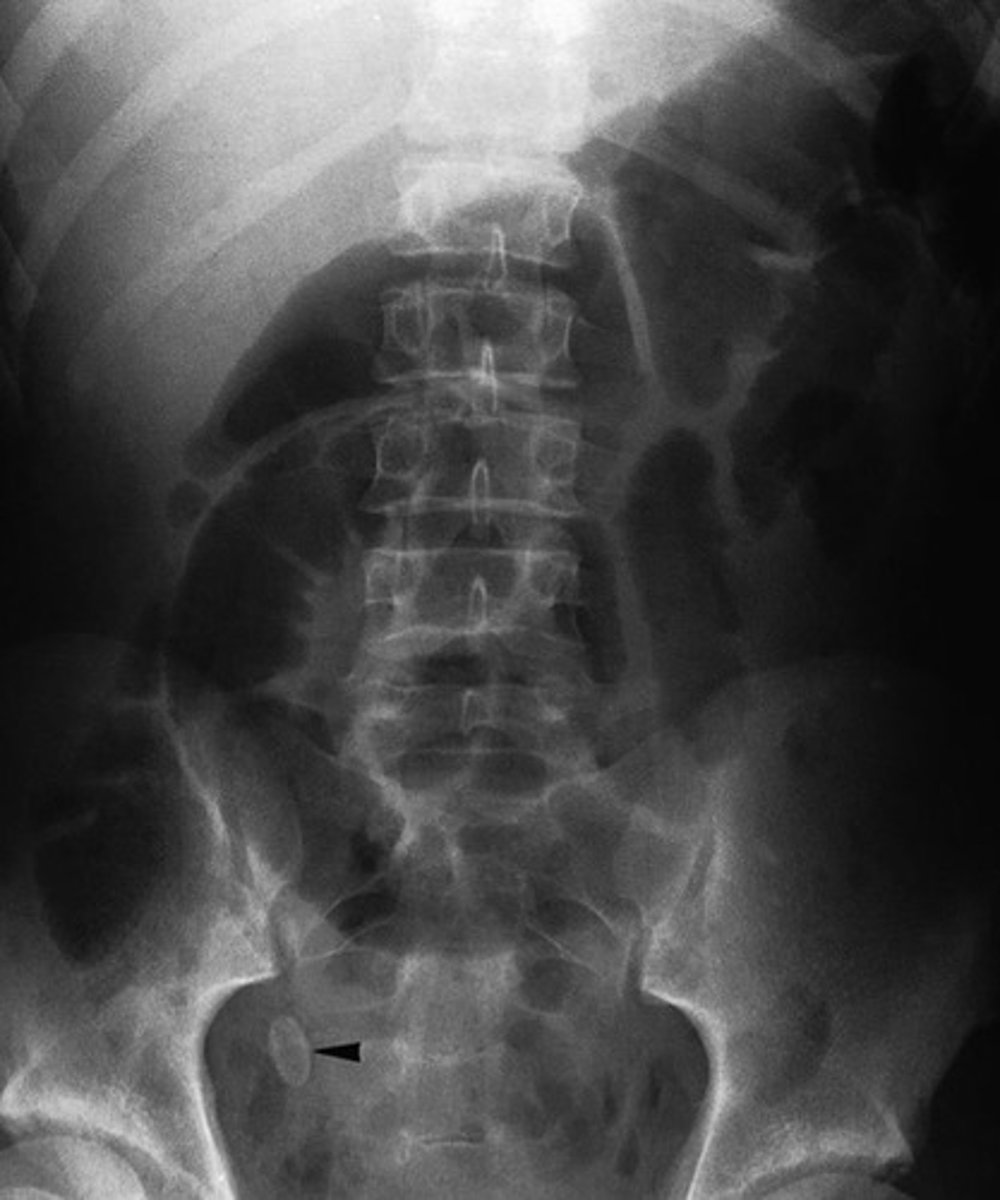
What do pelvic vein phleboliths look like on an X-ray?
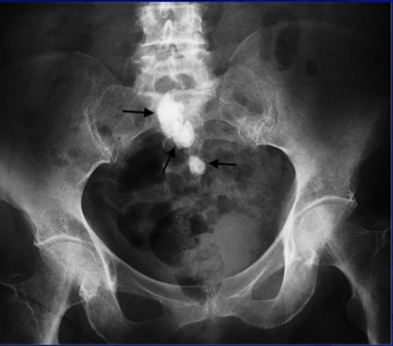
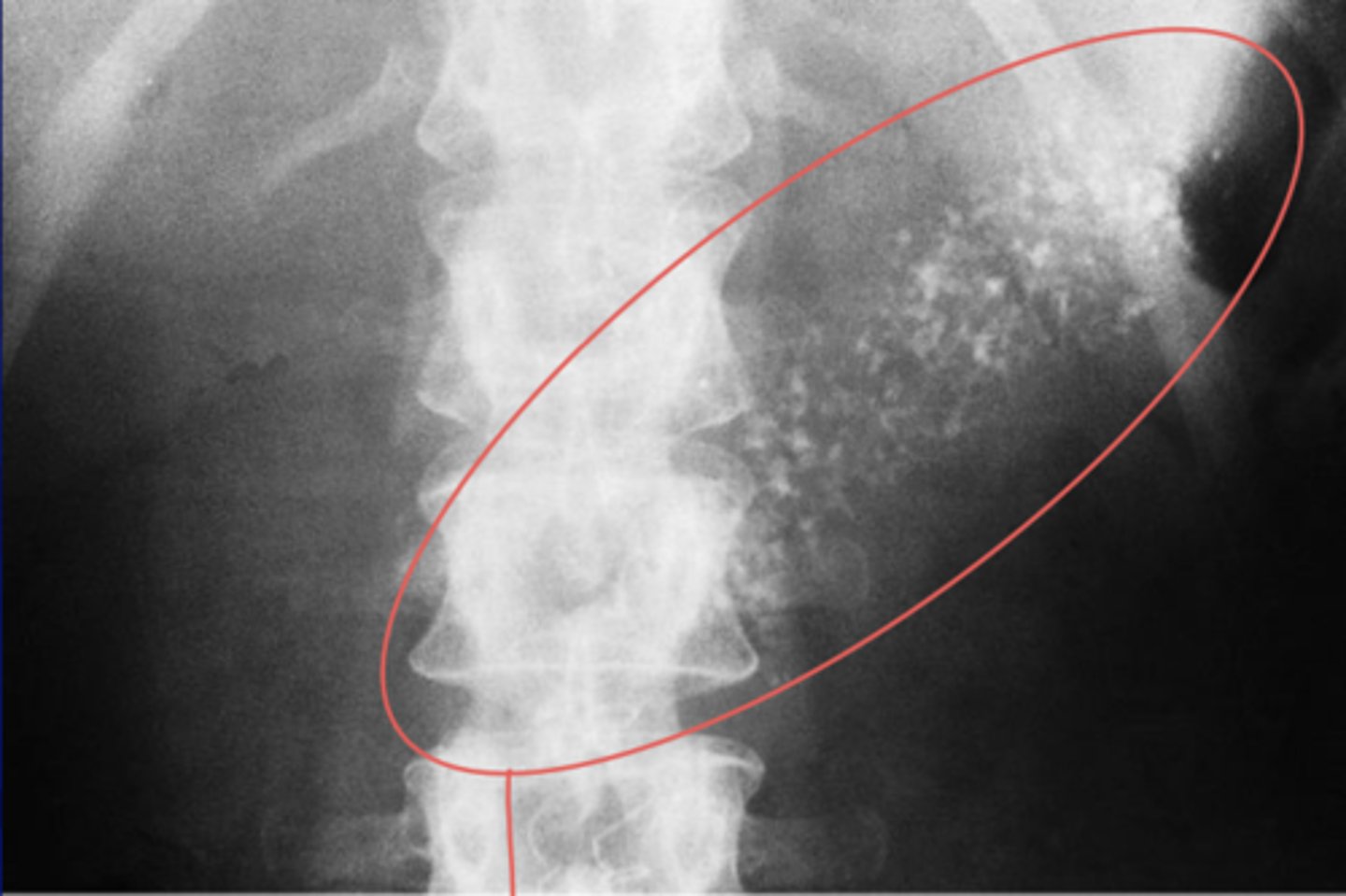
What do calcified mesenteric lymph nodes look like on an x-ray? And what can cause it
Tuberculosis
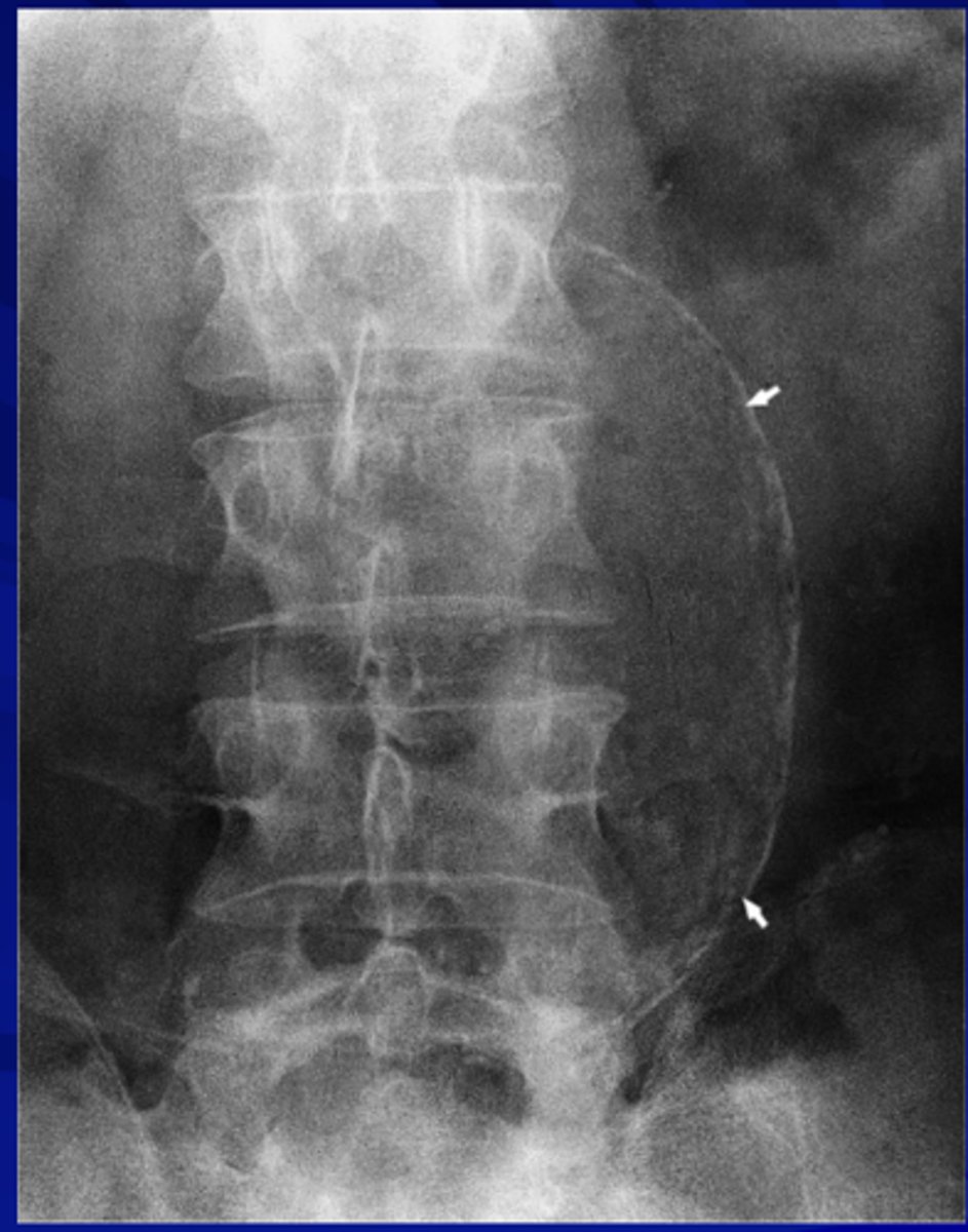
What does an aortic aneurysm with calcification look like on an x-ray?
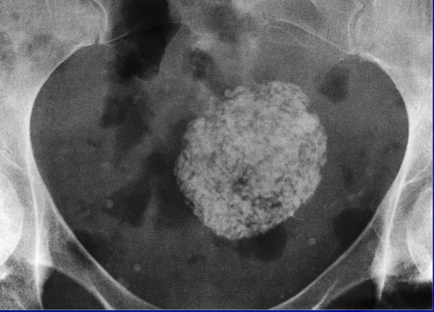
What does a uterine fibroid look like on an x-ray?
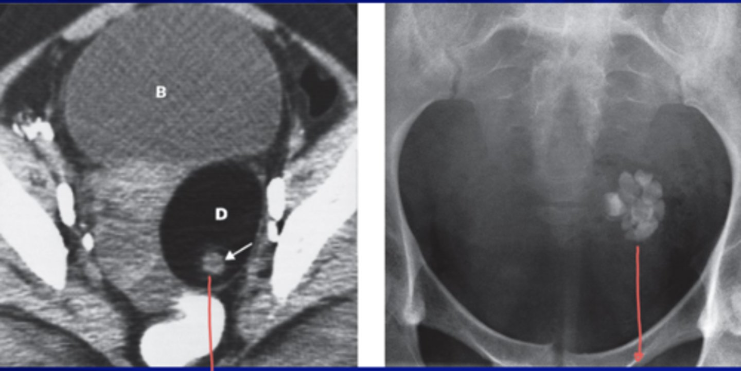
What does a dermoid cyst look like on an x-ray?
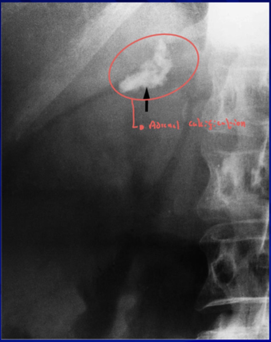
What does an adrenal calcification look like on an x-ray and what can cause it?
- Adrenal hemorrhage
- Tuberculosis
What do pancreatic stones look like on an x-ray?
What does an Appendiceal fecaliths look like on an x-ray and what can it cause?
What does an abdominal mass look like on an x-ray?
Pericardial effusion
-waterbag appearence
-greater than 250mL to dtx on radigraph
-visible on CT, but procedure of choice is ECHOcardiography
Constrictive pericarditis MC due to
-TB and Viral pyogenic infx
-Can also be due to radiation thx
-most will have pericardial calcification ± visible on CT others will have plueral effusion
Cardiomegaly causes?
-Valvular disease, cardiomyopahthy, congenital heart dz, pericaridal effusion, mass lesions
Symmetric englargement seen in?
-cardiomyopathy and pericardial effusion
Specific chamber enlargement seen in?
Congenital and valvular Dz
Dilated cardiomyopathy caused by
-ineffective contraction during systole
-MC from infection or metabolic d/o
-Collagen vascualr dz
-Toxic agents like alcohol* and chemo drugs (Doxorubicin, Adriamycin)
Heart measurement most reliable on?
Upright posteroanterior (PA) chest image to assess cardiac size
Left atrial Enlargement
-Isolated MC in mitral stenosis*
-Earliest sign displacement is esophagus posteriorly
-nL inferior carinal angle shouldnt be over 75 degrees
Rheumatic heart dz mostly efx
Mitral sometimes aortic valve
L atrial enlargement also seen in
congenital cardiac lesions w intracardiac shunts
pts who have L ventricular failure
Isolated R.ventricular enlargement
-difficult to appreciate b/c overlaps r atrium and l ventricle
On lateral view
both R ventricular and R atrial enlargment will cause filling in anterior clear space behind sternum
L ventricular enlargement due to?
-CAD
-Aortic stenosis
-Aortic regurgitation
LVH
difficult to detect bc may look normal but if suspected do Echo