The Brain Lab pt 1
1/53
There's no tags or description
Looks like no tags are added yet.
Name | Mastery | Learn | Test | Matching | Spaced | Call with Kai |
|---|
No analytics yet
Send a link to your students to track their progress
54 Terms
Cerebral cortex
the wrinkled, outermost layer of gray matter in the brain’s cerebrum
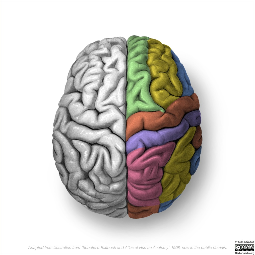
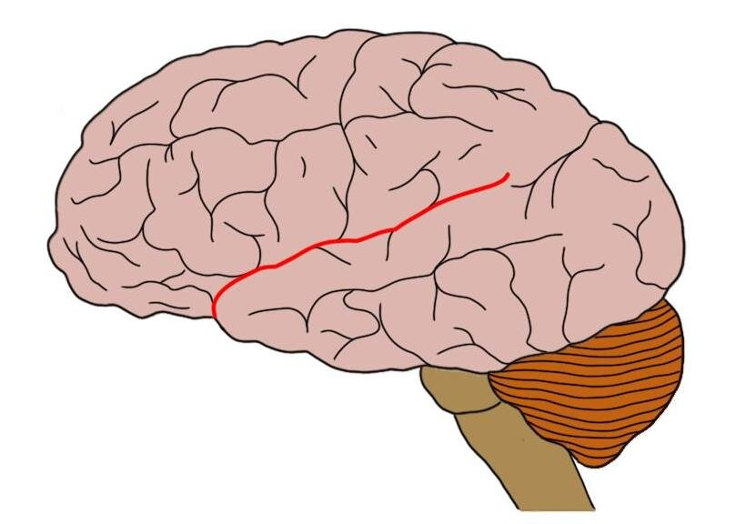
Motor area
Red line (long strip directly behind the frontal cortex)

Sensory area
The strip behind the motor area (yellow)

Frontal lobe
The brown bit, the front bit
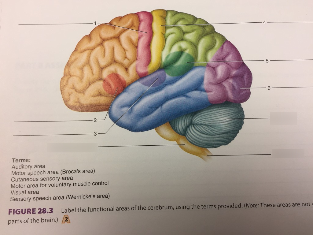
Temporal lobe
The blue bit, the part that is by the temporal bone

Parietal lobe
The part behind the sensory area
The green bit

Occipital lobe
Base of the brain
The purple bit

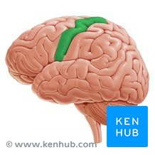
Central sulcus
the line between the motor and sensory areas
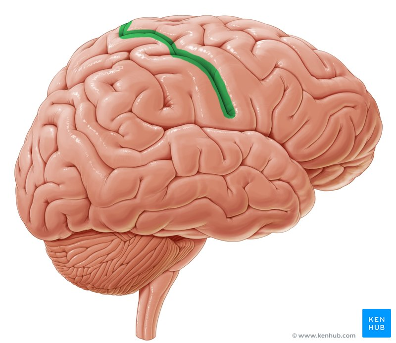
Precentral gyrus
literally the motor area
Postcentral gyrus
Literally the sensory area

Lateral cerebral sulcus (sylvian fissure)
Separates the parietal and frontal from the temporal
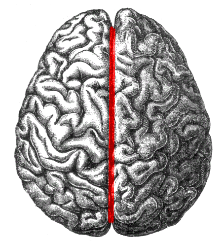
Longitudinal cerebral fissure
Midline of the brain, separates the right and left
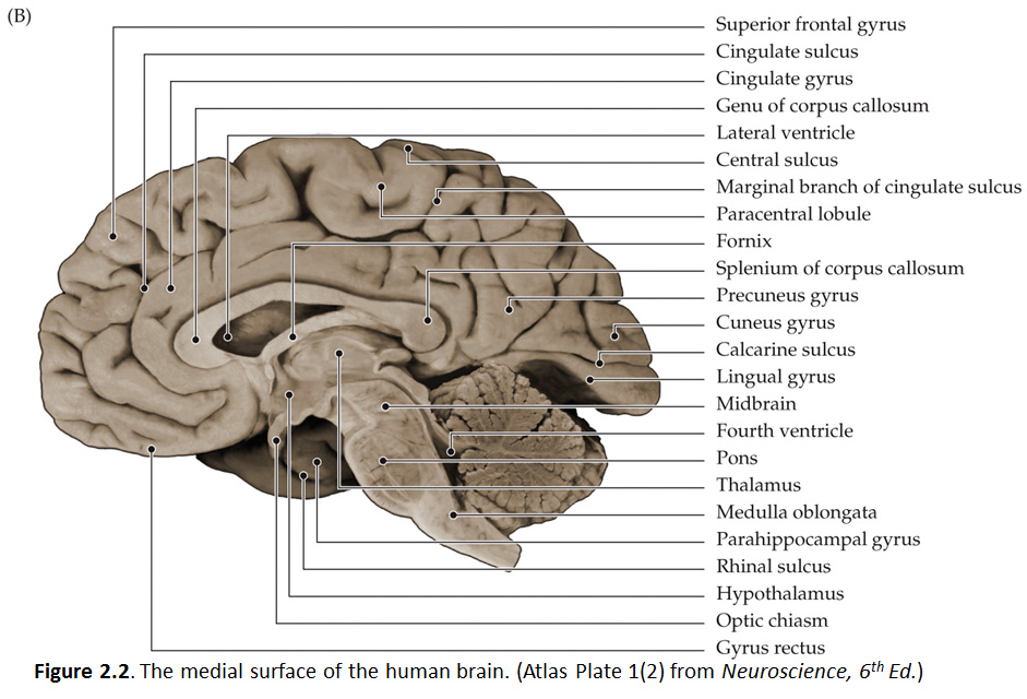
Medial side of the brain
The ___________ of the brain, viewed via a midsagittal cut, reveals inner structures connecting the two hemispheres and controlling vital functions
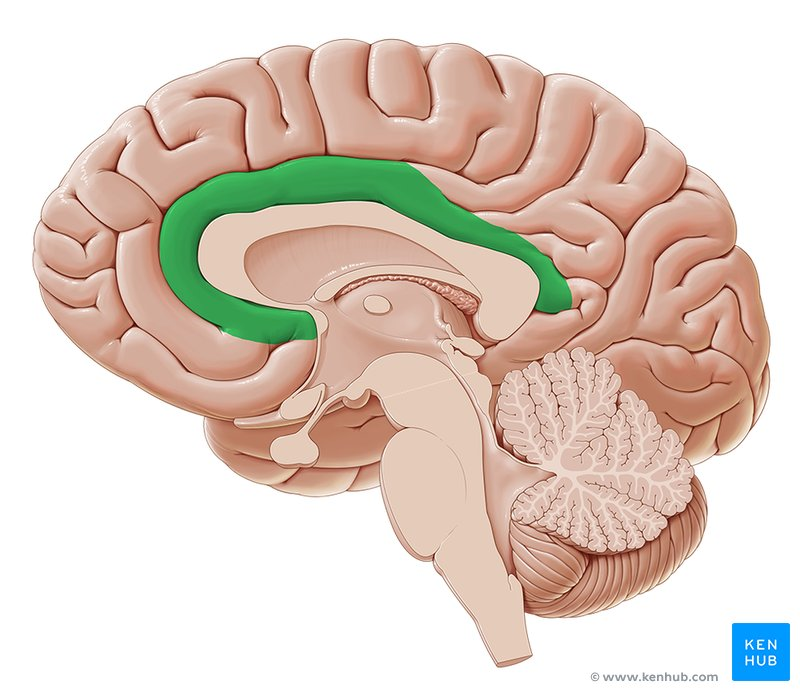
Cingulate gyrus
limbic cortex, the part under the brain surface
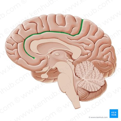
cingulate sulcus
the ridge above the cingulate gyrus
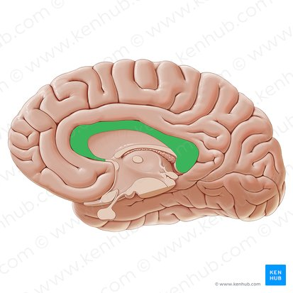
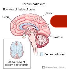
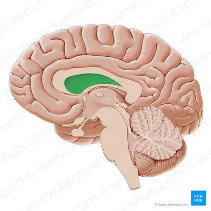
Corpus callosum
the long grooved curve under the limbic cortex
body of the corpus callosum
The more posterior long bit (basically just it fr)
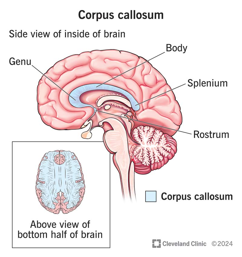
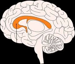
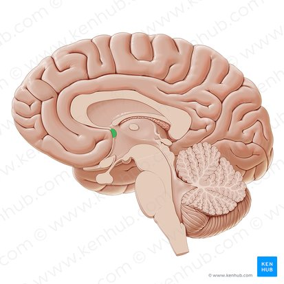
Genu of the corpus callosum
what the black arrow points to, the most anterior part of the corpus callosum
Splenium of corpus callosum
what the red is covering
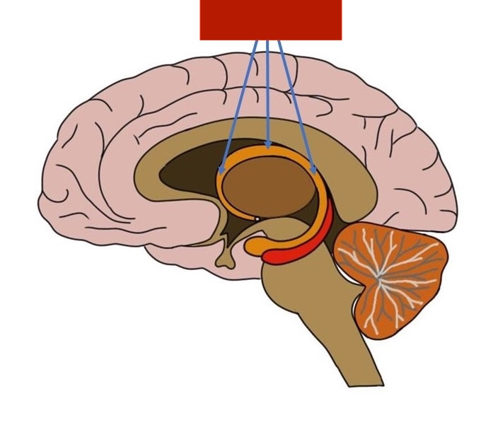
Fornix
the curved swirly bit underneath the corpus callosum
Septum pellucidum
the space above the fornix
Anterior commissure
Posterior commissure
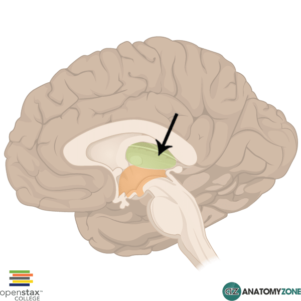
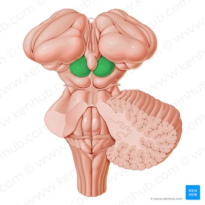
Thalamus
the whole most central structure below the fornix
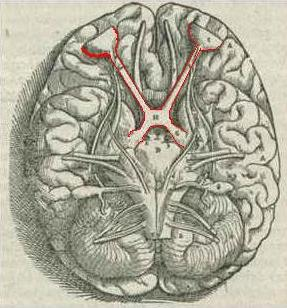
Optic chasm
x-shaped at the base of the brain, where optic nerves cross
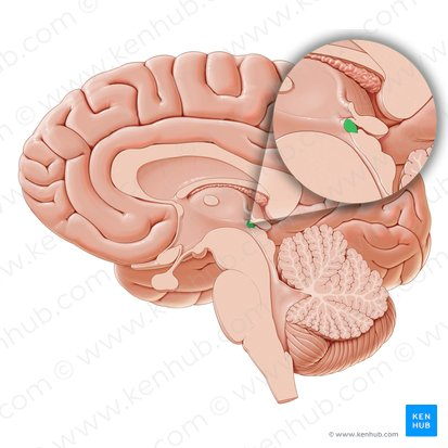
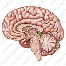
Pineal gland
the posterior pointy bump
Superior colliculus
bumpy bit in back that is above the cerebellum
Inferior colliculus
The bottom bump
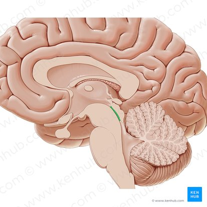
Sylvian aqueduct
The line beneath the inferior colliculus
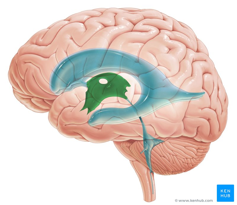
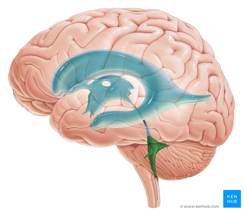
Third ventricle
Gap at the thalamus
Fourth ventricle
Between the pons and the cerebellum
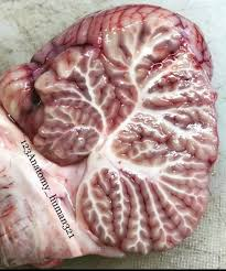
Arbor vitae
The little tree in the cerebellum
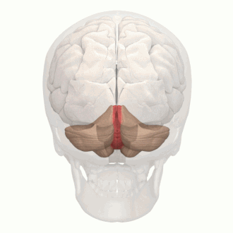
vermis of cerebellum
the middle of the two haves of the cerebellum
Medulla oblongata
The (purple) line
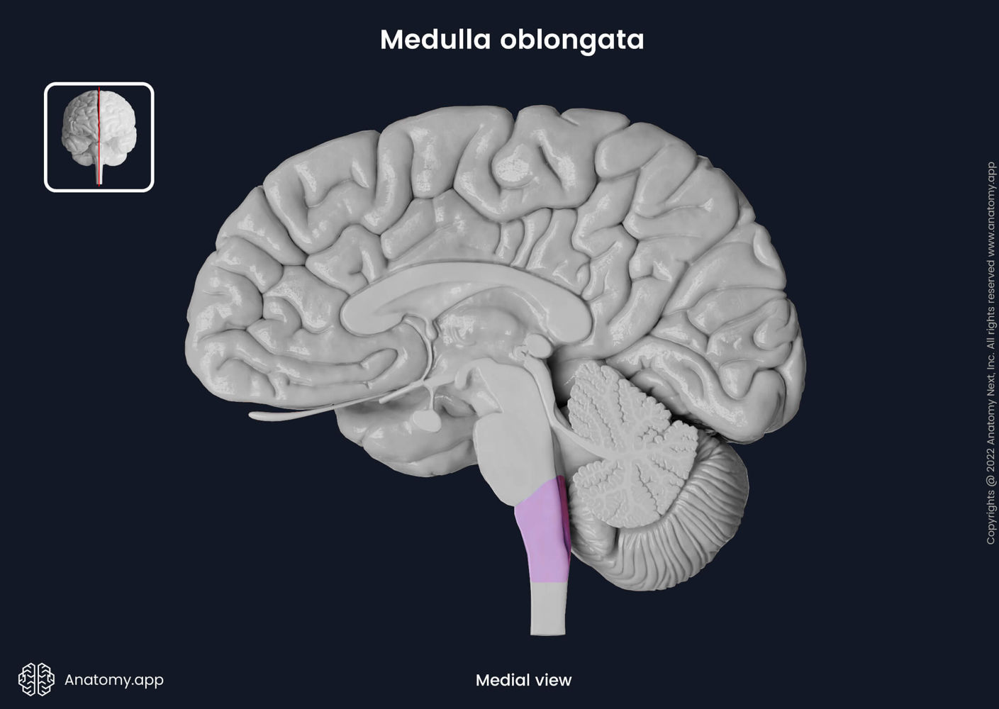
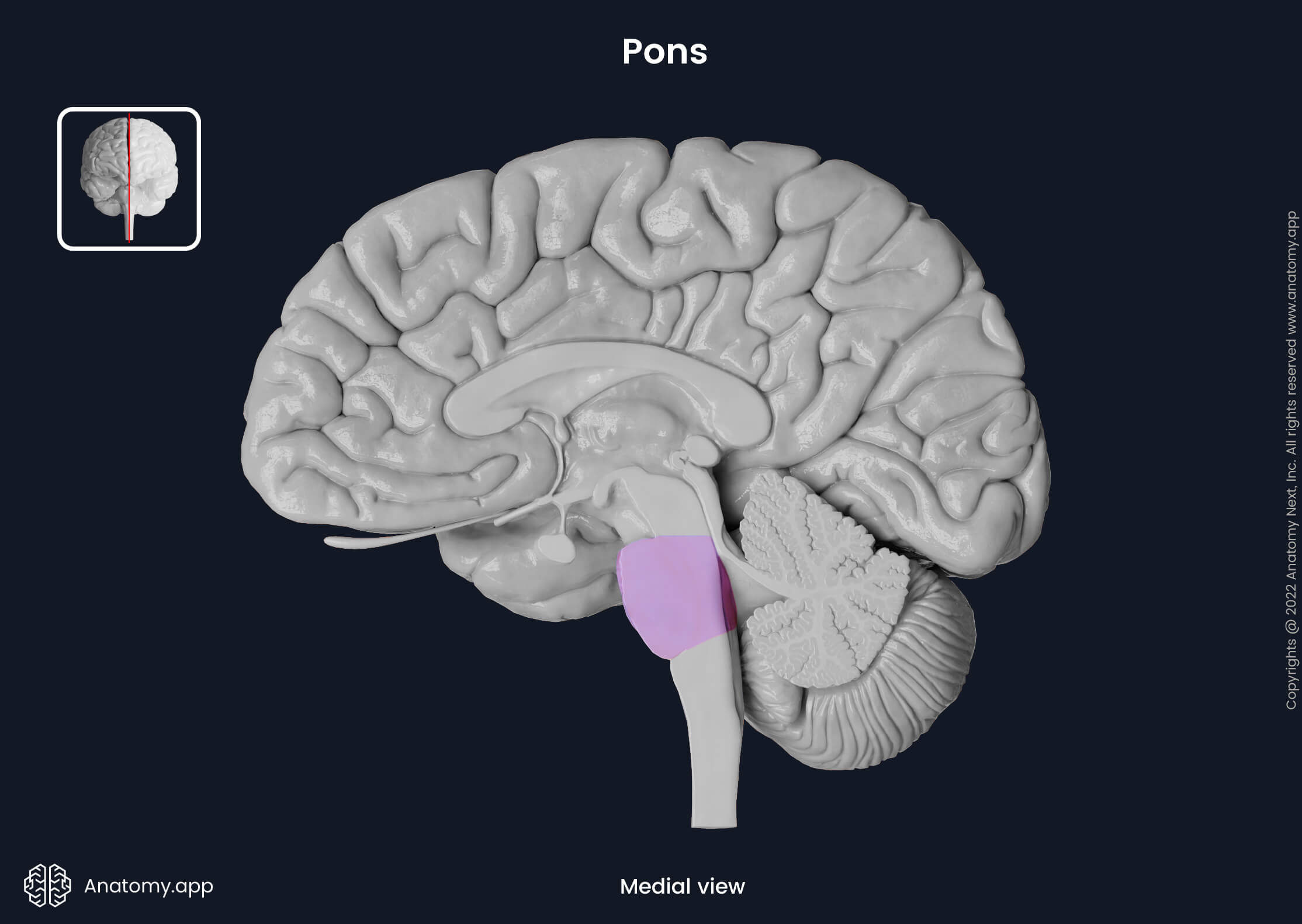
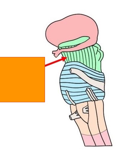
Pons
Blob at the top of the medulla oblongata
Cerebral peduncle
ridges between cerebellum
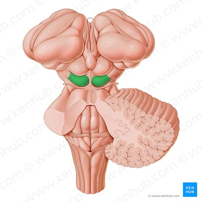
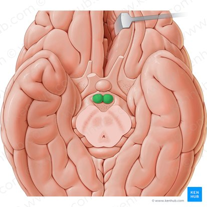
Mammillary body
two little bumps above the cerebellum
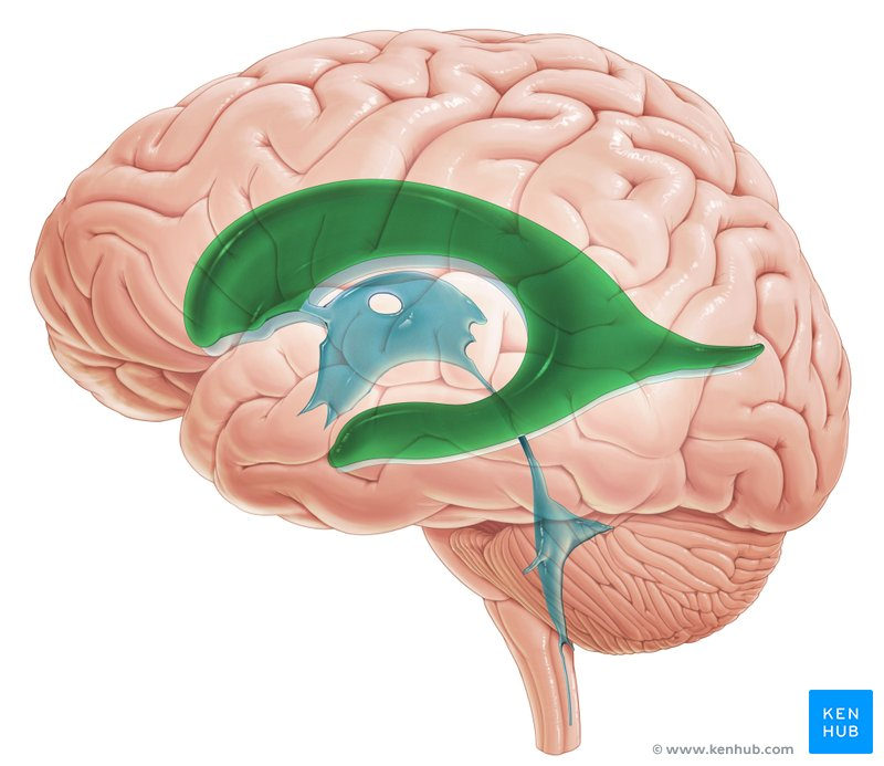
Lateral ventricles
space above the thalamus
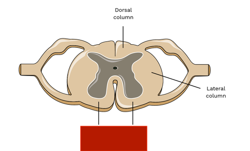
Dorsal column
white above the dorsal horn
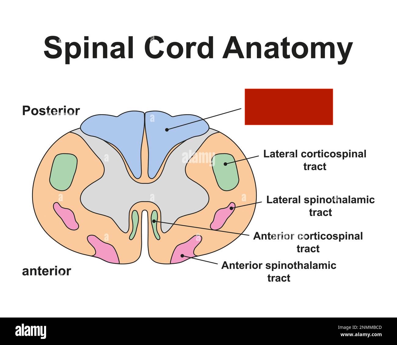
Lateral column
2 outer white parts
Ventral column
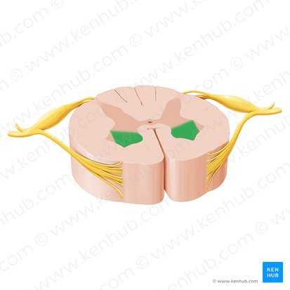
dorsal horn
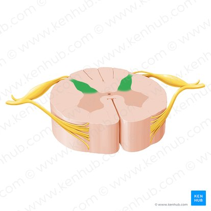
Ventral horn
Anterior horn
transverse commissure
line connecting the two halves
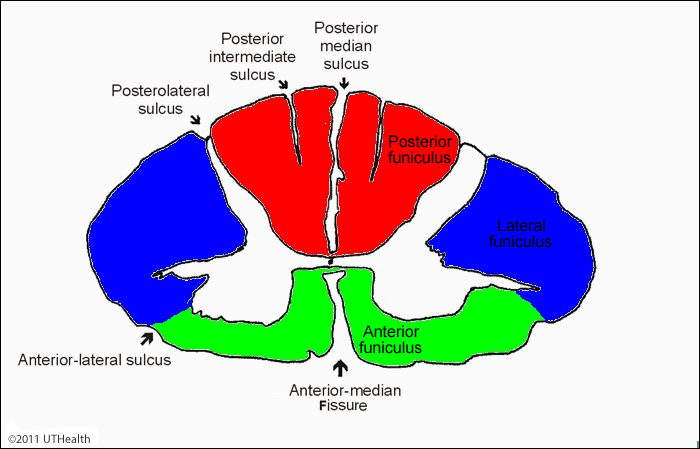
dorsal median sulcus
Red

Ventral median sulcus
Blue
Dorsolateral sulcus
Pink

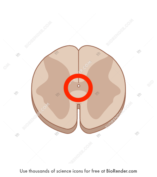
Central canal
hole in the very middle
ventrolateral sulcus
Antero-lateral sulcus
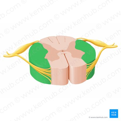
dorsal root
Green
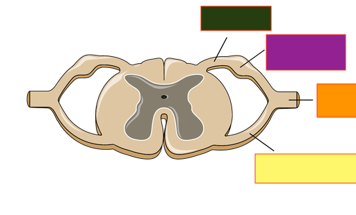
Ventral root
Yellow

Dorsal root ganglion
purple

Spinal nerve
orange
