Cerebral Hemi I + II
1/31
There's no tags or description
Looks like no tags are added yet.
Name | Mastery | Learn | Test | Matching | Spaced | Call with Kai |
|---|
No analytics yet
Send a link to your students to track their progress
32 Terms
Structure of the cerebral cortex
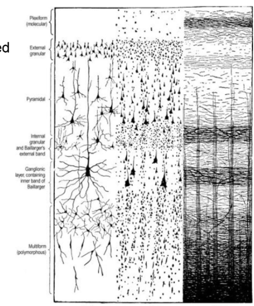
6 Layers of Neocortex (isocortex)
Structure of the cerebral cortex
~90% of the cerebral cortex consists of neocortex (remainder is phylogenetically older forms of cortex —> allocortex)
6 Layers of Neocortex (isocortex)
I - Molecular (plexiform) layer
II - Outer granular layer
III - Outer pyramidal layer
IV - Inner granular layer
V - Outer pyramidal layer
VI - Multiform layer
6 layers not uniform throughout
Primary motor and sensory areas provide the greatest contrast and are referred as hetereotypical.
Primary motor cortex (agranular cortex) —> dominated by pyramidal neurons
Primary sensory cortex (granular cortex) —→ dominated by smaller, stellate cells.
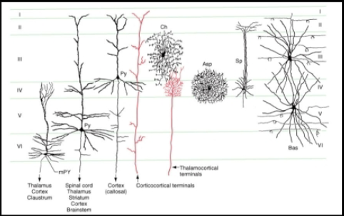
Cortical nuerons
Pyramidal
Intrinsic
Pyramidal
All players expect layer 1 (molecular); prominent in layers 2,3,5
Large apical dendrite —> extends towards molecular layer; Basal dendrites —> project horizontally
Major output pathway of the cerebral cortex (projection, association, and collosal fibers)
Giant pyramidal neurons of Betz (only motor cortex; in layer 5)
Intrinsic
Stellate (aspiny and spiny) neurons
Most numerous in layer 4 —> receives thalamacortical projections
Spiny stellate cells only excitatory interneurons (Glu)
Chandelier cells
Layer 3, dendrites in layer 4
Basket cells
Layer 3 and 5; dendrites in all layers
Cells of Martinotti
Found in deeper layers; multipolar neurons w/ short branching dendrites and an axon that projects to more superficial layers.
Note: Fusiform shaped modified pyramidal cells that project to thalamus are found in layer 6
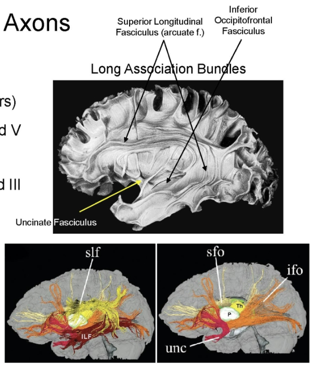
Axons
Intrahemispheric
Interhemispheric
Intrahemispheric (association fibers)
Long association (in layer 3 and 5) —> connects lobes together (distinct regions)
Short association (in layer 2 and 3) —> connect gyri together
Interhemispheric (Callosal fibers)
In layer 3; connects left and right hemispheres (corpus callosum) and temporal poles (anterior commissure).
Axons
Local intrinsic neuron
Corticofugal
Corticopetal
Local intrinsic neuron
Connects different layers together
Corticofugal
Goes to subcortical areas, brainstem, and spinal cord.
Types
Corticobulbar (layer 5 and 3)
Corticostriate and corticopontine (layer 5)
Corticothalamic (layer 6)
Corticopetal
From the thalamus (thalamocortical) to layer layer 5 (some to layer 3 and 6)
Noradrenergic, serotonergic, dopaminergic, cholinergic from other subcortical nuclei - diffuse inputs regulating cortical excitability
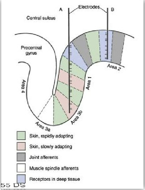
Cortical organization
A seconds vertical pattern of organization —> demonstrated physiologically
Exemplified via primary somatosensory cortex.
Cerebral cortex - Function areas
Structure and function of the cortex are heterogeneous, although it appears as homogenous sheets.
Structure (cytoarch.) differences are based on cortical thickness, width of individual layers, type, and number of cells per layer
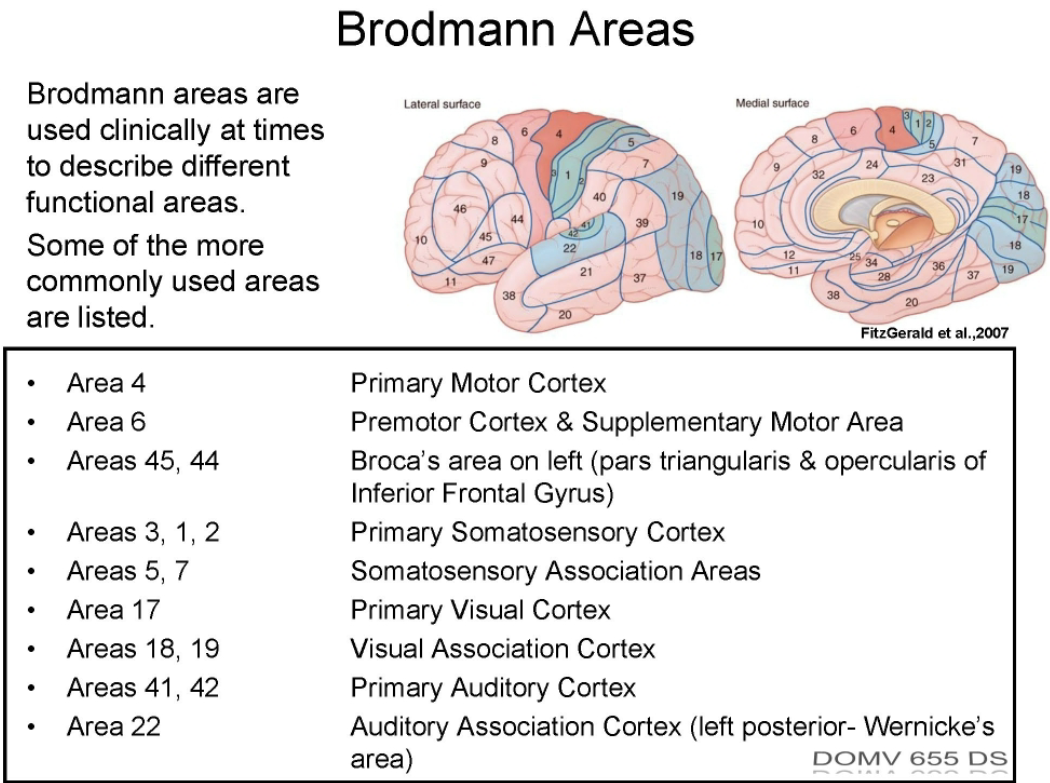
Korbinian Bordmann described 47 cytoarch. ares in man.
Brodmann’s areas used to describe function ares of cortex —> stoke and functional imaging have validity in these associations.
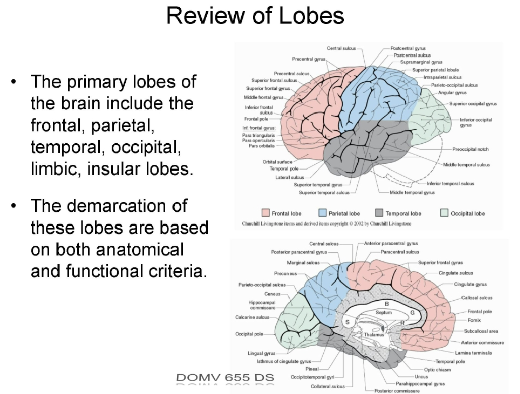
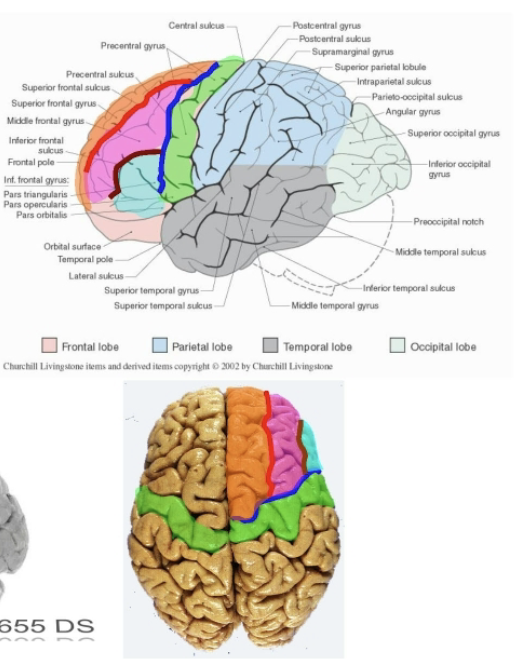
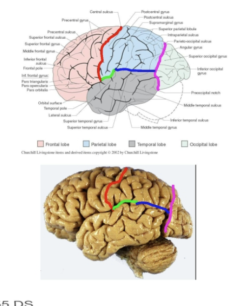
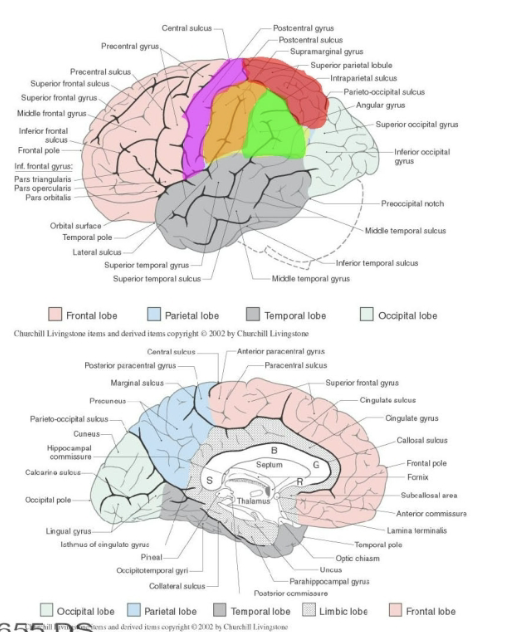
Review of lobes
Frontal lobe
Major functional components
Anatomical boundaries
Largest lobe in brain, has motor and cognitive functions
Major functional components:
Primary motor cortex
Supplemental motor areas
Frontal eye fields
Prefrontal cortex
Anatomical boundaries
Separated from the parietal lobes by the central sulcus and the temporal lobe by proximal portion of the lateral fissure.
Frontal lobe
Gyri and sulci
Primary gyri of the frontal lobe includes the precentral gyrus (primary motor cortex) and the superior, middle, and inferior frontal gyri.
Separated by the precentral sulcus and superior and inferior frontal sulci.
On medial surface, the precentral gyrus continues within the longitudinal fissure as the anterior paracentral gyrus (ant. portion of the paracentral lobule)
The superior frontal gyrus extends on the medial surface down to the cingulate sulcus (aka medial frontal gyrus)
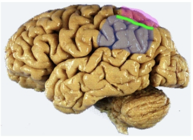
Frontal lobes
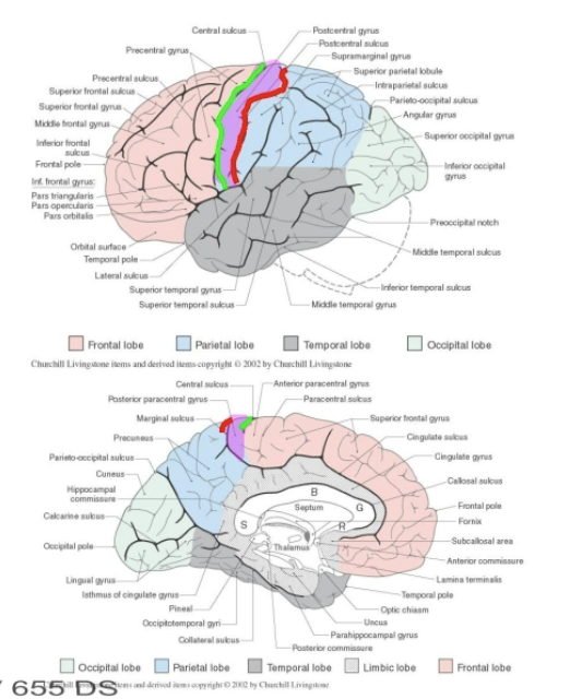
Paracentral lobule
Precentral gyrus
Paracentral lobule
Medial extension of both pre and post central gyri
Contains both primary motor (anterior) and primary sensory (posterior) functional areas.
Precentral gyrus
Part of the primary motor complex (broadmann area 4) —> major motor output register to spinal cord +brain stem
Somtatotopically organized
Legs located in anterior paracentral gyrus
Trunk, head, tongue located inferiorly and laterally
Tongue near lateral fissure
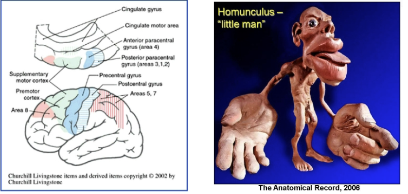
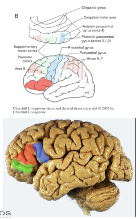
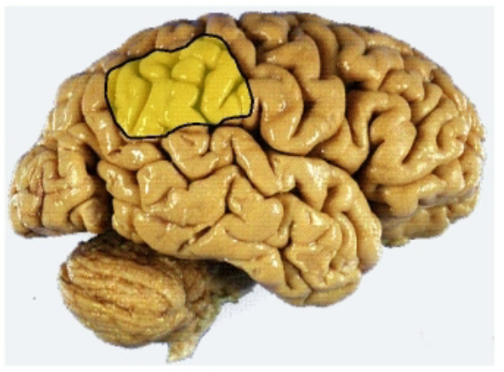
Supplementary Motor Areas
In front of the precentral gyrus —→ supplementary motor and premotor area related to planning of motor activities (area 6)
These areas communicate with area 4 and subcortical structures (basal ganglis, cerebellum, ect) in the planning of movements.
Frontal lobe
Functional area
Inferior frontal gyrus primary parts:
Anterior to premotor cortex in the frontal eye fields (area *)
These ares facilitate cortical (conscious) control of eye movements via connections to eye movments centers in brain.
Inferior frontal gyrus primary parts:
Pars opercularis (near lateral fissure)
pars trangularis
Pars orbitalis
Pars opercularis + trangularis correspond to Broca’s area (area 45,44), the motor area for speech in the dominant (usually left) hemisphere
These areas connect the brainstem nuclei for CN that control the motor output for speech.
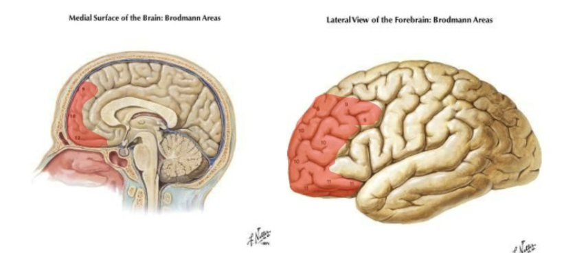
Prefrontal cortex
Much of the remainder of the frontal lobe (prefrontal lobe) classified as multimodal associational with a diverse cognitive functions
Judgment, foresight, a sense of purpose, responsibility, and social propriety.
Contains ~25 percent of the entire cortex of the human brain (primary broadmann areas 9-12)
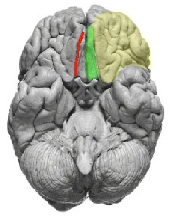
Frontal lobe
Ventral aspect
Ventrally, the prefrontal areas extend into the orbitofrontal gyri
The olfactory bulb and tract lie in the olfactory sulcus, forming the medial boundary of the orbitofrontal gyri.
Most medially, next to the longitudinal fissure is the gyrus rectus (extension of the medial aspect of the superior frontal gyrus)
Parietal lobes
Contents
Boundaries
Sensory and multimodal associative function
Contents
Primary somatosensory cortex (postcentral gyrus)
Sensory association areas w/ functions including understanding written language (usually left hemisphere)
Boundaries
Laterally, parietal lobes lie posterior to the central sulcus and anterior to the extension of the pariteo-occiptal sulcus to the pre-occipital notch.
Above the lateral fissure and a line between approx. the middle of the lateral fissure and the extension of the parieto-occipital sulcus.
Parietal lobes
Primary components
Primary components
primary somatosensory cortex (postcentral gyrus)
superior parietal lobe
inferior parietal lobe (supramarginal gyrus, angular gyrus)
Superior and inferior parietal lobules are separated by a intraparietal sulcus
Parietal lobes
Postcentral gyrus contents and location
Lies behind the central sulcus and anterior to the postcentral sulcus.
Contents
Primary somatosensory cortex, (SI-broadmann areas 3,1,2) on lateral surface of the hemisphere.
Posterior paracentral lobule on the medial surface of the hemisphere.
Parietal lobes
Somatotopic organization
Secondary somatosensory cortex (SII); location and features
Simular to motor cortex
Sensory areas from genitals, foot and leg are on the medial hemisphere in the post paracentral gyrus.
Tongue is located laterally
Neurons in postcentral gyrus respond to modality specific stimuli of discriminative touch, vibration, position, pain, and temperature.
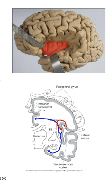
Secondary somatosensory cortex (SII) is located on the medial surface of the parietal operculum.
Has an additional crude somatosensory map
SII receives projections from SI and is also interconnected with SII of the opp. hemisphere.
Unlike SI, neurons in SII have larger bilateral receptive fields.
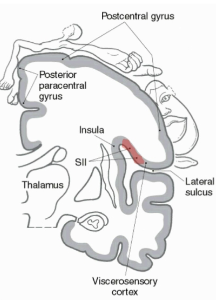
Insula
SII areas project top insular cortex —> distributes to limbic areas (tactile learning and memory)
The insula contains long and short insular gyri surrounded by boarders of the lateral fissure (circular sulcus).
Viscerosensory input (including taste) projects to the dorsal insula.
The anterior insula (near motor cortex) may also help to coordinate articulatory movements necessary for speech.
Parietal lobes
Superior and inferior parietal lobules; what are they separated by and associated areas
Superior and inferior lobules
Separated by the intraparietal sulcus.
Superior lobule (broadmann areas 5 and 7) integrates somatosensory input from multiple modalities —→ used in motor planning (kinesthetic sense, hand eye, coordination)
Project to supplementary motor areas in the frontal lobe.
Parietal lobes
Inferior parietal lobule
Angular gyrus
Supramarginal gyrus
The inferior parietal lobule contains the angular gyrus (area 39) and supra marginal gyrus *area 40)
General functions of this areas differ with the hemisphere involved. In the dominant hemisphere, the angular gyrus is a center for comprehension of written language.
Areas of the supramarginal gyrus and posterior superior temporal gyrus (Wernickes ares, broadmann areas 22) is the areas for comprehension of spoken language.
Parietal lobes
hemineglect syndrome
In the non dominant hemisphere the inferior parietal lobule modulates attention to stimuli both on the body and in the visual field (perceptrertual awareness)
Lesions to this areas are associated with hemineglect syndrome (failure to recognize the left side of the body as self).
Occipital lobes
Contents
Occipital lobes: visual function
Contains
Primary visual cortex and causal association (extrastriate) cortex
The primary identified guru are associated with the medial aspect of the hemisphere.
Occipital lobes
calcarine sulcus
extra striate cortex
Medially, the usually well defined parieto-occiptal sulcus separates the parietal and occipital lobes.
The calcarine sulcus divides the medial occipital lobe into the cunceus above and lingual gyrus below.
Remaining areas of the occipital lobe (extra striate cortex, areas 18 and 19) ae involved in the processing of visual data leading to the perception of motion, depth (binocular vision), color, and position of an object.
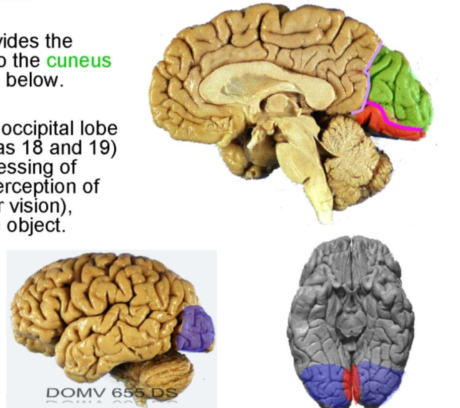
Occipital lobes
Primary visual (area 17) cortex
Primary visual (area 17) cortex is not he medial side of her ocicitla lobe on either side of the calacarine sulcus.
The retinal surface (therefore the visual field) is represented in a topographic (retinotopic) fashion int he areas around the calcimine sulcus.
Temporal lobes
Contents
Temporal lobes: integrative sensory, some memory, auditory, and olfactory function.
Contains
Primary auditory cortex
Nearby Wernickes areas that coordinates the understanding of spoke. language
Limbic areas including the hippocampus.
Temporal lobes
Basic anatomy
On the lateral surface the temporal lobe is composed of:
Superior, middle, and inferior temporal gyri
Separated but the superior and inferior temporal sulci.
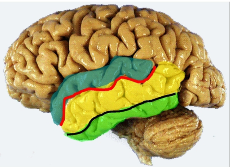
Temoral lobe
Inferior
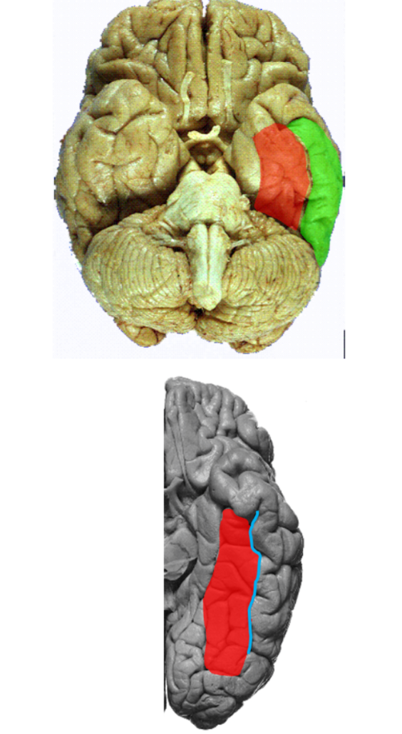
The occipitotemporal gyrus (fusiform gyrus) is located on the ventral aspect of the hemisphere medially adjacent to the inferior temporal gurus.
The occipitotemporal sulcus operates these structures.
These areas + adjacent areas of the ocecitpal lobe area involved in the recognition of objects and face.
Primary auditory cortex
transverse temporal gyrus
Function
Within the lateral fissure, the superior surface of the temporal lobe contains the transverse temporal gyrus (of Heschl, areas 41 and 42)
The primary auditory cortex is responsible for audition, receiving information. from both ears
Together with the immediately surrounding association cortex, its is generally involved in the process the association and recognition of sounds.
Limbic lobe
Anatomy
Limbic lobe
Defining sulci
Hippocampus and cingulate gyrus