cell structure combined: introduction to cell structure , domain bacteria, domain archaea, domain eukarya
1/184
There's no tags or description
Looks like no tags are added yet.
Name | Mastery | Learn | Test | Matching | Spaced |
|---|
No study sessions yet.
185 Terms
characteristics of all cells
-cell membrane
-cytoplasm
-ribosomes
-enzymes
-RNA
-DNA
two prokaryote domains
bacteria and archaea
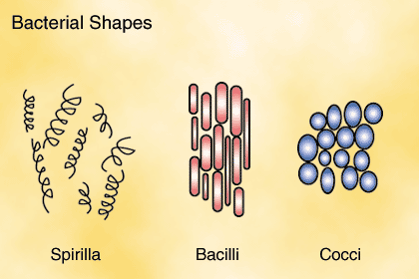
major cell morphologies
-coccus (spherical or ovoid)
-bacillus (rod/cylindrical)
-spirillum (spiral)
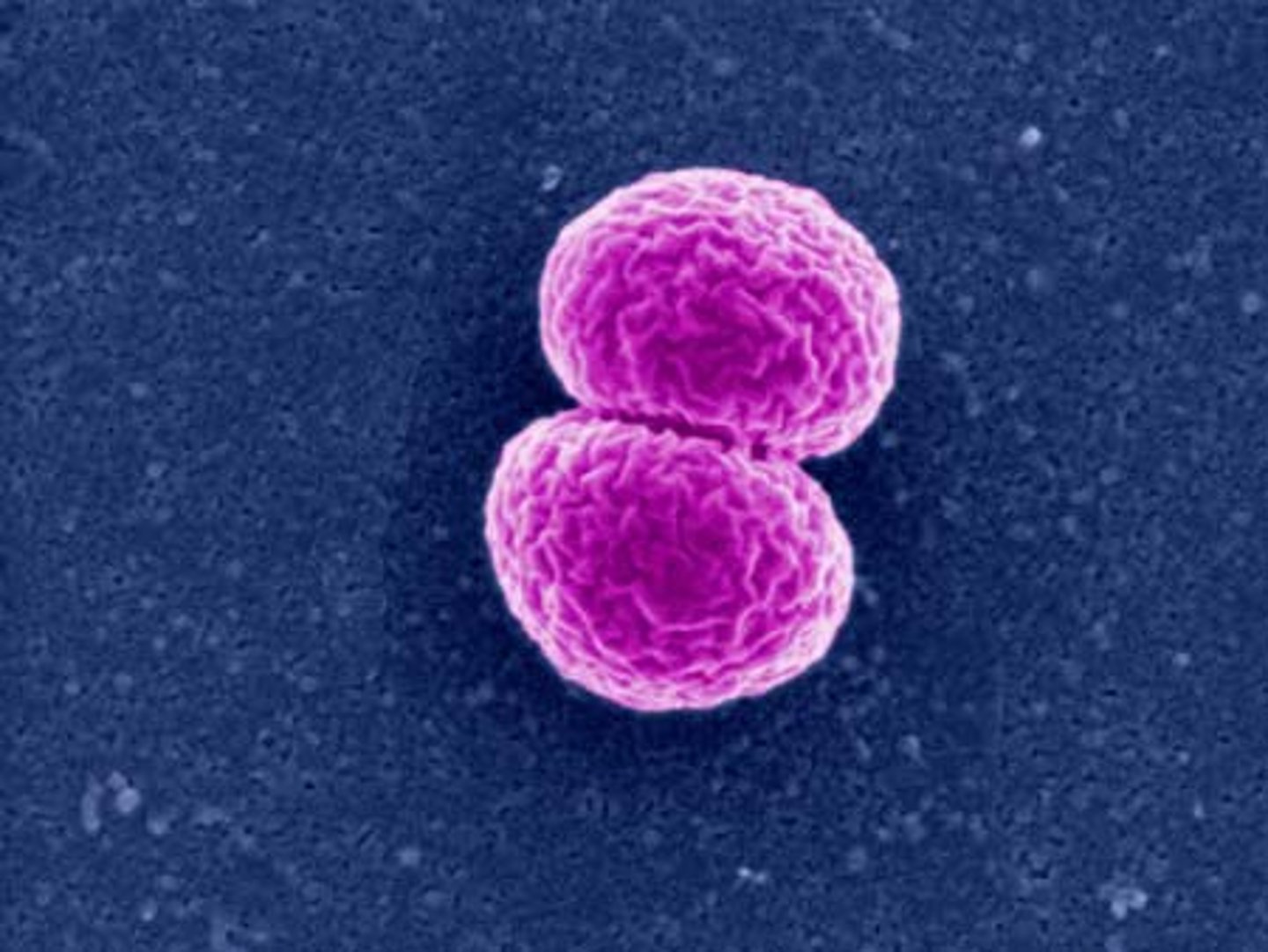
diplococci
pairs of cocci
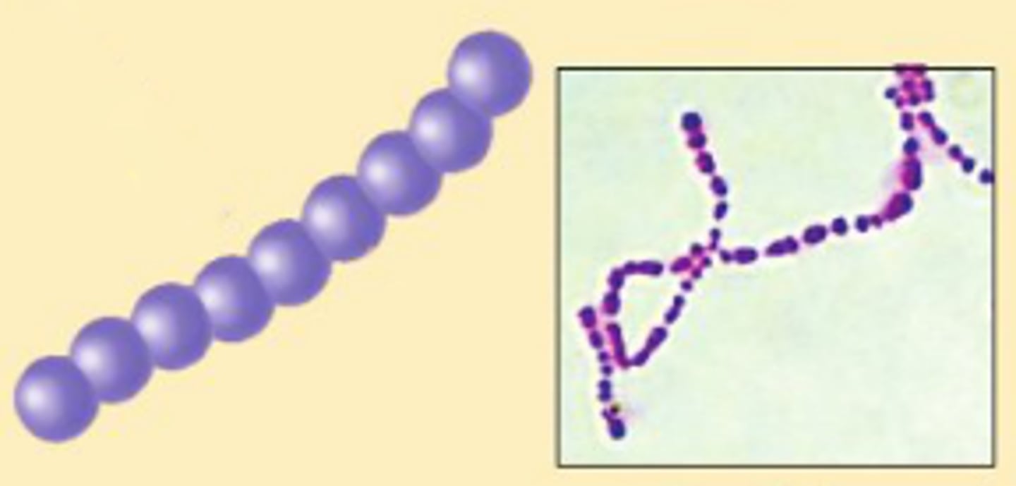
streptococci
chains of cocci
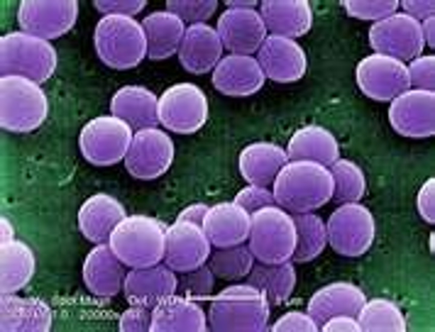
staphylococci
grape-like clusters of cocci
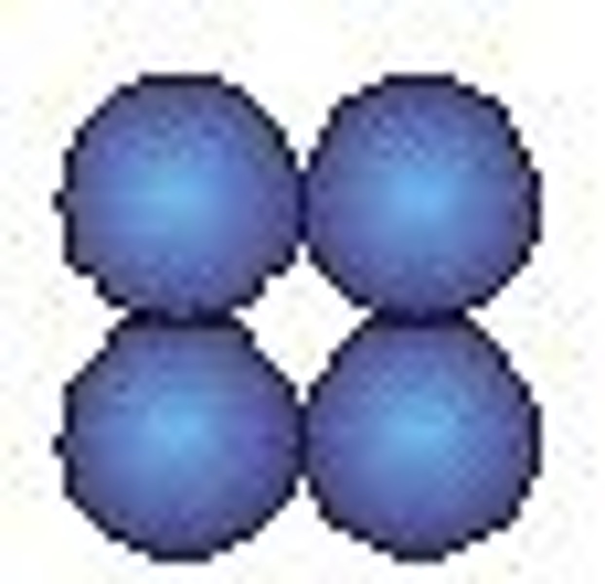
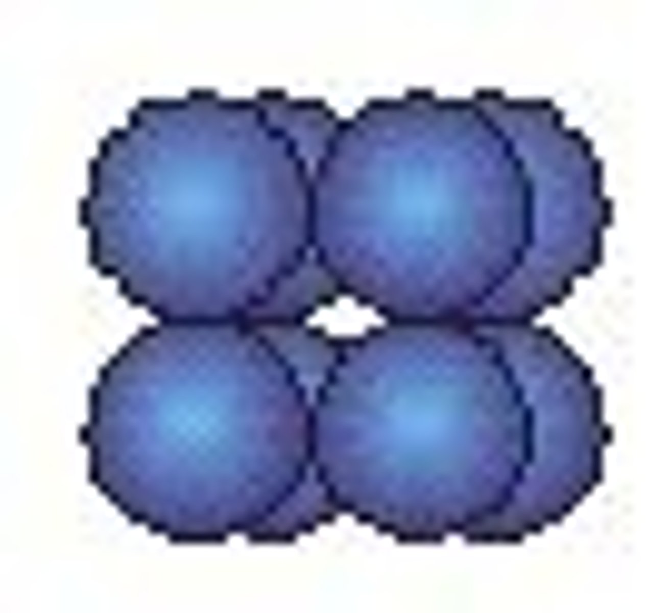
cocci tetrads
4 cocci in a square
sarcinae cocci
cubic configuration of 8 cocci all perpendicular to each other
diplobacilli
pairs of bacilli
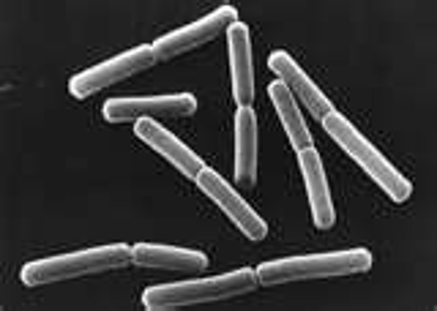
streptobacilli
chains of bacilli
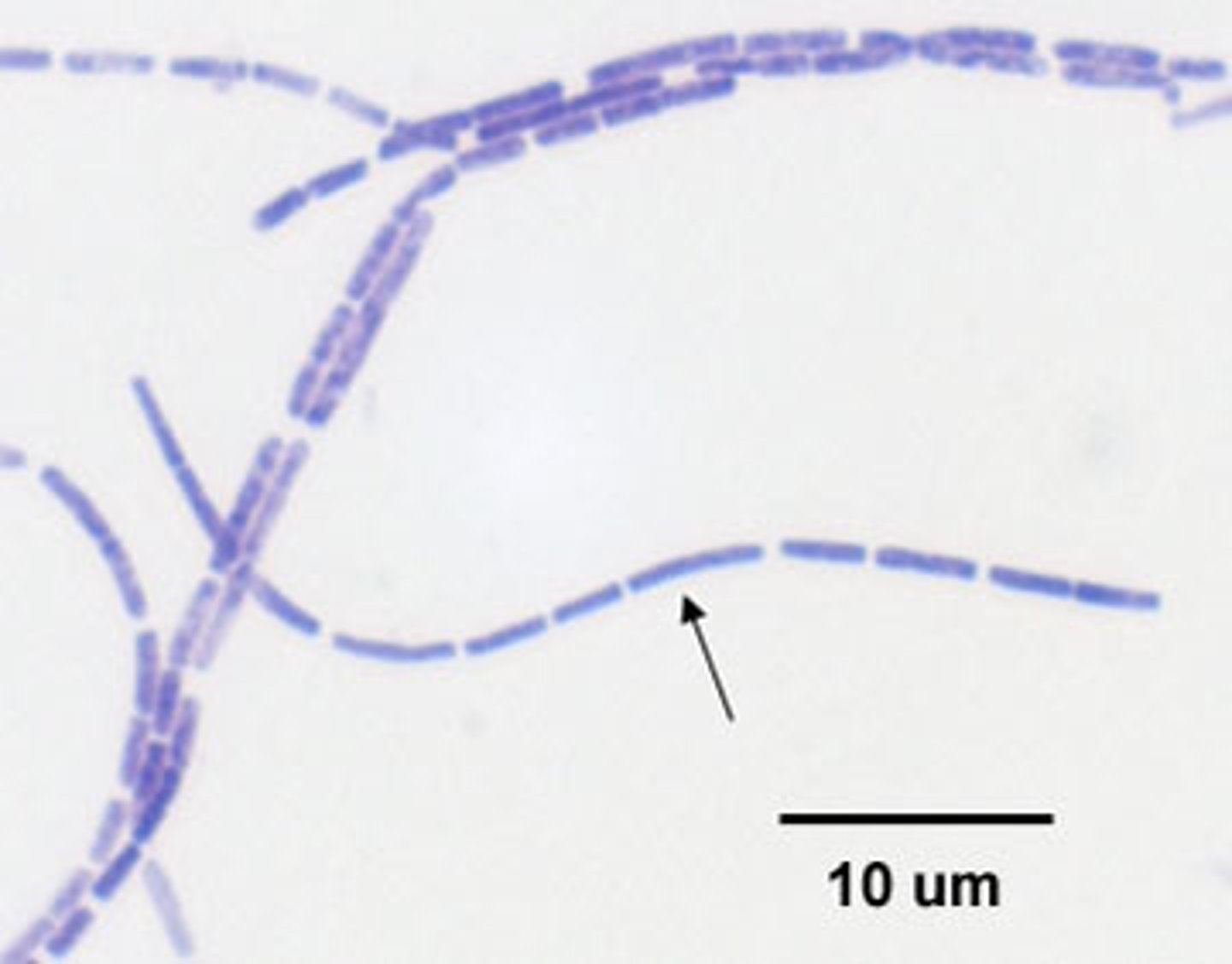
palisade bacilli
several parallel cells along a long axis

coccobacilli
very short rods
vibrios
resemble rods, comma shaped
spirilla
rigid helices
spirochetes
flexible helices
mycelium
network of long, filamentous cells
pleomorphic
variable shapes
archaea
unique shapes (branched flat square)
prokaryote size
0.2 um-700um diameter
eukaryote size
10 um-200 um
surface to volume ratio
as volume increases the SA:V ratio decreases
advantages to being small
- higher surface to volume ratio
- nutrients and waste can be transferred into and out of the cell more easily
- higher metabolic rate
- supports faster growth rate, faster evolution
cell membrane functions for all domains
-requirement for all organisms
-separates cytoplasm from environment
-regular transport
-energy metabolism (prokaryotes)
-protein attachment
-receptors
fluid mosaic model of membrane structure
membrane somewhat fluid, somewhat solid (liquid crystal)
basic structure: lipid bilayer with floating proteins
phospolipid bilayer
double layer of phospholipids that have a hydrophilic head and two hydrophobic tails
peripheral proteins
-loosely connected to membrane proteins on cytoplasmic side
integral proteins
-amphipathic
-embedded withing membrane
-project outward or inward
transmembrane proteins
completely cross membrane
membrane strengthening agents
sterols and hopanoids
sterols
-rigid lips that strengthen and stabilize membranes
-all eukaryotes
hopanoids
-structurally similar to sterols
-present in membranes of many Bacteria
membrane fluidity
temperature dependent
too cold: solidification (gelling)- due to vanderwaals forces
too hot: thermal lysis (cell death)
how to maintain correct membrane fluidity
-adjust fatty acid composition
cold: more unsaturated fatty acids which minimize van der Waals forces
hot: more saturated fatty acids which maximize van der Waals forces
-adjust ratio of sterols, hopanoids, or other lipids
transport
how a molecule gets across a membrane
depends on size, shape, charge of molecule
passive transport
transport which does not require cellular energy
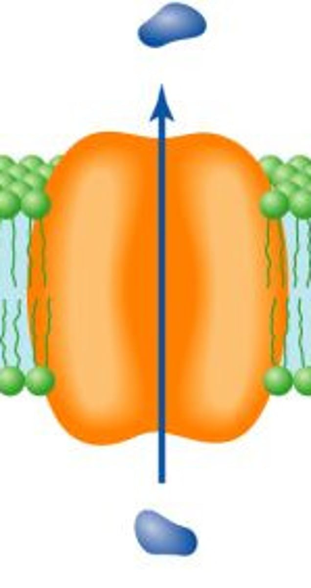
simple diffusion
Movement of molecules from high to low concentration
Non polar molecules, water
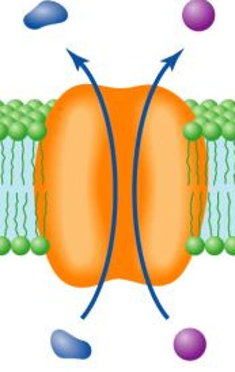
facilitated diffusion
transport which requires a transport protein
ex: ions and polar molecules
osmosis
diffusion of water across a membrane
active transport
transport which requires the cell to expend energy
always protein mediated
moves molecules against concentration gradient
uniporters
transport one types of molecules in one direction across the membrane
symporters
function as co-transporters of 2 or more molecules in the same direction
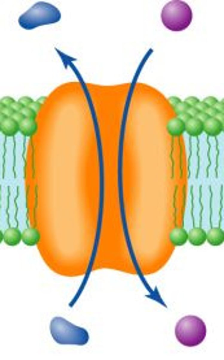
antiporters
transport a molecule across the membrane while simultaneously transporting another molecule in the opposite direction
cell net charge
negative
receptor-mediated transport system
ligand binds specific protein receptors on cell surface
-show saturation effect
-highly specific (lock and key)
group translocation
simultaneous transport and chemical modification of transported substance (only prokaryotes)
osmotic lysis
pressure from water entering cell causes a rupture of the cell membrane and death of the cell
tonicity
solute concentration of a solution
isotonic
solute concentration on both sides of membrane are equal
hypertonic
higher solute concentration therefore, lower water concentration
(cells want to be this!!)
hypotonic
means lower solute concentration therefore, higher water concentration
hypertonic environment on the outside of the cell
-solute concentration higher on outside
-water LEAVES cell
-plasmolysis occurs
hypotonic environment on the outside of the cell
-solute concentration lower on outside
-water ENTERS cell & cell swells
-cell wall protects from lysis
water moves towards...
higher solute concentration
plasmolysis
cell shrinks and cell membrane pulls away from wall
cells without cell walls
-genus mycoplasma
-cell membrane is stronger (more sterols)
-stays slightly hypertonic
gram positive
-stain purple
-thick layer of peptidoglycan
gram negative
-stain pink
-thin layer of peptidoglycan and outer membrane
peptidoglycan
the structural polysaccharide in cell walls of bacteria
peptidoglycan structure
-mesh-like
-identical subunits
-strands are composed of structural polysaccharides with amino acids
peptidoglycan subunits
-disaccharide of two alternating modified sugars joined by beta glycosidic bonds
peptidoglycan strands
-helical shape
-crosslinked by covalent bonds
-in gram positive, there is an inter-bridge of additional amino acids
-in gram negative, crosslinks are directly between amino acids
gram positive cell wall
-90% peptidoglycan
-contain teichoic acids
-layer of proteins on surface
-lipoteichoic acid
-mycolic acid
teichoic acids
negatively charged
functions: protection from environment binding to host cell storage of PO4
lipoteichoic acids
anchors wall to membrane
mycolic acids
found in acid fast bacteria ex. mycobacterium
periplasmic space of gram positive bacteria
-lies between cell membrane and cell wall
-smaller than gram negative
-secretes enzymes called exoenzymes
exoenzymes
produced by enzymes in periplasmic space of gram positive bacteria
aid in degradation of large nutrients
gram negative cell walls
-more complex
-5-10% peptidoglycan
-thin layer of peptidoglycan surrounded by an outer membrane
-no teichoic acid
-periplasmic space much larger
lipopolysaccharide (LPS)
3 parts: lipid A, core polysaccharide, O side chain
importance of LPS
-contributes to negative charge
-protection from viruses
-act as endotoxin
periplasmic space of gram negative bacteria
-may constitute 20-40% of cell volume
-many enzymes present in periplasm (hydrolytic enzymes, transport proteins, and other proteins)
glycocalyx
polysaccharide rich material exterior to cell wall which as a capsule and slime layer
glycocalyx functions
-attachment to solid surfaces
-protection
S-layer
-regularly structured layers of protein or glycoprotein that self-assemble
-in gram negative -> adheres to outer membrane
-in gram positive -> associated with peptidoglycan
s-layer functions
-protect from ion and pH fluctuations, osmotic stress, and enzymes
capsule layer
a glycocalyx that is highly organized, tightly attached to cell wall
slime layer
a glycocalyx that is unorganized and loosely attached to the cell wall
bacteria
single-celled organisms that lack a nucleus prokaryotes
bacteria cell size
0.2 um - > 700 um
what are the major cell shapes?
cocci, bacilli, spirillum
external structures beyond the cell envelop
fimbriae, pili, and flagella
functions of external structures
protection, attachment to surfaces, horizontal gene transfer, cell movement
fimbriae
short, thin, hairlike appendages made of protein (up to 1,000 per cell)
-mediate attachment to surfaces
-some are required for motility, DNA uptake
sex pili
similar to fimbriae but longer, thicker, and less numerous (1-10 per cell)
-gene transfer between bacteria
flagella
threadlike locomotor appendages extending outwards from the plasma membrane and cell wall
3 parts: filament, hook, basal body
functions of flagella
-motility and swarming behavior
-attachment to surfaces
patterns of flagella distribution
monotrichous, polar flagellum, amphitrichous, lophotrichous, peritrichous
monotrichous flagella
one flagellum
polar flagellum
flagellum at end of cell
amphitrichous flagella
flagella at both ends of the cell
lophotrichous flagella
cluster of flagella at one or both ends
peritrichous flagella
spread over entire surface
structure/ parts of flagella
filament, hook, basal body
filament
extends from cell surface to the tip
hollow, rigid cylinder composed of flagellin
some bacteria have a sheath
hook
links filament to basal body
made of protein
basal body
series of rings that drive flagellar motor
flagellar, spirochete, twitching, gliding
types of motility
Taxis
directed cell movement in response to stimuli such as chemicals, temperature, light, oxygen, osmotic pressure, and gravity
Chemotaxis
movement toward a chemical attractant (positive) or away from a chemical repellent (negative)
flagellar motility
-flagella rotate like a propeller, reaching speeds up to 1100 revolutions per second
-structure: rotor and stator
-function: proton motive force drives protons through channels, generating torque that powers rotation