Biology Module 1
 Call Kai
 Learn
Learn Practice Test
 Spaced Repetition
 Match
Match Flashcards
 Knowt Play
1/272

There's no tags or description
Looks like no tags are added yet.
Name | Mastery | Learn | Test | Matching | Spaced | Call with Kai | Chat |
|---|
No analytics yet
Send a link to your students to track their progress
273 Terms
what are the types of cells?
prokaryotic: primitive, simple
bacteria, archaea
eukaryotic: more evolved
animal, plant, fungi, protista
describe prokaryotic cells
0.1-0.5 micrometres
no nucleus; no membrane surrounding genetic material
most genetic material forms large loop called bacterial chromosome
rest in small circular rings called plasmids
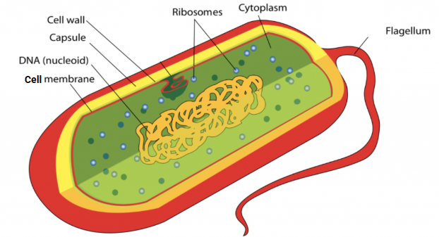
what are the main structures of prokaryotic cells?
cell membrane
cytoplasm
ribosomes
genetic material → in large loop called nucleoid
the structures inside prokaryotes:
nucleoid
DNA is a single loop
not surrounded by membranes
float around in fluid-like cytoplasm
structures present only in some prokaryotes?
cell wall
pili
flagella
capsule
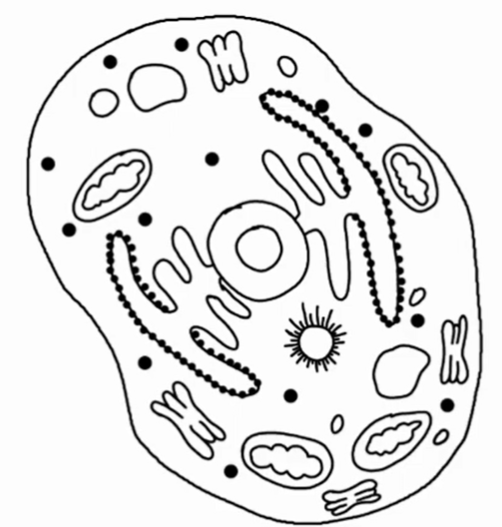
describe eukaryotic cells
10-100 micrometres
characterised by membrane-bound nucleus containing genetic material
internal structures are membrane-bound organelles
both unicellular & multicellular
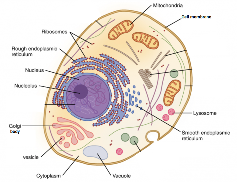
what are the main structures of eukaryotic cells?
double-membrane-bound nucleus
mitochondria
golgi apparatus
chloroplasts
what are the structures common to both prokaryotic & eukaryotic cells?
ribosomes
cell membrane
cytoplasm
genetic material
draw a prokaryotic cell

label the diagram of a prokaryotic cell
draw a eukaryotic cell
label the diagram of a eukaryotic cell
classify the prokaryotes (groups)
bacteria
cyanobacteria
stromatiles
nitrogen fixing bacteria
archaea
methanogens
thermophiles
halophiles
deep sea bacteria
why is methane an increasing threat to environment?
increases more rapidly than any other gas
where are methanogens found?
marine & freshwater sediments, intestinal tracts of herbivores (e.g. cows), sewage treatment facilities
what is methane produced by?
methanogens
how do microorganisms produce methane?
from other gas present during fermentation
methanogens are anaerobic
methanogens use H2, S & CO2 in inorganic reactions for growth & chemical energy
produces CH4 in process
where are halophiles found? give an example
environments where salt concentration is very high (e.g. Dead Sea in Middle East) & evaporating ponds of saline water
what eats halophiles?
filter feeders
are halophiles aerobic or anaerobic?
all aerobic but have another system of producing energy
what is the red colour in halophiles caused by?
unique pigment called bacteriorhodopsin
enables them to photosynthesise & produce energy without using oxygen
what do thermophiles need for growth?
high temperatures (80-105C)
where are thermophiles found? give examples
areas of volcanic activity
e.g. hot springs, geysers, hydrothermal vents/cracks in ocean floor
what are thermophiles also called?
ones that live in hydrothermal vents in depths of ocean sometimes called deep-sea bacteria
what do thermophiles use sulfur for?
energy source
what are thermophiles in deep-sea food web?
primary producers (chemoautotrophs)
where is deep sea bacteria found?
bioling undersea vents of volcanoes
what are deep sea bacteria in food webs?
basis of food web in undersea vents
how do deep sea bacteria obtain energy?
uses sulfur compounds from volcanoes
where do cynaobacteria live & with what?
marine environment
some free living, some exist in association with fungi (mutualism)
where do ancient forms of cyanobacteria exist?
in stromatolites
what is cyanobacteria also known as & why?
blue-green algae because they are aquatic & photosynthesise
what do cyanobacteria do for the soil?
many fix nitrogen thus enriching soil they live in
how are stromatolites formed?
water colonies of cyanobacteria trap calcium carbonate & grow upwards in columns towards the sun
where can people visit to see stromatolites?
Western Australia at Shark Bay
what is the growth rate of stromatolites?
1mm per year
diameter of 200cm
height of 50cm
where are nitrogen fixing (changing) bacteria found?
nodules living on roots of legume (pea plants) plants or in soil
what do nitrogen fixing bacteria do?
convert:
nitrogen gas from atmosphere → ammonium ions → amino acids in plants → plants grow in low-nitrogen soil
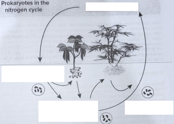
label the nitrogen cycle
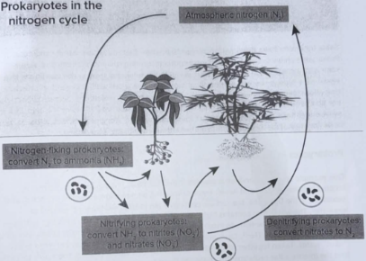
where is bacteria found & what can they be?
in all types of environments, harmful/beneficial
where is archaea found? give examples
in extreme harsh environments e.g.:
hot springs
very salty lakes
depe thermal vents of volcanoes
sewage lagoons
intestinal tracts of animals
what do some organisms develop & why?
develop strategies to survive harsh conditions
allows bacteria to be the most abundant life form in terrestrial & land ecosystem
what do almost all prokaryotes have?
cell wall
protective structure for survival in hyper- & hypo-osmotic coniditons
what do some soil bacteria form?
endospores that resist heat & drought
allows survival until favourable conditions recur
what is the Dead Sea & where is it located?
hypersaline basin located between Joran & Israel
what is the concentrations of the Dead Sea?
salt concentration 10 times higher than seawater
magnesium 40 times higher than seawater (toxic to most organisms)
what produces “hard” water?
elements that form divalent ions:
iron (Fe2+)
calcium (Ca2+)
magnesium (Mg2+)
what makes the Dead Sea unique & hostile?
high concentration of dilavent cations, acidic pH (6.0), intense solar radiation together
classify the kingdoms of eukaryotes
plant
algae
ferns
conifers
flowering plants
animal
invertebrates (i.e. worms, insects)
vertebraes (fish, amphibians, reptiles, birds, mammals)
fungi
mushrooms
yeast
protist
amoeba
paramecium
euglena
draw a eukaryotic plant cell

label the diagram of a eukaryotic plant cell
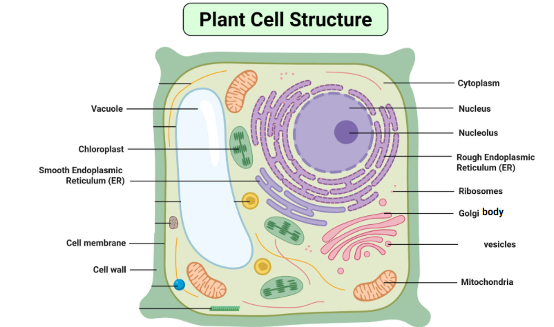
draw a eukaryotic animal cell
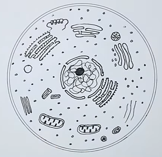
label the diagram of an animal cell
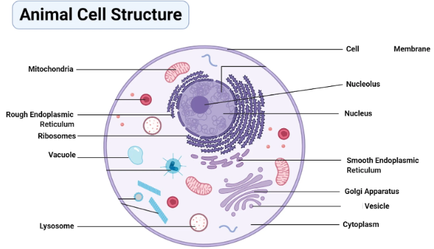
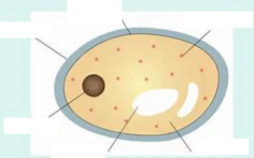
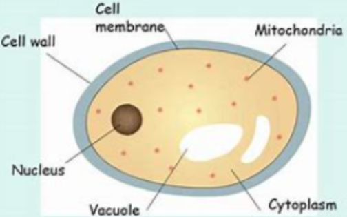
identify, draw, label
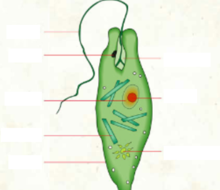
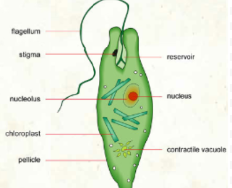
identify, draw, label

label the light microscope

function of the eye piece (barrel)
holds ocular lens
function of revolving nose piece
can be moved to change lenses
function of lens
magnifies samples/specimen
function of stage
holds sample
function of stage clip
holds slide in place
function of diaphragm
adjust light
function of light source
provides light
function of arm + base
holds microscope
function of focus knob
for resolution
what are the FOV’s of each lens on the microscope?
4x: 4.5mm or 4500 micrometres
10x: 1.8mm or 1800 micrometres
40x: 0.45mm or 450 micrometres
100x: 0.00018mm or 0.18 micrometres
400x: 0.000045mm or 0.045 micrometres
what is the proper procedure for using a light microscope?
Plug in microscope & turn on
Lower stage to lowest level using coarse focus knob
Turn revolving nosepiece until 4x lens (lowest magnification)
Put lens under stage at its highest position & adjust iris diaphragm
Prepare wet mount/slide
Place specimen slide on stage & centre it
Look through eyepiece & turn coarse focus knob until blurry image
Make image clearer using fine focus knob
Turn to 10x lens & only use fine focus knob
Draw scientific diagram of sample
define resolution
ability to see detail
define magnification
ability to make objects appear larger
how to calculate total magnification?
eyepiece mag x lens mag
what are the types of microscopes?
light & electron
what was discovered using the microscope?
cells & their structure
how do light microscopes work?
passing light through specimen & magnify light using lenses
how do electron microscope work?
passing beams of electrons through specimens & detect electrons by sensors which projects image onto screen
what organelles are visible with light microscope & which ones need staining?
nucleus (can be stained)
cytoplasm (needs staining)
cell wall
chloroplasts
vacuole
what organelles are visibe with electron microscope & which ones need staining?
cell membrane (needs staining)
cell wall
golgi (needs staining)
chloroplast
mitochondria
ER (needs staining)
ribosome
lysosome
cytoplasm (needs staining)
centrosome
cytoskeleton (needs staining)
identify the process of the development of the cell theory, include who & what year
roger bacon, 13th century → leonardo da vinci, 1485 → hans & zacharias janssen, circa 1600 → robert hook, 1665 → marcello malpighi, 1672 → anton van leeuwenhoek, 1676 → rene dutrochet, 1824 → robert brown, 1831 → matthias schleiden & theodore schwann, 1839 → rudolf virchow, 1858 → louis pasteur, 1862 → walter flemming, 1880-1882 → ernest ruska & max knoll, 1933
describe the ‘microscope’ & significance of Roger Bacon including what year
13th century
used convex lenses as magnifying glass
first to describe properties of convex lenses, hence inventor
describe the ‘microscope’ & significance of Leonardo Da Vinci including what year
1485
used glass lenses
1508: described & sketched first ideas for contact lenses
describe the ‘microscope’ & significance of Hans & Zacharias Janssen including what year
circa 1600
developed first compound microscope
used convex objective & concave eyepiece to enlarge
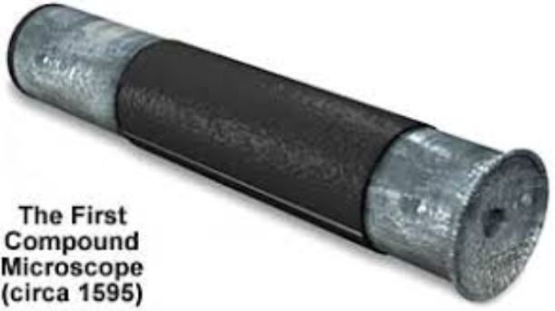
describe the ‘microscope’ of Robert Hooke including what year
1665
used bi-complex objective lens placed in the snout & two additional lenses, an eyepiece lens, a tube/field lens
what did Robert Hooke discover & in what year?
1665, discovered plant cells (more specific, the cell walls in cork tissue)
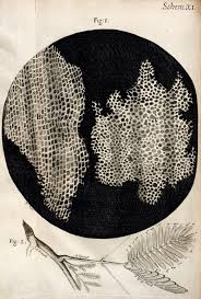
why did Robert Hooke call cells cells?
box-like cells of cork reminded him of cells in monastery
what did Robert Hooke report?
seeing structures in wood & other plants
describe the significance (what he discovered) of the microscope & Marcello Malpighi Including what year
1672
stated all plants built of chambers
credited with using microscope as a tool in organism study
founder of histology (study of tissue under microscope)
investigated microscopic anatomy of human body
discovered relationships of capillaries, veins, arteries
took tissues of tongue & discovered taste buds
describe the significance of the microscope & Anton van Leeuwenhoek including what years
1676: first encounter with microscopy: examining cloth samples with magnifying glass
1683: discovered bacteria from observation of saliva
describe the significance of Rene Dutrochet including what years
1863: discovered & named osmosis
1824
studied development of bird eggs
recognised the take-up of carbon dioxide by plant cells depended on their green pigment
stated all organisms are made of cells
describe the significance of Rober Brown including what year
1824
observed fertilisation mechanisms of plants
noted existence of a structure within cells of orchids
termed the “nucles” of cells
describe the significance of Matthias Schleiden & Theodore Schwann including what year
1839
proposed all organisms are made of cells
formulated early cell theory
Schwann discovered cells (Schwann cells)
forms sheath surrounding nerve axons
conducted experiments that helped disprove theory of spontaneous generation
coined “metabolism” to define chemical changes that take place in cells
demonstrated that yeast organisms cause fermentation of sugar solutions
describe the significance of Rudolf Virchow including what year
1858
“all cells arise from pre-existing cells”
rejected spontaneous creation of cells from non-living matter
describe the significance of Louis Pasteur including what year
1862
provided proof of Rudolf’s idea with classic swan neck flash experiment
describe the significance of Walter Flemming including what year
1880-1882
observed chromosomes during animal cell division
first to detail how chromosomes move during cell division
described whole process of mitosis (prophase, metaphase, anaphase)
describe the significance of Ernst Ruska & Max Knoll including what year
1933
created Transmission Electron Microscope
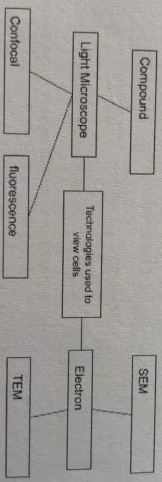
draw the flow chart of technologies used to view cells
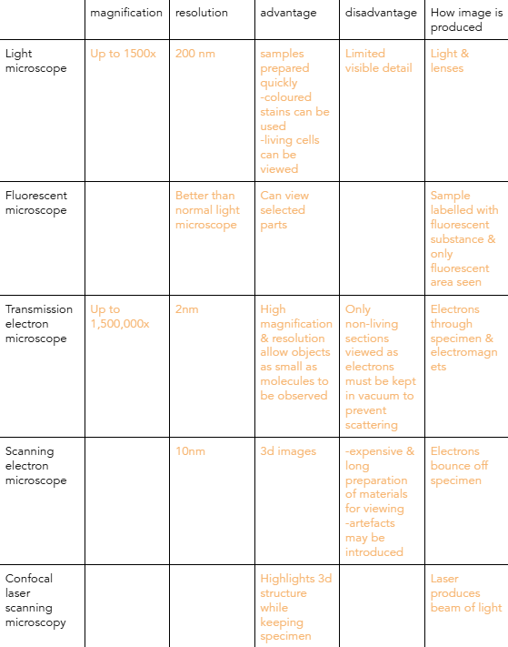
describe the different types of microscopes in terms of their magnification, resolution, advantage, disadvatage, how image is produced
define organelles & their function
identifiable bodies in cell
surrounded by membrane
specific functions within cell
carry out biochemical processes & reactions (e.g. photosynthesis, respiration
function of nucleus
contains genetic material
how big is a nucleus?
2-10 microns
what happens when nucleus is about to divide
chromatin arranges into chromosomes
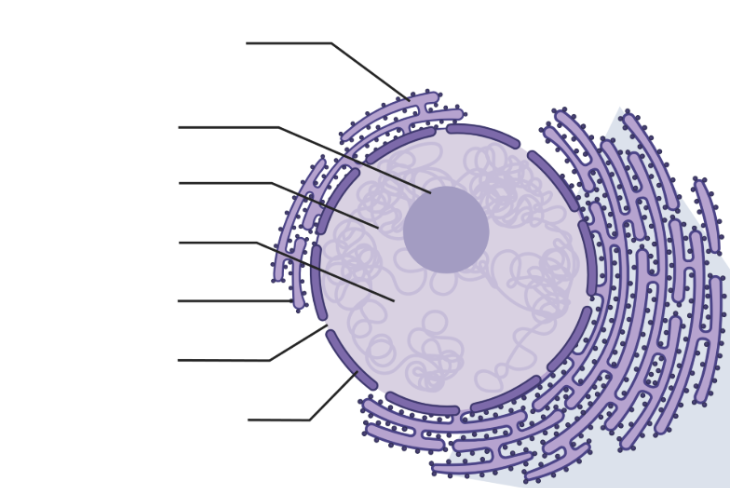
identify & label the organelle
