Heart & Borders
1/38
There's no tags or description
Looks like no tags are added yet.
Name | Mastery | Learn | Test | Matching | Spaced |
|---|
No study sessions yet.
39 Terms
Cardiac Output at Rest
5L/min
(7200 L/day)
Describe the blood flow route from body to heart to body
Blood enters heart through the superior & inferior vena cava → right atrium → tricuspid valve → right ventricle → pulmonary valve → pulmonary artery → lungs → pulmonary vein → left atrium → mitral valve → left ventricle → aortic valve → aorta → body
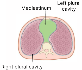
Where is the heart located
Located in thoracic cavity between 2 pleural cavities in the mediastinum
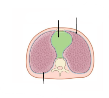
Transverse sections we are looking at it from the feet up to body (inferior view)
subdivisions of the mediastinum
Superior
Inferior
The inferior subdivision is further divided:
Anterior:
Middle
Posterior
Borders of superior subdivision of the mediastinum
Superior border: Superior thoracic aperture
Inferior border: Sternal angle
Borders of the anterior inferior subdivision of the mediastinum
Superior: T4/T5 (sternal angle)
Inferior: T12 (diaphragm).
Anterior: Posterior surface of sternum
Posterior: Pericardium
Borders of the posterior inferior subdivision of the mediastinum
Superior: T4/T5 (sternal angle)
Inferior: T12 (diaphragm).
Anterior: Pericardium and diaphragm.
Posterior: Vertebral bodies of T5–T12.
Blood vessels, Nerves & Structures of superior subdivision of the mediastinum
Arteries:
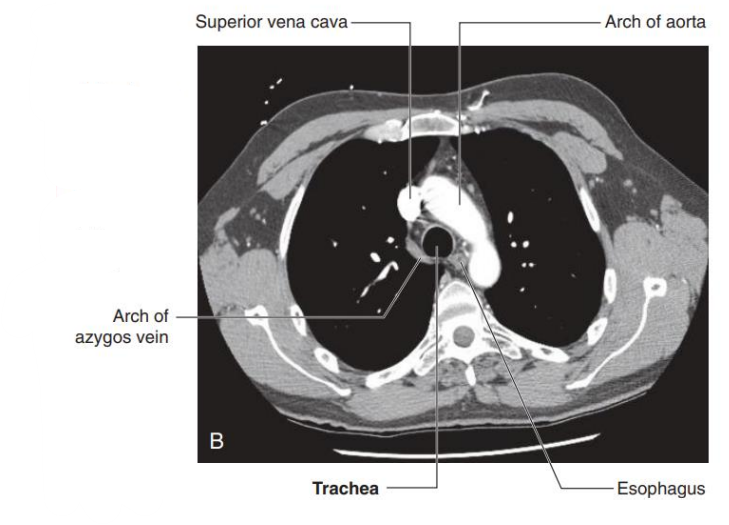
Arch of the aorta
Left common carotid artery & left subclavian artery (both come directly off aorta)
Right common carotid artery & right subclavian artery (come off the aorta through the brachiocephalic trunk)
Veins
Superior Vena Cava
Right & Left Brachiocephalic veins (come from SVC)
Left superior intercostal vein
Nerves
Phrenic nerves
Vagus nerves
Left recurrent laryngeal branch (of left vagus nerve)
Structures
Oesophagus
Trachea
Thymus
Thoracic duct
Contents of the anterior inferior subdivision of the mediastinum
Thymus gland
(Active in children - gradually regresses during childhood)
(Extends from cricoid to retrosternal area)
Contents of the middle inferior subdivision of the mediastinum
Pericardium
Heart
Roots of great vessels
Contents of the posterior inferior subdivision of the mediastinum
Thoracic aorta
Azygous system of veins
Oesophagus
Trachea
Thoracic duct
Sympathetic trunks
1st division off the aorta
Brachiocephalic trunk
Ligamentum arteriosum
Ligamentum arteriosum is a foetal blood vessel used when lungs weren't functional in utero (it is the remnants of a duct that connected the pulmonary trunk to the arch of the aorta)
Why don’t left & right vagus nerves have the same route
The aorta is in the left’s way
Which nerve is closely to the oesophagus
Vagus nerve
The vagus nerve goes under the _____vein
azygous
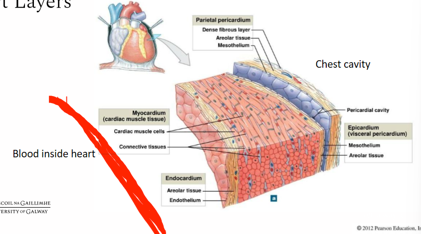
Layers to the pericardium
Outer fibrous layer
Inner double layer of serous membrane consisting of:
parietal layer (lines fibrous layer)
visceral layer (lines heart)(epicardium)
(outer fibrous layer & inner parietal layer are attached)
What is the fibrous pericardium attached to
Great vessels
Sternum (sternopericardial ligament)
Central tendon of the diaphragm
Parietal layer of serous pericardium
What is the space between the 2 internal serous pericardium layers called, filled with & used for
Called:
Pericardial cavity
Potential space
Filled with: Pericardial fluid
Used for: Facilitating gliding movements (beating of heart)
Cardiac tamponade
Excess fluid in pericardial cavity puts pressure on heart
Work through the layers between the chest cavity & the Blood inside the heart
How do pericardial sinuses come about
Naturally occurring open spaces in the pericardium caused by reflection of serous pericardium on the posterior surface of the heart
Name 2 pericardial sinuses
Oblique sinus
Transverse sinus
Where are each of the pericardial sinuses located
Oblique sinus is behind the heart (reflection surrounding veins)
Transverse sinus is between the great arteries & veins (posterior to the aorta & pulmonary trunk & anterior to the superior vena cava)
Lots of arteries supply the pericardium. Where are their branches from
branches from the internal thoracic, pericardiacophrenic, musculophrenic, inferior phrenic and the thoracic aorta
What veins drain the pericardium
pericardial veins drain into the azygos system of veins, the internal thoracic veins & the superior phrenic veins
What nerves supply the pericardium
the vagus nerve, sympathetic trunks and phrenic nerves
Why is pericardial related pain referred to the supraclavicular region or lateral neck
Pain from parietal pericardium is carried in somatic afferent fibres in phrenic nerves.
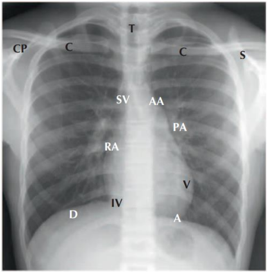
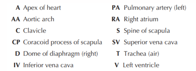
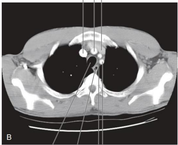
What can be seen in a T3 axial CT
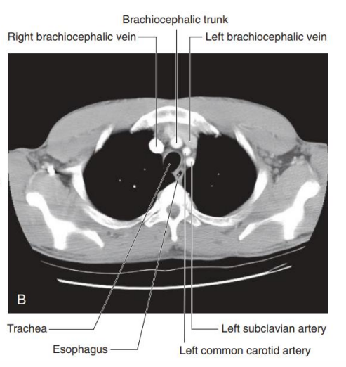
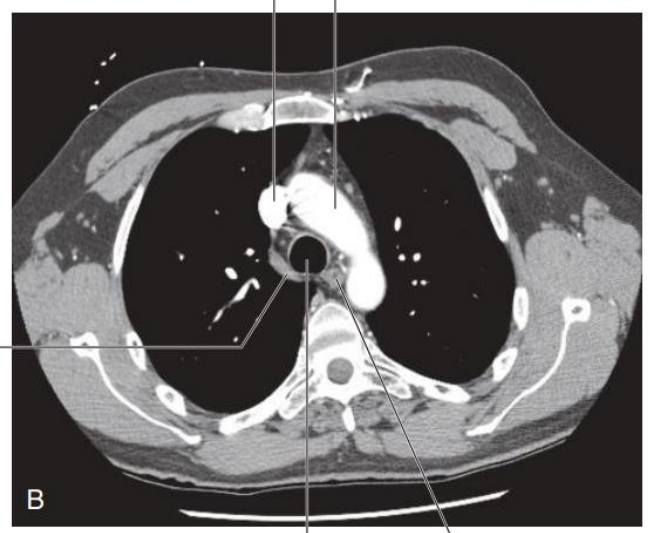
What can be seen in a T4 axial CT
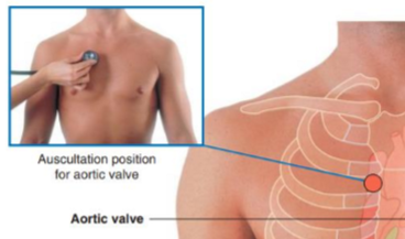
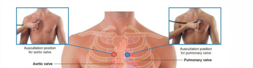
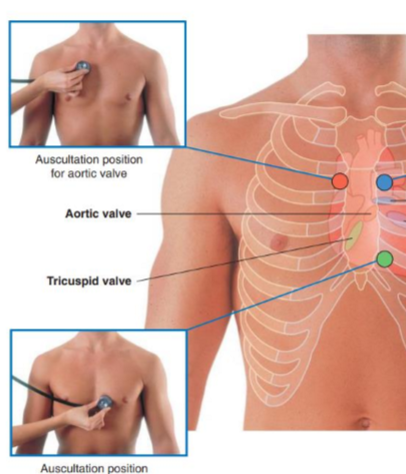
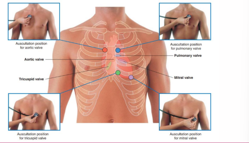
What is the auscultation position for the aortic valve
RHS 2nd intercostal space
What is the auscultation position for the pulmonary valve
LHS 2nd intercostal space
What is the auscultation position for the tricuspid valve
LHS 5th intercostal space at sternal border
What is the auscultation position for the mitral valve
LHS 5th intercostal space at the mid-clavicular line
What is the apex beat
The lowest and most lateral point on the chest wall where you can feel the heart beating
Where is the apex beat
LHS 5th intercostal space, mid clavicular line
Dextrocardia
Relatively uncommon embryological abnormality where the heart is positioned on the right hand side of the chest