Biopsychology
1/86
Earn XP
Name | Mastery | Learn | Test | Matching | Spaced |
|---|
No study sessions yet.
87 Terms
What is the nervous system?
1) System of nerve cells
2) Collects info from world
3) Then directs body organs / muscles via transmission of electrochemical messages
Divided into the CNS and the PNS
What is the central nervous system?
Complex processing
Brain - conscious and most unconscious processing
Spinal cord - messages to/from brain and body
What is the peripheral nervous system?
Messenger neurones
Somatic nervous system - voluntary conscious actions. Receives info from sensory (afferent) receptors and send info TO CNS
Autonomic nervous system - unconscious involuntary actions eg breathing. It has 2 main divisions: the sympathetic and parasympathetic nervous system. Only consists of motor neurones - info AWAY from CNS
Similarities and differences between the somatic and autonomic nervous system
Similarities
Both respond to external stimuli
Differences
Autonomic consists of 2 sub-systems, somatic consists of only 1
Autonomic only has motor pathways, somatic has sensory and motor pathways
The autonomic nervous system controls internal organs and glands, while the somatic nervous system controls muscles and movement
What is the sympathetic and parasympathetic nervous systems?
The sympathetic nervous system
Involved responses that prepares the body for fight or flight
Increases HR / BP
Slows less important functions
Activated during stress
Neurons from the SNS travel to every organ and gland to prepare the body for the rapid action when under threat
E.g. increased heart rate allows for better blood flow to muscles, increased pupil size to let in more light better vision and adrenal medulla stimulated to release more adrenaline
The parasympathetic nervous system
Opposite functions of the SNS
Returns body to normal state
What is a neuron?
Neuron - the basic building blocks of the nervous system. They are specialised cells that carry neural information throughout the body. Nerve cells that process and transmit messages through electrical and chemical signals. 3 main types: sensory, relay and motor

What are the features of a neutron?
Dendrites send and receive information to from and to other neurons. Branch-like structures found at the end of neurons.
Axons transmit the info it receives down its body to dendrites.
The myelin sheath is an insulating layer and increases amount of electrical impulses to transmit quickly along nerve cells
The nucleus is responsible for cell function and regulation
The cell body contains genetic information
Axon terminals are at end of axons and transmit messages to other cells via use of neurotransmitters at synapses
Nodes of ranvier allow ions to diffuse in and out the neuron, transferring the electrical signal down the axon
What is the structure of a neuron?
The cell body includes a nucleus (contains genetic material), dendrites (carry nerve impulses from neurons to cell body)
The axon impulses away from cell body down the neuron. Covered in fatty layer of myelin sheath (protects axon + speeds up electrical transmission of impulse)
The myelin sheath is segmented by gaps called nodes of Ranvier (speed up transmission of impulse by forcing it to jump across the gaps along the axon
At the end of the axon are terminal buttons that communicate with the next neuron in the chain across a synapse
Location and features of 3 types of neurone
Motor neurons (efferent) - (CNS+PNS). They have short dendrites and long axons. These connect the CNS to effectors such as muscles and glands. Carries info away from CNS. Multipolar - sends and receives messages from many sources
Sensory neurons (afferent) - are located outside of the CNS in the PNS in clusters known as ganglia (in PNS only). Carry messages TOWARDS BRAIN, they have long dendrites and short axons. Connects CNS and the senses. Msgs to CNS via PNS. Unipolar - only transmits messages
Relay neurons (interneurons) carry nerve impulses between neurons. Make up 97% of all neurons and most are found within the brain and visual system (brain and spinal cord only). These connect the sensory neurons to the motor or other relay neurons. They have short dendrites and short axons. Multipolar - sends and receives messages from many source
S,R,M
What is a reflex arc?
Reflex arc - the pathway where impulses are carried from a receptor to an effector without involving any conscious thought
The impulse arrives along the sensory neuron, and passes through the dorsal root ganglion, into the spinal cord
The impulse is then passed to a relay neuron, in the spinal cord
The impulse spreads on to the motor neuron and along its axon
The impulse arrives at the effector, producing a response to a stimulus
This all takes less than 1 second
The response by the effector is called a reflex action, defined as a fast stereotypes response to a particular stimulus
Reflex actions help us to avoid danger, by allowing us to respond immediately to a potentially harmful situation without having time to think about it
What is a neurotransmitter?
chemical messengers released from synaptic vesicles that transmit signals across the synapse from one neuron to another during the process of synaptic transmission. Some perform excitatory function and some inhibitory function
Process of synaptic transmission
Synaptic transmission - a method of neurons communicating with each other, transferring info to the CNS across sensory neurons. Carries out responses dictated by the brain through sending info to effectors via motor neurons.
The process is as follows
An action potential arrives at the presynaptic membrane, causing depolarisation through the opening and inflow of calcium ions
The increased concentration of calcium ions in the membrane causes the vesicles (containing NT) to fuse with the pre synaptic membrane, and release their contents into the synaptic cleft through exocytosis
The NT diffuses across the synaptic cleft (down conc grad), and binds to complementary receptors on the postsynaptic membrane
Summation happens now. For an action potential to form in the postsynaptic cell, the electrical charge needs to pass the threshold. An excitatory or inhibitory effect happens on the NT. If excitatory, when detected by receptors, they make the electrical charge positive in the cell (process called depolarisation). This happens as receptors allow pos charged sodium ions into the cell. This makes the formation of a new action potential more likely. But some NT are inhibitory, when detected by receptors, hyperpolarise the postsynaptic neuron, making it negative by releasing potassium. Pushes it further from threshold, makes formation of new action potential less likely. Summation is the effects of all excitatory and inhibitory NTs influences on the postsynaptic cell. (These are added and subtracted, so summed!) If threshold met, new action potential formed then travels down next axon
Now the NTs been detected by receptors they detach. Some broken down, some reuptaken into the presynaptic cell (resets cell ready for next fire)
The action potential will then be transmitted along the axon of the following neuron
What is temporal and spatial summation?
Temporal summation - when there’s a number of action potentials needed before there is enough transmitter to initiate an action potential in the postsynaptic cell. Each action potential that arrives at the presynaptic membrane causes vesicles to release their neurotransmitter.
Spatial summation - summation is the effects of all excitatory and inhibitory NTs influences on the postsynaptic cell. A number of presynaptic neurons may form synapses with postsynaptic neurons. Action potentials arriving in each presynaptic neuron will release a NR and build up the threshold level until it triggers a postsynaptic impulse
What is excitation and inhibition?
For an AP to form - electrical charge must pass threshold. NTs either have excitatory or inhibitory effect
Excitation - Detected by receptors —> make electrical charge more pos in cell —> called depolarisation (receptors allow pos sodium ions in cell) —> makes the postsynaptic cell more likely to fire an impulse and the message is continued
Inhibition - such as GABA. detected by receptors —> hyper polarise post synaptic neuron —> more neg thru potassium release —> postsynaptic cell less likely to fire an impulse
Summation - Combined effect of all exc / inhib NT influences of post synaptic neuron. Added and subtracted, if threshold reached, AP forms
NTs now detach. some broken donna and some reuptake into pre synaptic cell - reset cell ready for next fire
How do synapses ensure one way transmission?
Signals can only pass in one direction across a synapse so they go to a specific target
This is because the synaptic vesicles are ONLY present in the presynaptic membrane
And the receptors sites for the NTs are ONLY present on the post synaptic membrane
Diffusion of the NTs mean they can only go from high to low concentration, so can only travel from the presynaptic to the postsynaptic
membrane
How do synapses increase the possible range of actions in response to a stimulus?
By allowing the interconnection of many nerve pathways
The nervous system receives info from various sources about different situation e.g. receptors in eyes give details about person you see
All the pieces of info will produce action potentials in many neurons in your nervous system
Explain the process of synaptic transmission (4) Brief
Impulses reach presynaptic terminal
Impulses trigger release of NTs
NTs cross the synapse from vesicles
NTs combine with receptors on the postsynaptic membrane
Stimulation of postsynaptic receptors by NTs results in either excitation or inhibition
What is the endocrine system and its function?
Control vital physiological processes in the body
A network of glands that instruct glands to secrete chemical messages called hormones directly into bloodstream
These hormones then bind to specific receptors to regulate the activity of cells/organs in body
Instead of using nerves to transmit messages, it uses blood vessels (chemical system)
Different hormones produce different effects (behaviours)
Define gland, hormone, adrenaline and fight or flight response
Gland - an organ in the body that secretes hormones for specific purposes
Hormone - chem messengers that travel thru bloodstream to reg certain bodily processes
Adrenaline - a hormone produced by the adrenal glands which is part of body’s immediate stress response system
Fight or flight response - the sympathetic response that increase activity to areas designed to help you escape or prepare for a fight. The body becomes physiologically aroused in readiness to fight an aggressor or, in some cases, flee
Name the parts of the endocrine system and their functions
Hypothalamus - control system that regulates the entire endocrine system. It’s responsible for stimulating or controlling the release of hormones from the pituitary glands. Sends the PG a signal for it to secrete a hormone into the bloodstream
Pituitary gland - known as the master gland because hormones released by it control and stimulate the release of hormones from other glands in the endocrine system. Divided into anterior (front) and posterior (rear), which release different hormones
The anterior releases adrenocortical trophic hormone (ACTH) which stimulates the adrenal cortex and the release of cortisol
The posterior releases oxytocin which is responsible for uterus contractions during childbirth. Levels increase when you cuddle someone you love
Adrenal gland - Divided into the adrenal medulla which releases adrenaline and noradrenaline which plays a key role in fight or flight response. And the adrenal cortex which releases cortisol which stimulates the release of glucose while suppressing the immune system
Pineal gland - releases melatonin, responsible for important biological rhythms including the sleep-wake cycle
Thyroid gland - releases thyroxine which regulates metabolism. Metabolism involves the chemical processes of converting food into energy, so people with high metabolism find it hard to gain weight
Ovaries - releases hormone oestrogen which controls the regulation of female reproductive system, including the menstrual cycle and pregnancy
Testes - releases androgens which includes testosterone. Testosterone is responsible for the development of male sex characteristics during puberty and muscle growth
An outline of the fight or flight response
HPA pathway (chronic)
hypothalamus → pit gland → adrenal cortex → ACTH → corticosteroids → cortisol → suppress immune system / LT release glucose
SAM pathway (acute)
hypothalamus → sympathetic branch → adrenal medulla → adrenaline/noradrenaline → BF to muscle/brain, inc HR/alertness, inc pupil dilation
(The hypothalamus can activate either of these two pathways depending on the situation)
Threat has passed → the parasympathetic nervous system returns to the body's resting state
Different types of stress
Acute stress (short term/sudden) e.g. when a dangerous dog is running at you, public speaking
Chronic stress (long term/ongoing) e.g. when you have an exam coming up soon, job issues, grief
What is homeostasis?
The body’s steady state. Maintaining the body’s internal environment at a constant in spite of large changes in the external environment. The antagonistic nature of the ANS ensures the body returns to its normal state asap (goes up then goes down). The sympathetic branch produces arousal to deal with the emergency and the parasympathetic branch controls the normal body state by storing and conserving energy
Evaluation of fight or flight
There may be gender differences in behavioural responses to stress: Taylor et al suggest that for females, behavioural responses to stress are more characterised by ‘tend and befriend’ behaviours than fight or flight. Women have evolved a different system for coping with stress because their responses evolved in the context of being the primary carer of children. For example, fleeing at the first sign of danger would be putting their children at risk, so they are less likely to do this. This involves protecting themselves and their children through nurturing behaviours (tending) and forming alliances with other women (befriending), because when in a stressful situation they may talk to others they haven't talked to before as they are in this together to try and get out of it. This finding, explained in terms of the higher levels of oxytocin in females, suggests that previous research, which has mainly focuses on males, has obscured patterns of stress response in females
Fight or flight doesn’t tell the whole story:
Gray - first phase reaction may not be to fight or flee → display the ‘freeze response’.
‘Stop, look and listen’ response, where they’re hyper-vigilant and alert to the slightest sign of danger
He said the first response is to avoid confrontation - an advantage
This creates best response poss
Assessing the situation before jumping straight to a conclusion that it is dangerous
There’s positive behavioural outcomes from stressful situations:
Dawans argues that acute stress can actually lead to more cooperative and friendly behaviour
As connections made with others
Explains human connections during times of crisis such as 9/11 terrorist attacks in NY
One reason why stress may lead to greater cooperative behaviour is because human beings are fundamentally social animals
Protective nature of human social relationships that has allowed our species to thrive
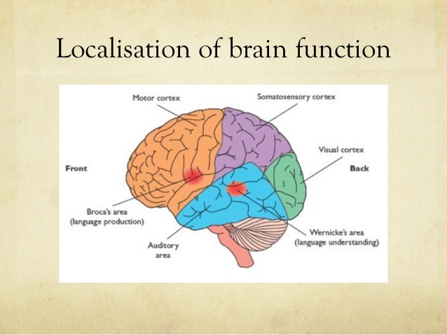
What is localisation of function in the brain?
The theory that different areas of the brain are responsible for specific behaviours, processes or activities
What is the motor area?
Frontal lobe - coordinates voluntary movement on the opposite side of the body. Damage to this may cause loss in control over fine movements
What is the somatosensory area
At the front of the parietal lobe that processes sensory info (touch, temp, pain)
What is the visual area?
Occipital lobe - receives and processes visual information
What is the auditory area?
Temporal lobe - processes sound - may produce partial hearing loss if damaged
What is Broca’s and Wernicke’s area?
Broca’s area - frontal lobe - speech production
Wernicke’s area - temporal lobe - language comprehension
What does ‘the brain is contralateral’ mean?
This means that the left side of the brain represents and controls the movement of the right side of the body and vice versa. Whatever happens on one side is processed by the other side of the brain. E.g. if a person has a stroke in the right hemisphere, the attack will be on the left side of the body.
What does lateralisation mean?
the main part of our brain (cerebrum) is divided in two symmetrical halves called the left/right hemisphere. Some of our psychological and physical functions are controlled by a particular hemisphere.
Describe the case of Phineas Gage
During the 19th century, Broca and Wernicke discovered that specific areas of the brain are associated with particular physical functions. Before their investigations, scientists generally supported the holistic theory of the brain - that all parts of the brain were involved in the processing of thought and action. Then came Phineas Gage. Broca and Wernicke argued for localisation of function (cortical specialisation) where different parts of the brain perform different tasks and are involved in different parts of the body. It therefore follows that if a certain area of the brain becomes damaged through illness or injury, the function associated with that area will also be affected
Phinneas Gage experienced a brain injury when an iron rod was driven through his skull; he somehow survived, but it destroyed much of his frontal lobe. However, his personality and behaviour changed so much that his friends described him as a different person entirely. The impact this accident had helped us understand the frontal lobe does, and its relation to personality.
However, his change in personality may not just be due to the accident. His personal life and appearance changed too, which meant his relationships with people changed, which may have changed his personality.
What is the brain’s structure?
Divided into 2 symmetrical halves called the left and right hemispheres. The outer layer of both hemispheres is the cerebral cortex (grey as lots of cell bodies). The cortex of both hemispheres is divided into 4 lobes: frontal, parietal, occipital and temporal lobe. They eahc have different functions.
What is meant by holistic V localisation
Holistic V localisation
In the 19th century scientists supported the holistic theory of the brain - that all brain parts were involved in the processing of thought and action
In contrast, Broca + Wernicke argued for localisation of function - the idea that different parts of the brain perform different tasks and are involved with different body parts
What are the language centres of the brain?
Language is restricted to the left side of the brain. Broca identified a small region in the left frontal lobe responsible for speech production and called it Broca’s area. Damage to Broca’s area causes Broca’s aphasia which means slow speech lacking fluency. And have difficulty with prepositions and conjunctions (a, the, and).
Around the same time, Wernicke identified a region called Wernicke’s area in the left temporal lobe that is responsible for language and understanding. Damage to this causes Wernicke’s aphasia when people will produce nonsense words (neologisms). It was describing people who had no problem producing language but severe difficulties understanding it
Describe the Case Study Tan
Epilepsy = lost the ability to speak, apart from to say tan
He was treated by Broca and after death he did a post mortem on him. He found a lesion (an area of damage) on the left temporal lobe
He concluded this was the only visible area of damage and it was also responsible speech production
The term Broca aphasia is now used to describe people with speech difficulties like slow speech
This suggests functions are localised in in these areas
HOWEVER: Recent MRI scan on his brain showed damage far beyond Broca’s area suggesting other areas could be responsible for his speech problems
Evaluation for localisation of function in the brain
RS: the case study of Tan + modern CR
Challenge to localisation: equipotentiality. Not everyone agrees that functions are localised in the brain. A conflicting view is the equipotentiality theory, positing that all areas of the brain are equally able to perform a task. Lashley RS 50 rats ran maze before and after areas of their brain destroyed. Found ability to successfully re run the maze depended on how much of the brains cortex destroyed, not which areas. Suggests higher cognitive functions not localised but distributed across the brain (basic motor and sensory functions still may be localised)
Idea of localisation fails to take individual differences into account. Herasty found that women have larger Broca and Wernicke’s areas than men, explaining the greater ease of language used by women. This however, suggests a level of beta bias as the differences between men and women are ignored, and variations in the acitivty/ size of of areas observed during language activities are not considered
What is hemispheric laterislation?
Lateralisation is the fact that the two halves of the brain are functionally different and that each hemisphere has functional specialisations, e.g. the left is dominant for language, and the right excels at visual motor tasks
What is split brain research?
Studies how hemispheres function when they can’t communicate with each other.
A series of studies which began in the 60s involving people with epilepsy who had had a surgical separation of the hemispheres to reduce epilepsy.
This let researchers test lateral functions of the brain in isolation. Corpus callosum is cut in patients with severe epilepsy, allowing researchers to investigate the extent to which brain function is lateralised
Split brain research is severing the connections between the RH and LH, mainly the corpus callosum. Sperry tested the capabilities of the separated hemispheres, if they were able to send visual info to other hemispheres. During an epileptic seizure, the brain experiences a lot of electrical activity which travels from one hemisphere to the other. To reduce these fits, the brain is cut into 2 halves.
Sperry and Gazzaniga’s split brain research
They devised a study into how 2 separated hemispheres deal with speech and vision. They took advantage of the fact that info from the left visual field goes to the right hemisphere and vice versa. Because the corpus callosum is cut, info presented to one hemisphere has no way of travelling to the other
Procedure
the patient fixates on a dot in the centre of a screen whilst info was presented to either the left or right visual field. They would then be asked to make responses with either their left hand (controlled by RH) or their right hand (controlled by the LH), or verbally (controlled by left HS).
11 people who had split-brain operations were studied where an image was projected to their RVFs (processed by the LH), and the same/different image could be projected to the LVF (processed by the RH). In a ‘normal’ brain, the corpus callosum would immediately share the info between both hemispheres giving a complete picture of the visual world. However, presenting the image to one hemisphere of a split brain participant meant the info cannot be shared to the other hemisphere
Findings
when a picture of an object was shown to their RVF, they could describe what was seen. But they could not do this if the object was shown to the LVF they said nothing was there. This is because language is controlled by the LH, they couldn't send messages from the RH to the LH. Also, they could only select a matching object out of sight using their left hand (linked to RH as RH controls vision), they couldn’t give verbal info/signals
Difference between the right and left hemispheres
The left hemisphere makes up the 2 main centers, Broca’s and Wernicke’s areas, so we say language is laterislised (performed by one hemisphere)
The right hemisphere only produces basic words/phrases but contributes to emotional context to what is being said
This suggests that the LH is the analyser and the RH is the synthesiser
What are some functions that are not lateralised?
Like vision. The motor and somatosensory areas appear in both hemispheres
Further twist: in the case of the motor area the brain is cross-wired (contralateral wiring), the RH controls movement on the left side and vice versa for the LH
Vision is complex. It is contralateral and ipsilateral (opposite and same-sided). Both eyes receive light from the left visual field (LVF) and the right visual field (RVF). The LVF of both eyes is connected to the RH, and the RVF of both eyes is connected to the LH. This enabled the visual areas to compare the slightly different perspectives from each eye and aids depth perception
Evaluation hemispheric lateralisation
Sperry and Gazzaniga’s split brain research
The main advantage of brain lateralisation is that it increases neural processing capacity (the ability to perform multiple tasks simultaneously). Rogers et al. (2004) found that in a domestic chicken, brain lateralisation is associated with an enhanced ability to perform two tasks simultaneously (finding food and being vigilant for predators). Using only one hemisphere to engage in a task leaves the other hemisphere free to engage in other functions. This provides evidence for the advantages of brain lateralisation and demonstrates how it can enhance brain efficiency in cognitive tasks —> chickens better survival techniques —> live longer lives. However, ungeneralisable to humans
Research challenges the popular idea that individuals possess a dominant brain hemisphere that shapes their personality traits or skills.
Nielsen et al. conducted a large-scale analysis of brain scans from over 1,000 participants, aged 7-29, to examine patterns of hemispheric lateralisation
Certain tasks activated specific hemispheres, supporting the concept of lateralisation
No evidence of an overall dominant hemisphere
This suggests that while lateralisation exists in terms of task-specific activity, the idea that people are inherently 'right-brained' or 'left-brained' is an oversimplification
The results indicate that both hemispheres contribute dynamically to various cognitive functions, rather than each hemisphere determining specific personality traits or abilities
Evaluation for split brain research
Limitation of Sperry's research is generalisation issues: Their research makes it hard to establish causal relationships. The behaviour of their participants was compared to a control group. An issue though is that none of the participants in the control group had epilepsy and this is a major confounding variable. So any differences that were observed between the two groups might be because of their epilepsy rather than the split brain. Is it the condition or the split brain treatment? A better control group may have been patients also with epilepsy so that a comparison could have been made to identify whether the epilepsy affected it or not. This means that some of the unique features of the split brain participants’ cognitive abilities might have been due to their epilepsy (through Fink’s research, above, supports Sperry’s conclusions
The experiments involving split-brain patients made use of highly specialised and standardised procedures ie strong scientific methodology. Sperry’s method of presenting visual information to one hemispheric field at a time was quite ingenious. Typically participants would be asked to stare at a given point , the “fixation point” whilst one eye was blindfolded. The image projected would be flashed up for one tenth of a second, meaning the split brain patient would not have time to move their eye across the image and so spread the information across both sides of the visual field and subsequently both sides of the brain. This allowed Sperry to vary aspects of the basic procedure and ensure that only one hemisphere was receiving information at a time. Thus he developed a very useful and well controlled procedure.
Sperry’s work prompted a theoretical and philosophical debate about the degree of communication between the two hemispheres in everyday functioning and consciousness. Some theorists for example Roland Pucetti 1977 have suggested that the two hemispheres are so functionally different that they represent a form of “duality” in the brain – that in effect we are all two minds. In contrast other researchers have argued that far from working in isolation the two hemispheres form a highly integrated system and both are involved in most everyday tasks
What is plasticity of the brain?
Brain's ability to change and adapt (functionally and physically) as a result of experience and new learning. Involves the growth of new connections
During infancy, the brain grows thousands of synaptic connections, about 15,000 per neuron at 3 years old. Twice the amount in the adult brain. As we age, synaptic pruning occurs, which enables lifelong plasticity/ change of the adult brain, which used to be thought to not be able to change once adult - where new neural connections are formed to meet the demands of the brain. Plasticity happens all throughout our lives
Define synaptic pruning, axonal sprouting and recruitment of homologous areas
Synaptic pruning - As we age, rarely used connections are deleted and frequently used connections are strengthened.
Axonal sprouting - Undamaged axons grow new nerve endings to reconnect neurons whose links were injured or severed
Recruitment of homologous areas - Regions on opposite sides of the brain take on functions of damaged areas
What is some research into plasticity
Maguire et al (2000) studied the brains of 16 male London taxi drivers and found significantly more volume of grey matter in the posterior hippocampus than in a matched control group who weren’t London taxi drivers but taxi drivers somewhere else (still male/same age). Difference is London ones have taken this knowledge test.
This part of the brain is associated with the development of spatial and navigational skills in humans and other animals.
As part of their training London cab drivers must take a complex test called ‘The Knowledge’, which assesses their recall of the city streets and possible routes. It appears that this spatial learning through their experience alters the structure of the taxi drivers’ brains.
This suggests brain plasticity as the physical structure of their brains changed due to the intense demands placed on them during the test. They found the longer they have been a taxi driver, the bigger the posterior hippocampus (pos correlation)
This implies the experience of being a taxi driver changes the brain/hippocampus
What does functional recovery of the brain mean?
A form of neural plasticity. Following damage through trauma such as a stroke, it is the brain’s ability to adapt and compensate for those areas that are damaged. It can redistribute or transfer functions usually performed by damaged areas to an undamaged area. Healthy areas take over the functions of damaged or even missing areas. Neuroscientists suggest that this process can occur quickly after trauma (spontaneous recovery) and then slow down after several weeks or months. At this point the individual may need rehabilitative therapy
What happens to the brain during recovery?
The brain is able to rewire and reorganise itself by forming new synaptic connections Liken this to avoiding roadworks on the way to school by finding a different route.
Secondary neural pathways that would not typically be used to carry out certain functions are activated or ‘unmasked’ to enable functioning to continue, often in the same ways as before (Doidge, 2007).
This process is supported by a number of structural changes in the brain. These structural changes are:
1) Axonal sprouting (growth of new nerve endings which connect with other undamaged nerve cells to form new neural connections)
2) Reformation of blood vessels
3) Recruitment of homologous areas (when a homologous/similar area of the brain on the opposite side of the brain is used to perform a specific task e.g. equivalent to broca's area on right side would carry out its function. Functionality shifts back to normal after time).
Plasticity evaluation
Negative plasticity:While brain plasticity allows for remarkable adaptation and recovery, it can also lead to negative behavioural consequences, a phenomenon known as negative plasticity. Research by Medina et al. has shown that prolonged drug use, such as marijuana, can result in maladaptive changes in brain structure, leading to poorer cognitive functioning in later life and an increased risk of dementia. Furthermore, between 60-80% of amputees experience phantom limb syndrome, where they continue to feel sensations in the missing limb. These sensations are often unpleasant and painful, and are thought to be caused by cortical reorganization in the somatosensory cortex following limb loss. Although this cortical reorganization reflects the brain’s adaptive capacity, it highlights that not all changes in brain structure yield positive outcomes. Therefore, while plasticity demonstrates the brain's resilience, it also indicates that neuroadaptive changes may, under certain circumstances, produce detrimental effects on cognition and well-being
It’s a life-long ability. A strength is that brain plasticity may be a life-long ability. In general, plasticity reduces with age. The brain has a greater ability to reorganise in childhood as it is constantly adapting to new experiences and learning. But Bezzola et al demonstrates that it persists in later life by at showing 40 hours of golf training produced changes in the neural representations of movement in participants aged 40-60. Using FMRI, the researchers observed increased motor cortex activity in the novice golfers compared to a control group, suggesting more efficient neural representations after training. This indicates that the adult brain retains a capacity for plasticity, showing that neural adaptations can continue beyond the developmental years. This finding is significant as it highlights that brain plasticity, although reduced with age, remains functional across the lifespan, supporting lifelong learning and adaptationResearch support for brain plasticity: Maguire et al - in flashcard above
Functional recovery evaluation
Real life application. Research and understanding into plasticity and functional recovery has contributed to the field of neurorehabilitation. Simply understanding that axonal growth is possible encourages new therapies to be tried. E.g. constraint induced movement therapy is used with stroke patients whereby they repeatedly practice using the affected part of their body (such as their arm, while their unaffected arm is restrained). This forces patients to use their affected limbs, promoting axonal growth and improving motor function. This shows that research into functional recovery is useful as it improved rehab outcomes, improving QOL and reducing healthcare costs
Cognitive reserve. A limitation of functional recovery is that level of education might influence recovery rates. Schneider et al found that the more time people with a brain injury spent in education (their cognitive reserve), the greater their chances of a disability-free recovery. He found that 40% of those who achieved a disability-free recovery had spent more than 16 years in education, however only 10% had a disability-free recovery with less than 12 years of education. This implies that people with brain damage who have insufficient disability-free recovery are less likely to achieve full recovery, so functional recovery does not work the same on everyone and to the same extent depending on other factors like education
Research supports Danelli et al. Shows how resilient the brain is to extreme damage. Infant EB had massive brain tumour, so at age 2 had a hemispherectomy (remove entire left side of brain). So he lost the language centres\Broca’s/Weernicke’s areas. After surgery, lost all speech, but over two years recovered his ability to talk and developed relatively normally as a teen. FMRI scans showed right hemisphere functioned like a typical left hemisphere. This case study suggests that even extreme trauma can be compensated for by the function of other structures in the brain, especially if that trauma occurs early in life. Only case study tho :(
Name the 4 ways of studying the brain
FMRIs
EEG (electroencephalogram)
ERPs (event-related potentials)
Post-mortem examinations
Define spatial and temporal resolution
Spatial resolution - level of accuracy in identifying the exact location of a brain structure (where activity happened)
Temporal resolution - level of accuracy in identifying the exact location of brain activity in time (when activity happened/how accurate in time)
What are FMRIs?
Large machine taking up an entire room, and a bed that retracts into a hole in the middle of the machine
It works by using large magnets to detect the blood flow in the brain, whilst a person is performing a task in specific areas by detecting magnetic variations between oxygenated and deoxygenated haemoglobin
This allows researchers to detect which regions of the brain are rich in oxygen and are thus active. When a brain area is more active, it consumes more oxygen and to meet this increased demand, blood flow is directed to the active area (known as haemodynamic response)
FMRI produces 3-dimensional images (activation maps) showing which parts of the brain are involved in a particular mental process
FMRI Evaluation
High spatial resolution. It produces images that have very high spatial resolution, meaning it’s composed of more pixels so the image is higher quality (it depicts detail by the milimeter). It provides a clear image of how brain activity is localised. Spatial resolution refers to the smallest feature that a scanner can detect and is an important feature of brain scanning techniques. The higher this measure is, the more accurately psychologists can discriminate between different brain regions. For this technique, the spatial resolution is around 1-2 mm which is significantly greater than other alternatives such as EEG/ERP
Expensive compared to other neuroimaging techniques and poor temporal resolution. It is costly compared to other alternatives, which means it is less likely to be chosen as a number one option for most cases. Also, it has poor temporal resolution because there is a 5 second time lag behind the image on the screen and the initial firing of neuronal activity. This means this technique may not represent moment to moment brain activity, making this a less valid method
Does not rely on use of radiation, non-invasive and straightforward to use. If this technique is done correctly, it is virtually risk free, non invasive, and straightforward to use. Unlike the PET scanning technique, FMRI does not use radiation or involve inserting instruments directly into the brain, making it mainly risk free. Consequently, this should allow more patients/participants to undertake fMRI scans which could help psychologists to gather further data on the functioning human brain and therefore develop our understanding of localisation of function.
What is an EEG
A skull cap you put on your head with 22-34 electrodes + make contact with your scalp
.
Record of the tiny electrical impulses produced by brain’s activity. The recording shows the brainwave patterns that are generated from the action of thousands of neurons =overall account of brain activity
The read out from each electrode is the sum total of activation of the brain cortex under the electrode. Displayed as a series of lines showing distinct patterns called brainwaves. Amplitude (size of waves) shows brain wave intensity, and frequency (distance between each wave) shows the speed of activation. Brainwaves include alpha, beta, theta and delta
EEG Evaluation
Cheaper than most other brain scanning techniques such as FMRI, which machines cost millions of pounds compared to EEG. It is also able to be used in an experiment where the participant can move, whereas other brain scanning techniques like FMRI require the participant to remain very still in a tight enclosed space. This technique is also portable so it is easier to use in a range of places and is not restricted to one room in a hospital
It has good temporal resolution. It measures the electrical activity of the brain in a resolution of milliseconds. You can see exactly the instance of when the activity takes place. However the main criticism is the poor spatial resolution, the EEG cannot identify the exact location of the brain activity, but only assume brain activity from a general region under each sensor. OIt can only sense activity on the outside of the brain but struggles deep within the brain.
What is an ERP?
Isolating specific responses of neurons to specific stimuli or tasks
.
Same equipment as an EEG, but approach the data produced in a very different way
.
EEG = general message brain activity, ERP = teasing out and isolating responses to specific events (hidden within the EEG)
.
It presents a stimulus hundreds of times → brain activity wave is recorded → statistical averaging → brain activity not associated w/ the stimulus (recorded by chance) removed
.
What’s left is a smooth curve of activity called an event related potential (this is the brain's response to just that stimulus)
.
These complex waveforms can be interpreted in detail, like detecting exactly when cognitive processes happen in the brain after the stimulus is shown
.
When interpreting these waveforms, the peaks show either a P or N, followed by a number showing how long after the stimulus the peak happens.
ERP’S Evaluation
Strength is that it brings much more specificity to the measurement of neural processes than could ever be achieved using raw EEG data. As ERPs are derived from EEG measurements, they have excellent temporal resolution, especially when compared to other techniques like FMRI. This means that ERPs are frequently used to measure cognitive functions and deficits such as the maintenance of working memory. It allows researchers to isolate and study how individual cognitive processes take place in the brain, whilst EEGs record general brain activity.
Poor spatial resolution, which means they only detect activity in more general regions of the brian, decreasing the accuracy of the results
In order to establish pure data in ERP studies, background noise and extraneous material must be completely eliminated. This is a problem because that is not always easy to achieve
What is a post mortem examination?
The brain is analysed after death to determine whether certain observed behaviours during the person’s lifetime can be linked to structural abnormalities in the brain. Those subject to one usually had a rare disorder. May also include the comparison with a neurotypical (healthy) brain in order to ascertain the extent of difference. Correlating behaviours before death with brain structures after death
study the brain after death to try and correlate structural
abnormalities to behavioural change
Post mortem evaluation
High spatial resolution allows the study of microscopic brain structures down to the neuronal level. Even studying individual nerve cells, which is simply not possible with other techniques. Post mortem exams have been significant in the historical development of psychology’s understanding of brain functioning, such as the discovery of the language sectors
An obvious weakness is that the brain is studied after death, so there is no way of seeing the brain live and in action. It is hard to tell the exact cause of brain condition by looking at the brain after death because we cannot see it in action. Even damage revealed in post mortem may not be the true cause of the observed unusual behaviour. However, discoveries of abnormalities could lead to the generation of hypotheses that are tested with other measures
What is a biological rhythm?
Distinct patterns of changes in body activity that conform to cyclical time periods. All biological rhythms are governed by 2 things:
by internal body clocks that regulate bio rhythms (endogenous pacemakers)
By external stimuli/changes to the environment involved in control of bio rhythms (exogenous zeitgebers)
Some of these rhythms occur many times in the day (ultradian rhythms - rhythms of less than 1 a day. E.g. animal feeding patterns)
Others take longer than a day to complete (infradian rhythms - rhythms that last for longer than 1 day. E.g. menstrual cycle)
And in some cases much longer (circannual rhythms - yearly rhythms like migration and hibernation)
All living organisms are subject to biological rhythms which can influence how our bodies' systems behave.
What is a circadian rhythm?
Biological rhythms, they last for around 24 hours (subject to a 24hr cycle), which regulate a number of body processes such as the sleep/wake cycle and changes in body temperature
What is the sleep/ wake cycle?
Governed by external changes in environment like daylight (exogenous zeitgeber), which is why we feel drowsy at night and alert in the day
Also governed by an internal (endogenous) pacemaker - a biological clock called the suprachiasmatic nucleus (SCN), it provides info from the eye about light. Exogenous zeitgebers (daylight) can reset the SCN.
Researchers have tried to find out what would happen if the biological clock was left to its own devices, without the influence of external stimuli like light? If we had no idea whether it was day or night would we still fall asleep and wake up at regular times? This led to the research down below…
Siffre Cave Study
Spent several long periods of time underground in cave to study his own biological rhythms
Aimed to see if we have an internal body clock and if so, to see if it can work independently
He deprived himself of all natural light, clocks calendars and sound, but still had food/drink
His free running biological clock settled down to one that was just beyond the usual 24hr one (extended to a 24.5 hr one). This suggested he has an internal clock independent of the natural day/night cycle.
He continued to fall asleep and wake up on a regular schedule
His perception of time was completely distorted. It took him 5 minutes to count to 120 (trying to do 1 count per second). When his team alerted him it was time to come out on 14th Sept, he thought it was only 20th Aug. “My psychological time had compressed by a factor of two”, - interview
He caught a 48 hour cycle a few times. With 36 hours of continuous wakefulness, followed by 12 hours of sleep. Sometimes he slept 2 hours or 18 hours, but he couldn't tell the difference, and that's the problem with psychological time
He found that the 24 hour sleep wake cycle extended to near 25 hours when there was a lack of external cues like light or a clock
Evaluation of Siffre’s research
Controlled variables:
Siffre controlled artificial light, only using it when necessary. This allowed him to isolate the endogenous pacemaker(internal body clock) and examine how it works without exogenous zeitgebers (external time cues like light and social cues). Detailed data collection:
Siffre kept detailed logs of his sleep/wake cycles and other bodily functions, producing rich, qualitative and quantitative data on circadian rhythmsSmall sample. Case study → cannot say one person’s cycle can represent the rest of the population → ungeneralisable. Czeisler - he had dim lighting so and exo zeit had an impact - confounding v - lower internal val, AND found individual differences in sleep/wake cycles varying from 13-65 hrs ++++ Duffy found some people have a natural preference to go to bed early and waking early (known as larks), and some people the opposite (owls). Even Siffre in 1999 in a later study observed that his own sleep/wake cycle had changed and slowed down from when he was younger. Data drawn from one point in their life may not be applicable later on. So because of individual differences and the challenges in establishing the ‘norm’, Siffre’s results have limited applicability to real life
A strength of research into circadian rhythms is that it’s been used to improve medical treatments. Circadian rhythms coordinate body processes like heart rate and digestion, these rise and fall during the course of the day which led to the field of chronotherapeutics which is about timing medical treatments to match a person's biological rhythms. For example, aspirin as a treatment is most effective if taken last thing at night because it reduces blood clotting and this can reduce the risk of a heart attack. Heart attacks are more likely to occur early in the morning, so the timing of taking aspirin (late at night) is important. This shows that circadian rhythm research can help increase the effectiveness of drug treatments so they work better and possibly help to save more lives
One strength of circadian rhythms research is the real life application that shows fixed working hours produce better productivity than uneven night hours, which decreases productivity. This clearly shows the negative consequences that occur when circadian rhythms are disrupted. For example, people who work night shifts often experience reduced concentration around 6am (a circadian trough) meaning mistakes and accidents are more likely (Boivin et al). Research has also pointed to a relationship between shift work and poor mental health - shift workers are 3 times more likely to develop a heart disease than people who work typical patterns (Knuttson). This shows research into the sleep/wake cycle may have real world economic implications in terms of how to best manage worker productivity. However, studies investigating the effects of shift work tend to use correlational methods. This makes it hard to establish whether desynchronisation of the sleep/wake cycle is actually the cause of the negative effects like reduced concentration and poor mental health. There may be other factors like family life or money. This suggests it may not be biological factors that create the neg/adverse consequences associated with shift work
Another example of a circadian rhythm - core body temperature
Research into this
Contradictory research
📚 Key Research: Folkard et al. (1977)
Aim:
To investigate how core body temperature relates to cognitive performance.Method:
Participants were asked to read stories aloud and recall details of them at different times of the day. Their core body temperature was recorded throughout.Findings:
Participants showed better recall and cognitive performance at 3pm, when their body temperature was higher( typically is in the afternoon). vs 9am (lower cbt)
This suggests that body temperature may have a direct effect on mental alertness and memory.
Conclusion:
Core body temperature is a reliable circadian rhythm that correlates with cognitive efficiency, supporting the idea that biological rhythms influence behaviour and mental performance.
Buhr (CR)
🌡 Main idea: Buhr et al. suggest body temperature may control the body clock, not just light.
💡 Light still matters: Light acts as a trigger; the SCN receives light info and adjusts body temperature accordingly.
🔄 Temperature rhythm: Body temperature naturally rises and falls over 24 hours (a circadian rhythm).
🧠 Powerful signal: Even small temperature changes can send strong signals to the body’s internal clocks.
Buhr is saying:
"Light may start the process, but it's the body temperature that actually keeps the cells in your body running on a 24-hour schedule.
What are endogenous pacemakers?
Internal body clocks that regulate our biological rhythms, such as the influence of suprachiasmatic nucleus (SCN, in hypothalamus, neurons fire with a 24hr pattern without any biological or external influence) on the sleep wake cycle.
They interact with exogenous zeitgebers
Also includes the pineal gland which releases melatonin to make us drowsy on a 24hr cycle.
Pacemaker because it helps the organism anticipate and pace the events in a 24hr period e.g. the coming of the night.
Operates by instructing neural structures (SCN and pineal gland)
The clock has to be constantly reset, like with the changing seapsn/clocks going back, difference in lengths of light and darkness. This resetting occurs using exogenous zeitgebers, using environmental cues like light
Supporting research for endogenous pacemakers x2
Siffre’s research
Decoursey et al destroyed the SCN in 30 chipmunks brains, who were then returned to their natural habitat and observed for 80 days. Without a functioning SCN, the chipmunks lost their regular sleep-wake cycle, becoming active at inappropriate times, such as when they should have been resting and hidden from predators. It was found many were killed not long after. This demonstrates the essential role of the SCN as an endogenous pacemaker. Without the SCN to regulate circadian rhythms, the chipmunks could no longer maintain a sleep-wake cycle aligned with environmental dangers (e.g. predators), highlighting the SCN's critical role in regulating behaviours that promote survival. Evaluation: A strength is the manipulation of the SCN was conducted under controlled conditions so the results can be replicated, and show a clear cause and effect relationship i.e damage to SCN alters sleep/wake cycle. HOWEVER, issues generalising findings to higher order species like humans who have complex social cues which may alter sleep wake patterns. Considered unethical as many of the chipmunks were killed as a direct result of the experiment, which raises questions about the necessity and justification of such a study, particularly given the pain and distress inflicted on the animals.
Contradictory research to endogenous pacemakers x2
Campbell and Murphy demonstrated that light may be detected by skin receptor sites on the body even when the same info is not received by the eyes. 15 participants were woken at various times and a light pad was shone on the back of their knees and found that this exposure caused shifts in their circadian rhythms by up to 3 hours. This implies that the body can receive and process light cues from multiple sources, not just the eyes, and that these cues can serve as exogenous zeitgebers to alter circadian rhythms.This suggests that light is a powerful exogenous zeitgeber that need not necessarily rely on the eyes to exert its influence on the brain. Maybe the SCN and the eyes are not needed to control circadian rhythms.. This research provides strong evidence for the power of exogenous zeitgebers, particularly light, in influencing biological rhythms. It challenges the idea that endogenous pacemakers (like the SCN) alone are responsible for regulating circadian rhythms. Evaluation: practical applications in medical treatments that involve light therapy (e.g for seasonal affective disorder or sleep disorders). Replication issues: subsequent studies have struggled to replicate Campbell and Murphy's findings. This raises concerns about the reliability of the results, suggesting that more research is needed to confirm their conclusions. If these results cannot be reliably reproduced, it weakens the overall validity of the findings. Limited sample size of 15, limits generalisability. Ethics: sleep disruption - lack of protection from harm - stress/discomfort due to sleep deprivation
Damiola: reductionism, too simplistic to say the SCN is the only endogenous pacemaker regulating the sleepwalk cycle demonstrated that changing the feeding patterns in mice could alter the circadian rhythm of liver cells by up to 12 hrs. This shows there are many peripheral oscillators found in many other organs of the body including the adrenal gland, lungs, liver and skin that not only function in combination with the SCN, they also have the ability to work independently. This suggests it may be too simplistic to suggest that the suprachiasmatic nucleus is the only endogenous pacemaker responsible for regulating the sleep wake cycle
Role of SCN (suprachiasmatic nucleus)
Tiny bundle of nerve cells located in the hypothalamus in each hemisphere
Endogenous pacemakers - helps maintain sleep/wake cycle
Nerve fibres connected to the eye cross in an era called the optic chiasm on their way to the left and right visual area of the cerebral cortex
The SCN lies just above the optic chiasm (supra means above), it receives info about light directly from this structure (chiasm means cross)
This continues even when our eyes are closed, enabling the biological clock to adjust to changing patterns of daylight whilst we are asleep
Lack of light enters optic nerve
Info gathered from SCN and sends signals to pineal gland, (another endogenous mechanism gilding the sleep/wake cycle. During the night, the pineal gland increases melatonin production (induces sleep). Melatonin suggested as a causal factor in seasonal affective disorder
The lack of light causes the pineal gland to produce melatonin, which causes the onset of sleep
What are exogenous zeitgebers?
External factors in the environment that affect our biological clocks through a process called entrainment, like the influence of light on the sleep/wake cycle. In the absence of external cues, the free-running biological clock that controls the sleep/wake cycle continues to tick in a distinct cyclical pattern (Siffre’s study). This free-running cycle is then brought into line (entrained) by environmental cues, so there is an interaction of internal and external factors.
Light is a key zeitgeber in humans. It can reset the body’s main endogenous pacemaker (the SCN) and thus plays a role in the maintenance of the sleep/wake cycle. Light also has an indirect influence on the key processes in the body that control functions like hormone secretion and blood circulation
Supporting research for exogenous zeitgebers x1
Campbell and Murphy
Burgess: found that exposure to bright light prior to an east-west flight decreased the time needed to readjust to local time on arrival. Volunteers participated in one of three treatments (continuous bright light, intermittent bright light, dim light). Each of which shifted their sleep/wake cycle back by 1 hour a day over 3 days. Participants exposed to continuous bright light shifted their circadian rhythm by 2.1 hrs over the course of the study. Those exposed to intermittent bright light shifted their rhythm by 1.5 hrs, a the dim light group shifted theirs by just 0.6hrs
Contradictory research to exogenous zeitgebers x1
Siffre
Decoursey
Ralph et al bred mutant hamsters with a 20 hour sleep/wake cycle. When SCN cells from the foetal tissue of mutant hamsters were transplanted into the brains of normal hamsters, the cycles of the second group defaulted to 20 hours too, indicating that the SCN had overridden the normal cycle and "reset" it to the mutant timing. This strongly supports the idea that the SCN is a crucial endogenous pacemaker because the rhythm was dictated by the transplanted SCN, rather than the environment or other external cues. This also shows that the SCN is responsible for controlling circadian rhythms in a way that is intrinsic to the organism, independent of external zeitgebers like light. Evaluation: ethical issues of using invasive procedures like genetic manipulation on animals, brain transplants could cause long term distress and pain, the negatives outweigh the positives
What is an infradian rhythm?
A type of biological rhythm with a frequency of less than one cycle in 24 hrs, menstruation and seasonal affective disorder (SAD)
What is the menstrual cycle (infradian rhythm)
The function of each cycle is to ovulate.
Hormones stimulate a follicle in one ovary to ripen an ovum and to also release oestrogen
Once the ovum has ripened the follicle releases progesterone which causes the lining of the womb to prepare for pregnancy by increasing its blood supply
2 weeks after ovulation if there is no pregnancy, progesterone is reduced and this causes the lining of the womb to be shed and the ovum is absorbed into the body, causing a period
Research support for the menstrual cycle being endogenous
RS Adams
Aimed to show that female sexual behaviour is linked to the menstrual cycle
They conducted a pseudo-experiment with independent measures
The women were divided into those who were and were not taking birth control pills
They reported their levels of self-initiated sexual activity over a 28 day period
They found that amongst women not taking the pill, there was an increase in sexual activity around ovulation
Amongst women taking the pill, levels of sexual activity were constant throughout the study, bc the pill works by controlling hormone levels
However, the variations in sexual activity in women not on the pill suggests that hormonal fluctuations led to an increase in sexual activity just prior to ovulation. This makes sense, as the chance of conception is highest at this point
Research support for the menstrual cycle being exogenous x2
RS by Stern and McClintock
Influenced by factors like the cycles of other women
Studies 29 women with a history of irregular periods
Samples of pheromones were gathered from 9 of the women at different stages of their cycle, via a cotton pad placed on their armpit
The cotton pads were worn for 8 hrs to ensure pheromones were picked up. The pads were treated with alcohol and frozen, to be rubbed on the upper lip of other participants
On day 1, pads from the start of the menstrual cycle were applied to all 20 women, on day 2 they were all given a pad from the second day of the cycle, and so on
They found that 68% of women experienced changes to their cycle which brought them closer to the cycle of their ‘odour donor’ (synchronising with them)
What is SAD and RS for it
Seasonal Affective Disorder (SAD) - Depression associated with seasonal changes, usually the onset of winter and decreased darkness. A depressive disorder which has a seasonal pattern of onset. Symptoms are persistent low moods and a general lack of activity and interest in life. Triggered during winter/dark months when the number of daylight hours become shorter so often called the ‘winter blues’. This is a particular type of infradian rhythm called a circannual rhythm as subject to a yearly cycle. But could also be a circadian rhythm as it could be due to a sleep/wake cycle disruption, which can be attributed to prolonged periods of daily darkness during winter. Melatonin may cause this because the pineal gland secretes it at night and stops at sunrise, so the lack of light in the morning means it's secreted for longer. This has a knock-on effect of serotonin which causes depression
RS Terman: evidence supports the role of melatonin in SAD. Terman found that the rate of SAD is more common in Northern countries where the winter nights are longer. For example, Terman found that SAD affects roughly 10% of people living in New Hampshire (a northern part of the US) and only 2% of residents in southern Florida. These results suggest that SAD is in part affected by light (exogenous zeitgeber) that results in increased levels of melatonin
What is an ultradian rhythm? And an example of it
A type of biological rhythm with a frequency of more than one cycle in 24 hrs, such as the stages of sleep (sleep cycle). Or lasting less than 1 day
What is the sleep cycle?
Lasts 90 mins (60 in childhood) and continues throughout the night, we have 6 per night if we get 9 hrs of sleep. There are 5 stages. Each of these stages has a different level of brainwave activity, which can be monitored by EEG’s. Fluid, diff from person to person, an older person may have less stage 3 sleep as less growing
Stage 1 NREM - light sleep, easily woken, high frequency alpha brain waves with short amplitude.
Stage 2 NREM - alpha and moving into theta waves continue but there occasional random changes in pattern sleep spindles
Stage 3 NREM - deep sleep or slow wave sleep (SWS). Delta waves 20-50% of the time. Low frequency, high amplitude. Difficult to wake someone up. No muscle/eye movement. This is when you grow as growth hormones are released. Most likely sleepwalk/talk as some conscious
Stage 4 NREM - same as above but very deep sleep. SWS. Delta waves over 50% of the time. No muscle/eye movement.
REM sleep - the body is paralysed but the brain closely resembles that of an awake brain. During this time the brain produces theta waves and the eyes occasionally move around (rapid eye movement). Dreams most often experienced in REM sleep and sometimes in deep sleep
Place the following brain waves in order from highest frequency i.e. number of waves per second to the lowest frequency Delta, Theta, Alpha, Beta
Beta - highest frequency
Alpha - high frequency
Theta - low frequency
Delta - lowest frequency
So as the sleep stages go from alpha, theta, to delta, it means the frequency of brainwaves are decreasing because we are …
Why does the portion of REM sleep decrease in each cycle throughout the night?
I don’t know? Ask somebody
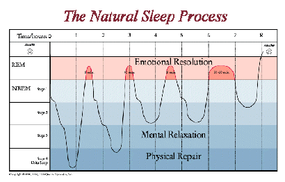
Portion of REM sleep increases throughout the night, which means our brain resembles that of an awake one, so that our brain is ready to wake up
As we go through the night and the cycles, we don’t go as deep into the stages before we go to REM sleep
Evaluation for ultradian rhythms?
Tucker RS for IDs in ultradian rhythms
Aim: find out whether there are differences in people's ultradian rhythm
Procedure: participants were studied over 11 consecutive days and nights in a strictly controlled lab environment. The researchers assessed sleep duration, time to fall asleep, and the amount of time in each stage
Findings: significant variations between apps in all these characteristics, particularly 3 and 4, which showed up consistently across 8 nights
Conclusion: suggested these differences are likely to be biologically determined. This makes it difficult to describe ‘normal sleep’ in any meaningful way
Kleitman - RS for different stages of the sleep cycle and REM sleep
Aim - to investigate brain activity change throughout sleep
Participants: 7 adult males and 2 adult females.
Procedure: participants were asked to report to the lab at bedtime and they were then connected to an EEG taking measurements all the way through the night
Findings: found that everyone had periods of REM sleep every night. Found high incidence of dream recall when the participants were awakened during REM periods of their sleep. Found that rapid eye movement during REM sleep varied depending on the dream type and mirrored their rapid eye movement
Concusion: it can be concluded that the stages of sleep follow a typical pattern throughout the night, and dreams mostly occur during REM sleep
.
Strength of research into ultradian rhythms is that it has improved understanding of age-related changes in sleep.
Sleep scientists have observed that SWS significantly reduces with age
The growth hormone is mostly produced during SWS and therefore this is reduced in older people, as they aren’t growing anymore they are no longer producing as much growth hormone as young people
May explain why older people experience issues like reduced alertness, impaired memory, and slower physical recovery
Understanding these changes has practical applications, as interventions like relaxation techniques and medication can be used to promote SWS in older adults. This not only improves their sleep quality but also potentially mitigates some of the associated cognitive and physical impairments. This suggests that knowledge of ultradian rhythms has practical value