3.1 - Exchange Surfaces
1/35
There's no tags or description
Looks like no tags are added yet.
Name | Mastery | Learn | Test | Matching | Spaced | Call with Kai |
|---|
No analytics yet
Send a link to your students to track their progress
36 Terms
Why have specialised exchange surfaces (SA:V)
Organisms need to exchange materials
Urea, carbon dioxide and heat out of body
Oxygen, glucose into
Occurs across plasma membrane
High SA:V
Diffusion of substances is fast
Generally for smaller organisms
Low SA:V
Diffusion is slower
Generally for larger organisms
Calculating
Calculate SA
Calculate V
SA:V with V = 1
Why specialised exchange surfaces in multicellular organisms
Require specialised surfaces unlike single celled organisms
Due to:
Cells are not in direct contact with external environment
Diffusion distances between cells and their environment are large
Larger organisms have higher metabolic rates so they need more oxygen and glucose
Key features of specialised exchange surfaces
Large surface area
Larger area across which substances can be exchanged
More substances can travel across per area
Root hair cells
Thin walls
Minimises diffusion distance
Alveoli
Good blood supply
Maintains steep gradient
Gills
Being surrounded by partially permeable membranes
Control what substances are exchanged
Lungs
Allow oxygen to enter the blood and carbon dioxide to leave
Uses exchange surfaces called alveoli
Is inside the body
Air is not dense enough to support and protect these delicate structures
The body would otherwise lose water and dry out
Pathway of air
Air enters the trachea
Travels into the two bronchi, with one bronchus going to each lung
Travels into smaller airways called bronchioles
Air travels into clusters of air sacs called alveoli
Airway tissue
Ciliated epithelium
Contains goblet cells and ciliated epithelial cells
Goblet cells produce and secrete mucus to trap dust and microbes
Cilia waft the mucus upwards to the mouth
Trachea
Large tube that carries air from throat to lungs
Rings of cartilage keep the airway open
Smooth muscle can contract or relax to open/close the airway and change airflow
Elastic tissue allow stretching and recoiling
Lined with ciliated epithelium
Bronchi
Two main branches extending from the trachea that carry air into each lung
Reinforced with cartilage to keep the airway open
Smooth muscle to contract/relax and change airflow
Elastic tissue allows stretching and recoiling
Lined with ciliated epithelium
Bronchioles
Two smaller airways branching from the bronchi
No cartilage to change shape
Smooth muscle to contract/relax and change airflow
Elastic tissue allows stretching and recoiling
Squamous epithelium
Alveoli
Gas exchange
Oxygen diffuses from alveoli into the pulmonary capillaries where it binds to haemoglobin
Carbon dioxide dissociated from haemoglobin and diffuses into the alveoli
Adaptations of alveoli
One layer of squamous epithelial cells
Large SA
Partially permeable
Surrounded by dense network of capillaries
Brings blood close to air for gas exchange
Ventilation of air
Maintains steep diffusion gradient
Elastic fibres
Allow stretching and recoiling
Collagen fibres
Prevents overstretching and bursting
Moist inner layer
Gases to dissolve
Ventilation
Is the constant movement of air into and out of the lungs
Consists of expiration and inspiration
Muscles involved in ventilation
Diaphragm
Sheet of muscles that moves the ribcage up and out when it contracts
External intercostal muscles
Found between the ribs and pull the ribcage up and out when they contract
Internal intercostal muscles
Found between the ribs and pull the ribcage down and in when they contract
Inspiration
External intercostal muscles contract while the internal intercostal muscles relax
Ribcage moves up and out
Volume of the thoracic cavity increases
Diaphragm contracts and flattens
Increases the volume of the TC
Lung pressure decreases below atmospheric pressure
Air flows into the lungs down the pressure gradient
Expiration
Normally a passive process however forced expiration can occur when playing wind instrument or after exercise
EI relax
Ribcage moves down and in
Volume of thoracic cavity decreases
Diaphragm relaxes and unflattens
Decreases the volume of TC
Lung pressure increases above atmospheric pressure
Air if forced out of lungs
Elastic fibres in alveoli also shrink when pressure decreases
Measuring ventilation
Ways to measure data on lung function, volume and capacity
Peak flow meter
Vitalograph
Spirometer
Measuring lung volume
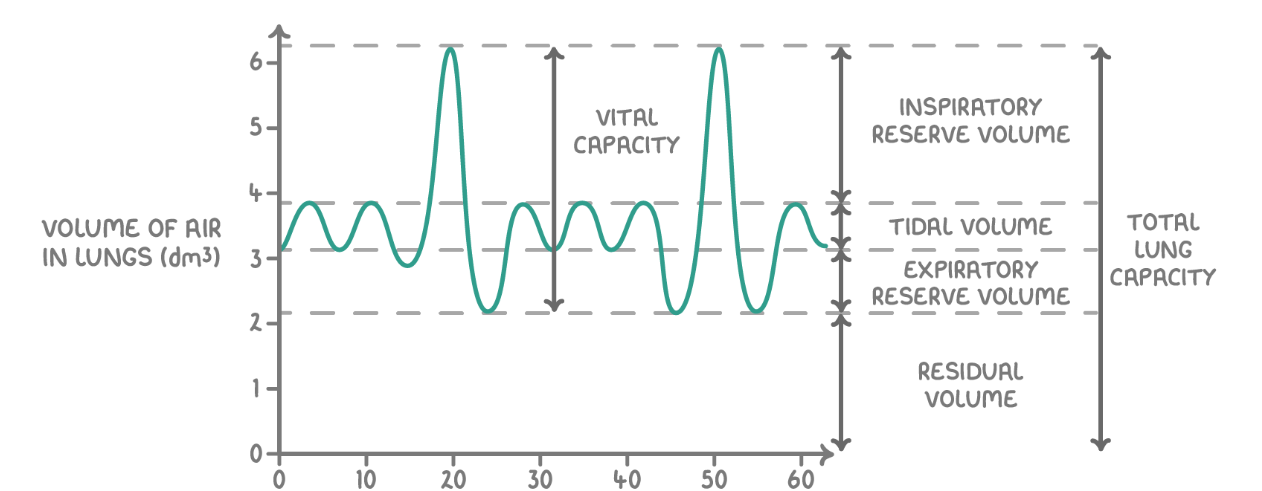
Definitions of: breathing rate, tidal volume, vital capacity
Breathing rate
Number of breaths taken per minute
Measured by counting the number of peaks in a minute
Tidal volume
Volume of air breathed in or out in an average breath during rest
Vital capacity
Maximum volume of air that can be inhaled or exhaled in one deep breath
Measured from max peak height
Definitions: inspiratory reserve volume, expiratory reserve volume, residual volume
Inspiratory reserve volume
Maximum volume of air that can be inhaled above normal inhalation
Expiratory reserve volume
Maximum volume of air that can be inhaled above a normal inhalation
Residual volume
Volume of air that remains in lungs after the largest possible exhalation
Calculating oxygen consumption
Slope of the spirometer trace
Ventilation rate equation
Ventilation rate = tidal volume x breathing rate
Why insects need gas exchange
Have chitin exoskeleton which prevents gas exchange
Covered in a waterproof cuticle to prevent water loss
To deliver oxygen to cells
Allows aerobic respiration to occur to release energy by hydrolysis of ATP
To remove carbon dioxide
Reduced pH which can denature enzymes
Tracheae
Air filled tubes branching through the body
Adaptations
Reinforced with spirals of chitin to prevent collapsing
There are multiple to increase SA
Tracheoles
Fine branches of tracheae that deliver gases to cells
Adaptations
Penetrate directly into tissues to reduce gas diffusion distance
Thin walls
High branched to maximise SA
Not reinforced with chitin to allow gas exchange
Fluid at ends (tracheal fluid) allows oxygen to dissolve to aid diffusion and reduce water loss
Spiracles
External opening of the tracheal system on exoskeleton along abdomen and thorax
Can be opened or closed to control gas exchange and minimise water loss
Process of gas exchange in insects
Air enters the tracheal system through open spiracles
Air moves into larger tracheae and diffuses into smaller tracheoles
Tracheoles branch out throughout body
Oxygen dissolves in tracheal fluid and diffuses down concentration gradient from tracheoles into body cells
Carbon dioxide diffuses out of cells into tracheoles
Air is then carried back to spiracles and released
How is concentration gradient maintained in insects
Cells using up oxygen for respiration
Keeps concentration low in cells
CO2 production in cells to keep concentration high
Continuous ventilation
Fresh air is supplied to tracheal system via spiracles
6 additional insect ventilation mechanisms
More spiracles open
Allows more oxygen to enter the tracheal system
Mechanical active ventilation
When muscles around tracheae contract and relax changing the volume and pressure on the abdomen and pumps air in and out the spiracles
Movement of tracheal fluid out of tissue
Increases diffusion rate and SA for gas exchange
Collapsable tracheae, accessory sac and air reservoirs
Inflate or deflate to ventilate and can increase the volume of air moved through the system
Movement of wing muscles connected to sacs
Pump air to ventilate tracheal system
Vibration of thoracic muscles
Pumps air to ventilate tracheal system
Lactic acid accumulation in insects
Can affect rate of gas exchange
Reduces the water potential in tracheal fluid at the end of tracheoles
Water leaves the tracheoles via osmosis
Higher SA for gas exchange
Respiratory system in bony fish
Have high oxygen needs
Live under water which is denser than air so slower diffusion of oxygen
Has lower oxygen concentration
Very active so high oxygen demands
Structure of gills
Covered by an operculum flap
Consists of stacked filaments containing gill lamellae
Gill lamellae are surrounded by extensive blood vessels
Adaptations of gills
Lamellae provide large SA
Lamellae membranes are thin to minimise diffusion distance
Gills have a rich blood supply to maintain steep diffusion gradients
Countercurrent flow of blood and water creates even steeper gradient
Overlapping filament tips increase resistance so water flow over gills more slowly
Counter-current
Blood and water flow over each lamellae in opposite directions
Means that oxygen rich blood meets water that is at its most oxygen rich when it first moves across gills
Maximising diffusion of oxygen
Oxygen poor blood returning from body tissue meets oxygen reduced water
Still allows diffusion of oxygen
Maintains a steep concentration gradient across the entire gill
Parallel flow
An equilibrium would be reached meaning less oxygen would diffuse
Less effective and efficient than counter-current
Ventilation: closed mouth
Floor of the mouth is raised so pressure increases and volume decreases
Means water is pushed over the gills and into the gill cavity
Oxygen is transferred into the blood
Ventilation: open mouth
When a fish opens its mouth, water enter buccal cavity
Floor of the mouth is lowered so volume increases and pressure decreases
Means water travels down the pressure gradient in the buccal cavity