Lecture 7: Parasitology III
1/67
There's no tags or description
Looks like no tags are added yet.
Name | Mastery | Learn | Test | Matching | Spaced |
|---|
No study sessions yet.
68 Terms
Plasmodium
a group of parasites that causes malaria
P. vivax, P. falciparum, P. ovale, P. malariae
Hepatocytes
main liver cells that perform metabolic functions
erythrocytes
red blood cells responsible for oxygen transport
Malaria location and reservoir
hepatocytes/erythrocytes and reservoir is humans
Malaria Transmission
spread by vector-borne female anopheles mosquitoes and also congenital & needle transfer
Malaria Epidemiology
affects worldwide, most fatalities in children, most morbidity and mortality due to P. falciparum, and increasing drug and insecticide resistance
Malaria life cycle alternates b/t
2 hosts:
female Anopheles mosquito vector sexual cycle
human host intermediate asexual cycle
Malaria life cycle Mosquito: first step of infection
Mosquito bites an infected human → picks up gametocytes. Gametocyte mature into gametes inside mosquito midgut, and then fertilization of microgamete and macrogamete becomes zygote
Malaria life cycle Mosquito: gametes fertilize
Zygote transforms into an ookinete that embeds in the outside of midgut wall and forms an oocyst that grows, divide, and produces thousands of sporozoites (sporogony)
Malaria life cycle Mosquito: final step to get ready to infect humans
Oocysts burst releasing sporozoites in mosquito’s body cavity, sporozoites migrate to salivary glands
They are now ready to infect humans
Malaria life cycle Human: mosquito infects humans
Mosquito injects sporozoites into human during blood meal, the sporozoites travel thru bloodstream to liver and inside become a liver schizont that produces thousands of merozoites
Malaria life cycle Human: RBCs invaded by merozoites
Schizonts rupture releasing merozoites into bloodstream, the merozoites invade RBCs, inside the RBC the parasite progresses thru 3 main stages; ring, trophozoite, and schizont
Ring stage
Earliest form inside RBC that looks like a ring and is the beginning of feeding
Trophozoite stage
The feeding and growing stage where the parasite digests hemoglobin and produces hemozoin (brown pigment) and it grows larger
Schizont stage
Sporozoite enters liver cell and become trophozoite that becomes a schizont that divides and forms 8-32 merozoites, when the merozoite mature the liver cell schizont rupture
Malaria life cycle Human: RBC ruptures
releases merozoites causing clinical symptoms such as fever, chills, anemia and then merozoites infect new RBCs
Sporozoite Role
parasites stored in salivary glands of mosquito that are injected into humans and start malaria by invading liver cells
Merozoite Role
Plasmodium parasites invade red blood cells, multiple, and cause symptoms of malaria
Liver to blood and infects RBCs and is asexual
Schizont Role
the asexual replicative stage that produces merozoites and causes RBC rupture, leading to fever and anemia
Trophozoite role
the feeding and growing form in RBCs that digests hemoglobin and matures toward division
Gametocyte Role
the sexual Plasmodium forms in human blood that are taken up by mosquitoes and begin the parasite's sexual cycle, enabling transmission to new hosts (infective to mosquito)
Ookinete Role
motile form that invades mosquito gut wall and becomes an oocyst
Oocyst role
In mosquito outside midgut wall that produces thousands of sporozoites
malaria species roles in symptoms
fever cycle with synchronous bursts of merozoites
P. vivax & P. ovale: 48 hr spike
P. malariae: 72 hr spike
P. falciparum: 48 hr broad
Malaria symptoms
chills, fever, splenomegaly, myalgia, headache, and anemia results from erythrocyte destruction
cerebral malaria
extreme result of P. falciparum only disruption of cytokines networks and IV quinidine for treatment
latent hepatic forms in
P. vivax & P. ovale
P. falciparum
multiple rings per RBC, no trophozoites or schizonts, and banana shaped gametocytes
Erythrocytic Stages of Human Malarias signs P. falciparum
Banana -shaped gametocytes, no circulating trophozoites or schizonts, and multiple ring stages per RBC
Malaria Immunity
slow to develop; requires multiple infection, short lived, and easily reinfected
Malaria Treatment
multidrug resistance common, especially chloroquine!
Malaria prevention
2 vaccines, avoid mosquitoes, and drugs like doxycicline
Phylum Platyhelminthes AKA flatworms
a diverse group of soft-bodied invertebrates that includes free-living and parasitic species
Phylum Platyhelminthes classes:
Cestoda and Trematoda
Class Cestoda
Tapeworms; flattened segmented bodies, no internal digestive system; nutrients absorbed across cuticle
Class Cestoda adult attach
by the anterior end (scolex) to the gut wall of definitive host
Class Cestoda transmission
ingestion of larval cysticerci (bad) or eggs (very bad), and their segments (proglottids) grow anterior to posterior
Class Trematoda
Flukes of the lungs, liver and blood and broad flattened bodies with a simple digestive system; a single opening serves as mouth and anus
Class Trematoda transmission
One or more intermediate host, one of which is a snail and transmission can be invasive or ingestive
Phylum nemahelminthes classes
Nematoda
Class Nematoda
Roundworms of the tissues and gut, has cylindrical body; well developed digestive and nervous systems.
Class Nematoda Transmission
Ingestive (no intermediate host) Ascaris, Trichuris, etc
Invasive (no intermediate host) Hookworms
Vector-borne (one intermediate host) Filarial Worms
Class Cestoda species
Taenia saginata (raw beef) and Taenia solium (raw pork) and both found in intestine
Class Nematoda (roundworm) species
Enterobius vermicularis found in lumen of intestinal/anal and transmitted by ingesting direct egg
Beef tapeworm
organism is Taenia saginata found in lumen of jejunum (upper small intestine) and humans are the only definitive host, cattle is intermediate host
Beef tapeworm epidemiology
World-wide and cosmopolitan in beef eating countries causes abdominal discomfort; nausea, vomiting and diarrhea and rarely serious
Beef tapeworm immunity and diagnosis
Humoral response to adults, reinfection is possible, and observation of proglottids or eggs in stool
Beef tapeworm treatment
drugs single dose is effective and prevented by good hygiene and well cooked beef
Egg / Oncosphere
host is cattle and is embryo in feces, eaten by cattle
Cysticercus
Larval stage (infective to humans)and found in cattle muscle
Adult tapeworm
Mature worm producing eggs and found in human intestine
Gravid proglottid
Segment containing thousands of eggs and found in human feces
Life cycle of Beef Tapeworm: Taenia saginata part 1
Adult Taenia saginata lives in the small intestine of humans, releases proglottids containing fertilized eggs passed in human feces, and cattles are infected by contaminated feed/water
Life cycle of Beef Tapeworm: Taenia saginata part 2
in cows intestine the oncosphere hatches and enters bloodstream to muscle tissue and develop cysticerci, and then humans eat undercooked beef containing cystercerci
Pork tapeworm
organism is Taenia solium, found in lumenal jejunum of adults and cysticeri found in any tissue
Pork tapeworm host
definite host are humans and intermediate host are pigs or humans
Pork tapeworm transmission
- Ingest cysticerci; definitive host (bad)
- Ingest eggs; intermediate host (VERY BAD)
Pork tapeworm lifecycle
like beef tapeworm except eggs are infectious for humans and cysticerci develop in any human tissue
Pork tapeworm diagnosis and prevention
Adults same as beef tapeworm and X-ray, calcified dead larva; CAT/MRI, viable cysticerci, prevented by good hygiene and well cooked pork, and freezing kills cysticerci
Pork tapeworm treatment
multiple treatment to kill cysticerci, consensus is to not treat cysticerci outside of CNS
Pinworm
Organism is Enterobius vermicularis and found in adults in colon
Pinworm transmission
Ingestion of eggs and definitive host is only humans
Pinworm epidemiology
World-wide and cosmopolitan; >20 million in USA and an equal opportunity parasite
Pinworm pathology immunity treatment
Intense perianal pruritis, bacterial infection, no immunity and treatment are drugs or multiple treatment for severe infection
Pinworm life cycle
infective eggs are ingested through contamination and hatch in the small intestine releasing larvae and mature into adult worms in the large intestine, migrate to perianal area and lay thousands of eggs on skin leading to scratching where eggs get into fingernails and become infective again
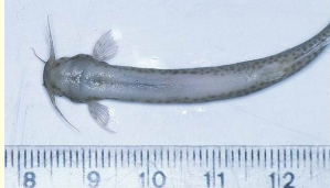
Candiru AKA vampire catfish
organism is Vandellia cirrhosa and are tiny parasitic catfish found only in the amazon/oranoco rivers of south america
Candiru food
Voracious appetite for blood; will parasitize fish, mammals, & HUMANS and it has no enemies
Candiru life cycle
Inserts itself inside the gill flap. Spines pierce the fish and draws blood while anchoring the candiru in place. It feeds on the blood using its mouth as a slurping apparatus and while rasping with the long teeth on its top jaw.