Oral Cavity and Salivary Glands
1/25
Earn XP
Description and Tags
go back over the muscles, TMJ
Name | Mastery | Learn | Test | Matching | Spaced |
|---|
No study sessions yet.
26 Terms
What are the two regions of the oral region?
Oral Region is subdivided into two parts
1) Vestibule - space between the lips and the teeth/gums
2) Oral Cavity Proper - space in between the teeth and the back part of your mouth
→ oropharynx is behind them
What are the parts of the Tooth?
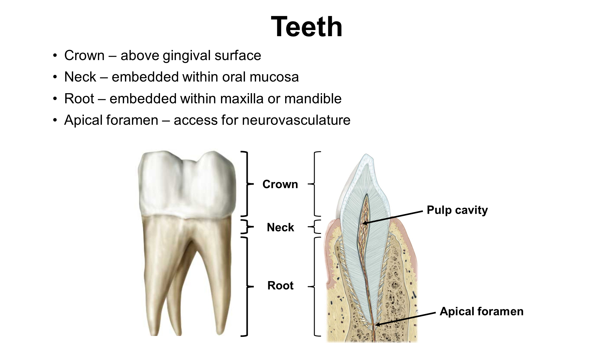
Crown - above gingival surface (gums)
Neck - part of the tooth embedded in the oral mucosa
Root - anchors that keep the tooth embedded within the maxilla or mandible
Apical Foramen - access for neurovasculature
What are Deciduous Teeth?
Baby Teeth
→ 20 teeth - with five on each side of the mandible and maxilla
→ when it is time for the teeth to come out, the roots of the teeth are eaten away at in order to make space for permanent teeth
Subdivided into three kinds of teeth
→ 2 incisors
→ 1 canine
→ 2 molars
What are Permanent Teeth? How are they innervated?
32 teeth » 8 on each side of the mandible and maxilla. Subdivided into four kinds of teeth
→ 2 incisors
→ 1 canine
→ 2 premolars
→ 3 molars
The teeth are innervated by the trigeminal nerves
→ the maxilla is innervated by CN V2 (used only for sensation) and the mandible is innervated by CN V3 (used also for motor and sensation)
What is the Hard Palate?
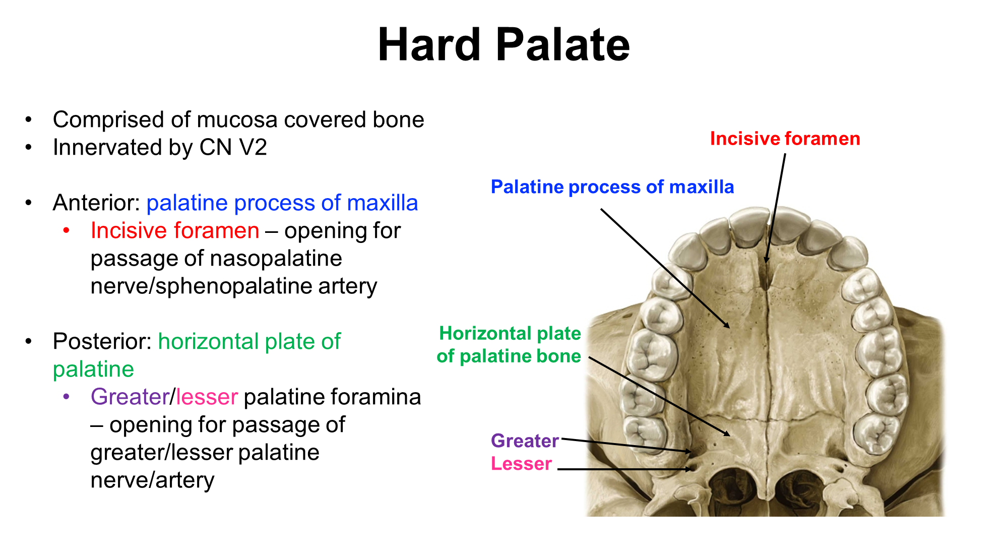
Hard palate is the mucosa that covers the bone on the top of your mouth and is innervated by the CN V2. Subdivided into two parts that each contain foramen.
1) Anterior: palatine process of maxilla
→ incisive foramen - the opening for nasopalatine nerve and sphenopalatine artery
2) Posterior: horizontal plate of palatine
→ greater/lesser palatine foramen - the opening for the passage of the greater/lesser palatine nerves/artery
What is the Soft palate? What are its structures
Mucosa covering skeletal muscle and aponeurosis on the top of the mouth. Lays behind the hard palate and is also innervated by CN V2
1) contains the uvula which is an extension that covers the nasopharynx when you swallow
2) Has two arches
→ Palatoglossal arch - connects the soft palate to the tongue
→ Palatopharyngeal arch - connects the soft palate to the pharynx
3) Palatine tonsils
→ tonsils that lay in between the palatoglossal and palatopharyngeal arch
What are the papillae of the tongue?
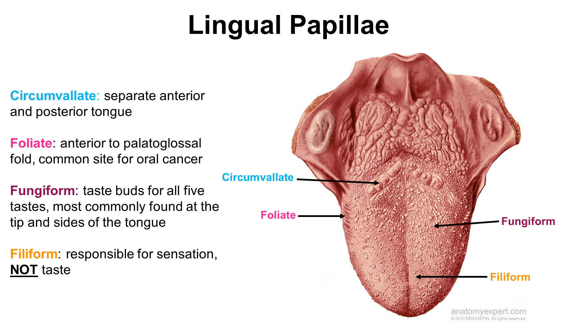
Lingual papillae are typically responsible for sensory and taste sensation on the tongue. There are four kinds
1) Circumvallate - the largest of the papilla
2) Foliate - anterior to the palatoglossal arch
→ tend to atrophy when you get older and are a site for oral cancer
→ columnar in shape
3) Fungiform
→ little white dots on the top of your tongue
4) Filiform
→ responsible for sensation and NOT TASTE the only one that is not used in taste
What are the surface structures of the tongue
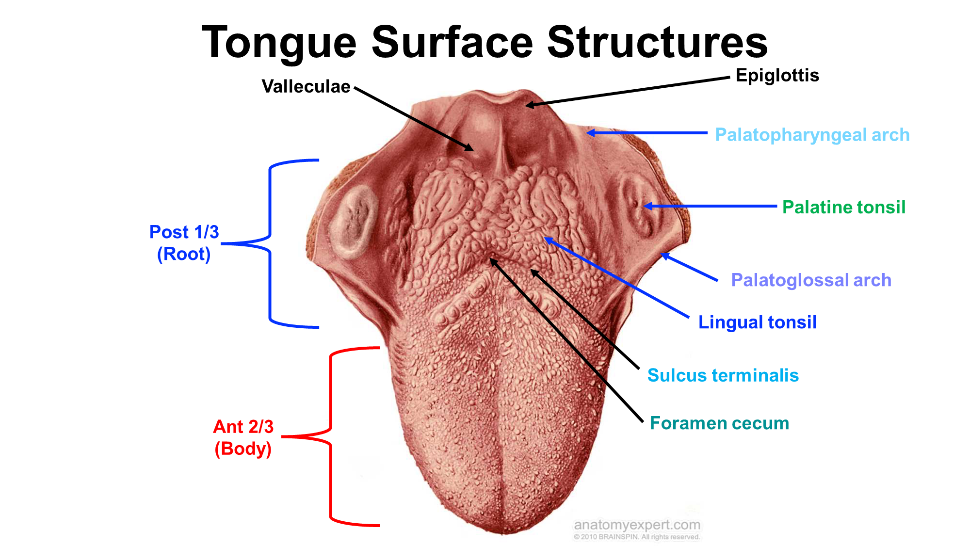
1) Anterior Portion (makes up 2/3 of the tongue)
2) Sulcus Terminalis
→ fold that divides the anterior and posterior portions of the tongue
→ where the sulcus comes together to a point it forms the foramen cecum an anatomic remnant
3) Lingual tonsils
→ lay on the posterior 1/3 portion of the tongue
4) Valleculae
What is the Thyroglossal Duct? What two issues are associated with it
Embryologic connection between the oropharynx and the neck
→ used in allowing for the thyroid to migrate from its initial location to the final location
persistent thyroglossal duct occurs when it fails to close
thyroglossal duct cyst - fluid collection within persistent duct
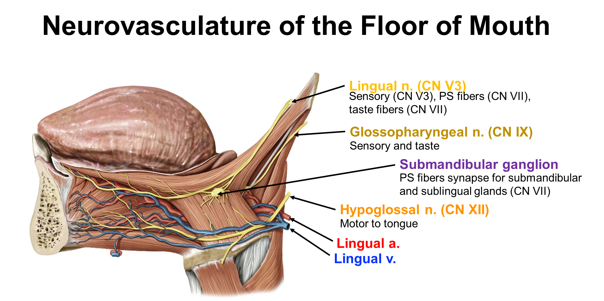
What is the General Sensation and Special Sensation (Taste) at the Tongue?
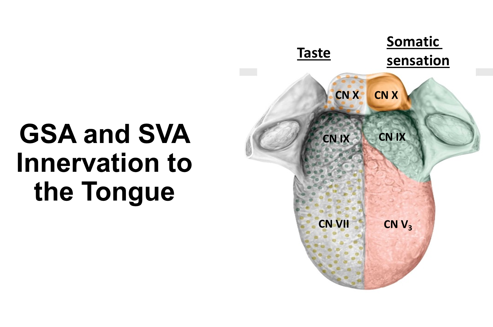
The first 2/3rds of the tongue has split innervation between general and special sensation
→ taste (special) is done by the facial nerve (CN VII)
→ general sensation is performed by the lingual branch of the trigeminal nerve (CN V3)
The posterior 1/3 of the tongue shares innervation for both special and general with the glossopharyngeal nerve (CN IX)
The very back, or the pharyngeal extremity is innervated by the vagus nerve or CNX
What are the two muscle groups of the tongue?
Intrinsic - muscles that provide shape to the tongue
Extrinsic - used to lift, point and move our tongue
→ Genioglossus
→ Hypoglossus
→ Styloglossus
→ Palatoglossus
What are the four extrinsic muscles of the tongue? (Origin, Insertion, Action and Innervation)
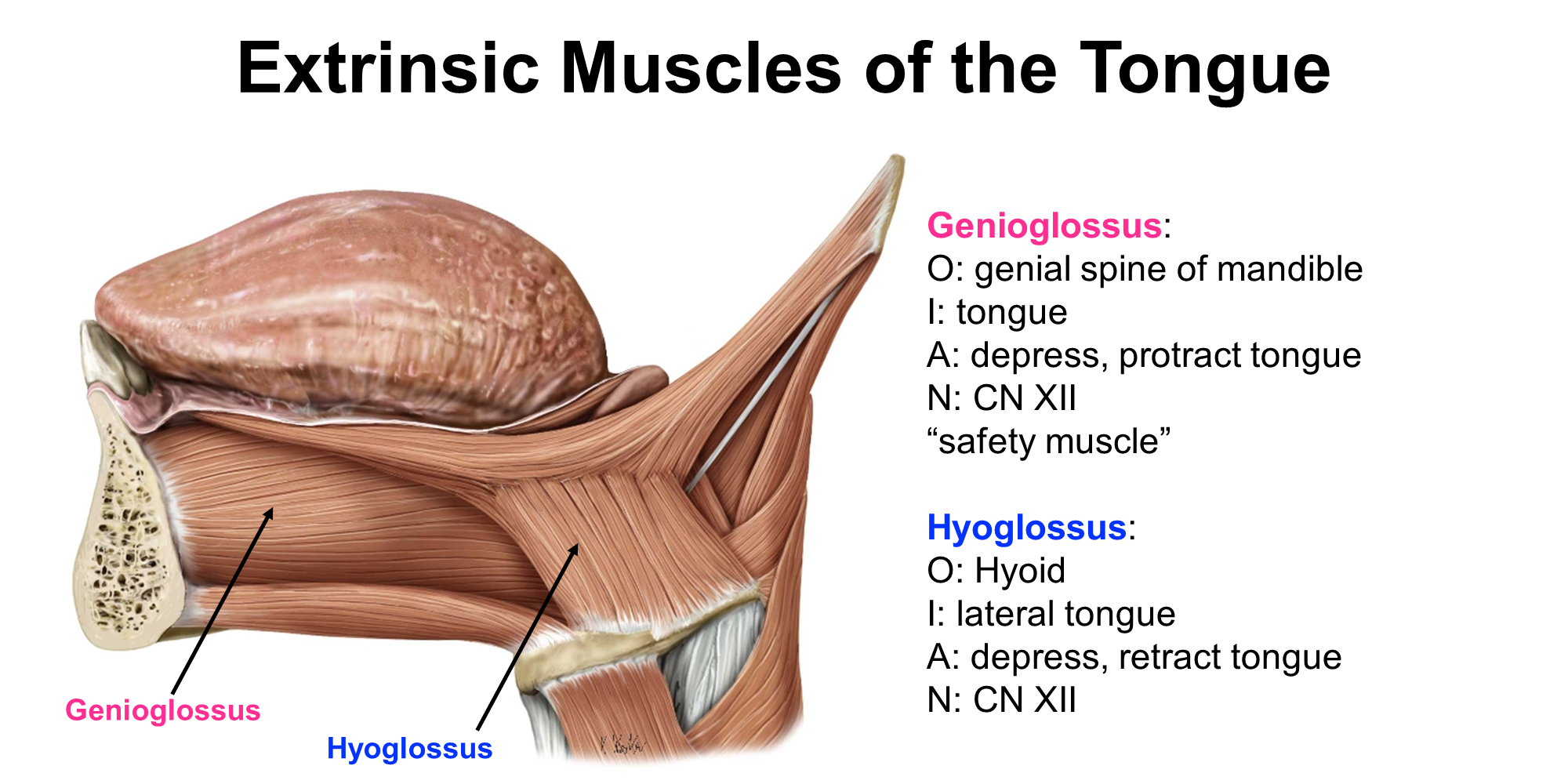
There are four muscles of the tongue
1) Genioglossus - originate in the genial spine of the mandible and inserts into the tongue
-> responsible for depressing and protracting the tongue
-> innervated by CN XII
2) Hyoglossus - originate in the hyoid bone and insert into the lateral tongue
-> responsible for depression and retraction of the tongue
-> innervated by CN XII
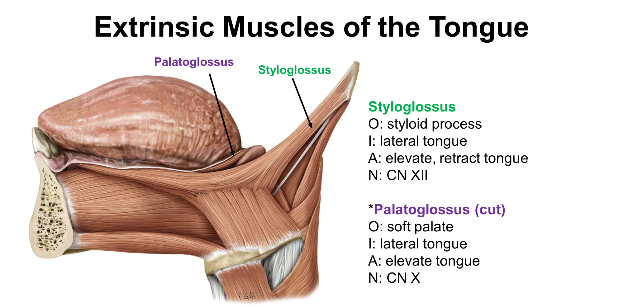
3) Styloglossus - originates in the styloid process and inserts into the lateral tongue
-> responsible for elevation and retraction of the tongue
-> innervated by CN XII
4) Palatoglossus - originates in the soft palate and inserts into the lateral tongue
-> responsible for elevation of the tongue
-> innervated by CN X
What is Hypoglossal Nerve Palsy?
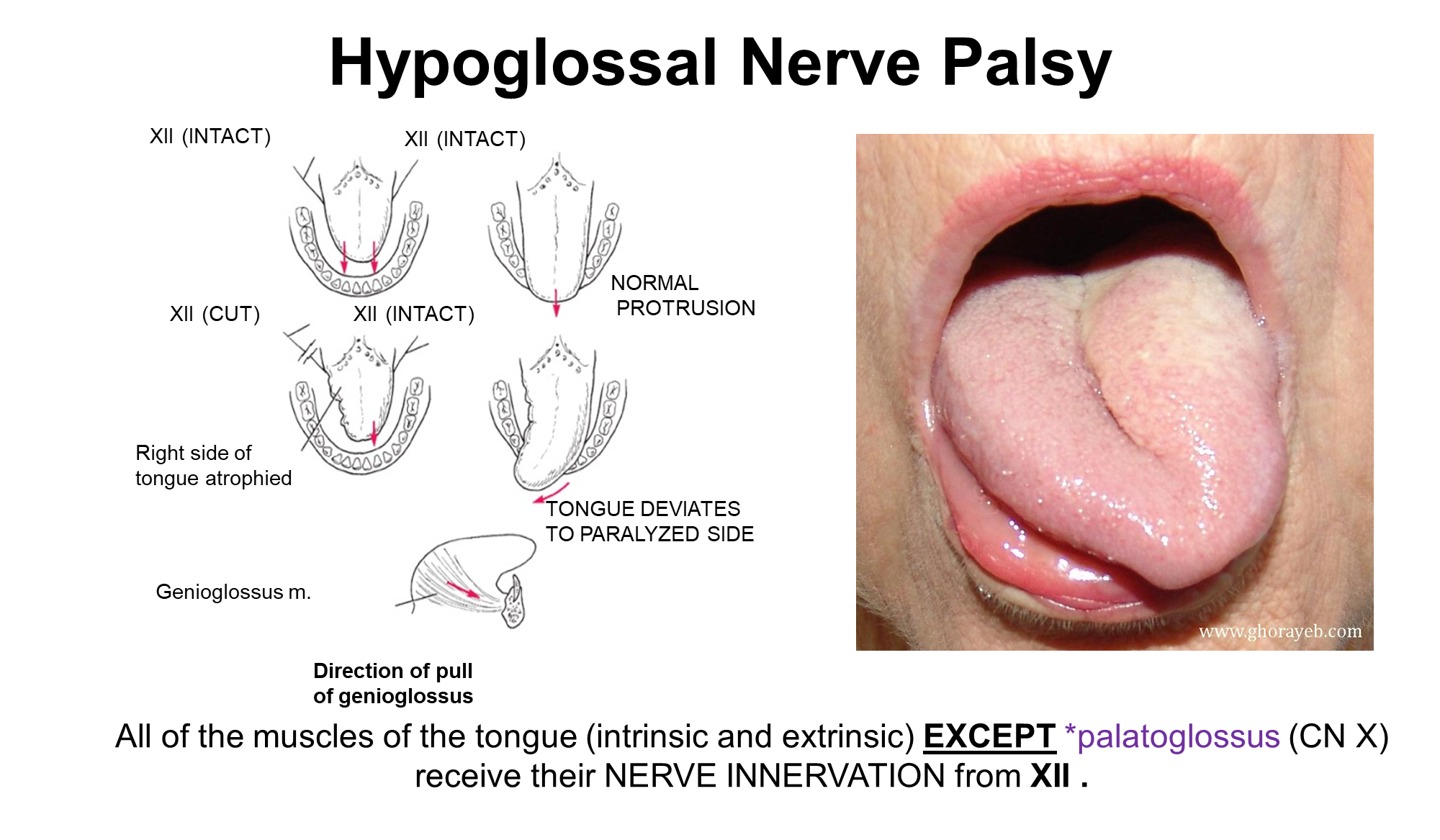
If your tongue deviates there may be a issue with Cranial Nerve XII
→ the tongue will deviate towards the paralyzed side
What are the Muscles of the Mouth Floor?
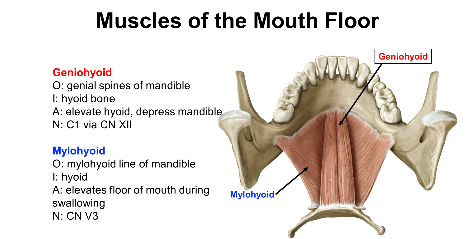
Not tongue muscles but aid in the movement of the mouth floor
What are the minor and major Salivary Glands?
Can either be minor or major
1) Minor glands - glands underneath the mucous membrane that produce saliva that keep our mouth moist when not eating
→ innervated parasympathetically from CN VII
2) Major Glands (3 of them) - the primary salivary sources when eating
→ Parotid
→ Submandibular
→ Sublingual
What is the Parotid Gland and what does it contain
Parotid
→ largest salivary gland but does not do most of the salivary production, innervated by cranial nerve IX
→ the parotid duct emerges from the anterior part of the parotid gland and inserts into the oral cavity on the side of your mouth (parallels zygomatic arch 1 finger down)
Contains the:
→ Parotid Gland and Duct
→ CN VII Motor Fibers
→ External Carotid Artery
→ Retromandibular Vein
What can a Parotidectomy Cause
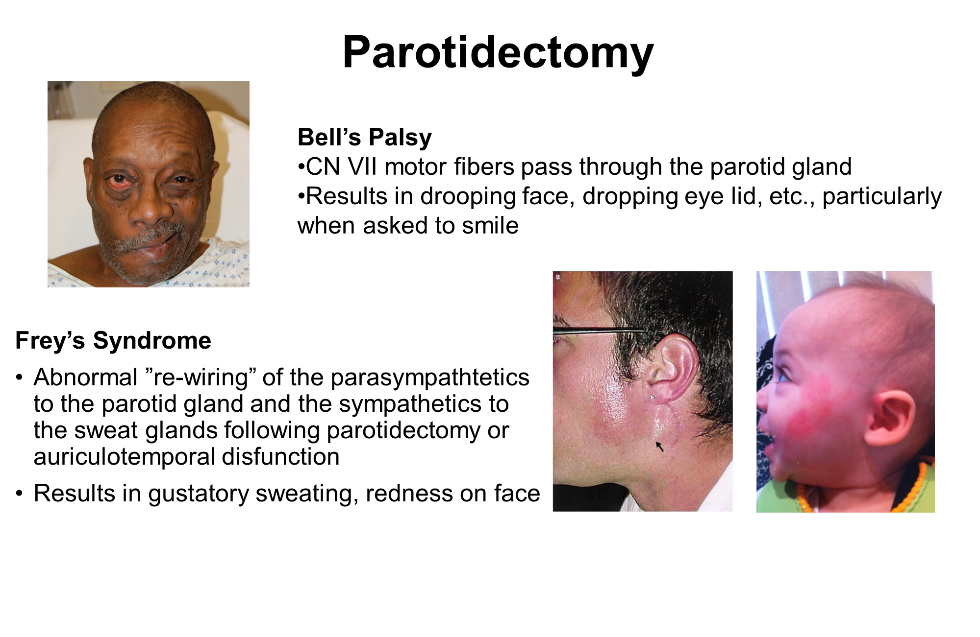
because removal of the parotid gland can remove the CN VII Motor Nerves, you can paralyze the face causing Bell’s Palsy
→ drooping face when asked to smile
Can also cause Frey’s Syndrome
→ Abnormal rewiring of the parasympathetics to the parotid gland can occur where they rewire to the sympathetic and sweat glands
→ causes you to sweat when you eat and have a red face
What are the Submandibular and Sublingual Glands
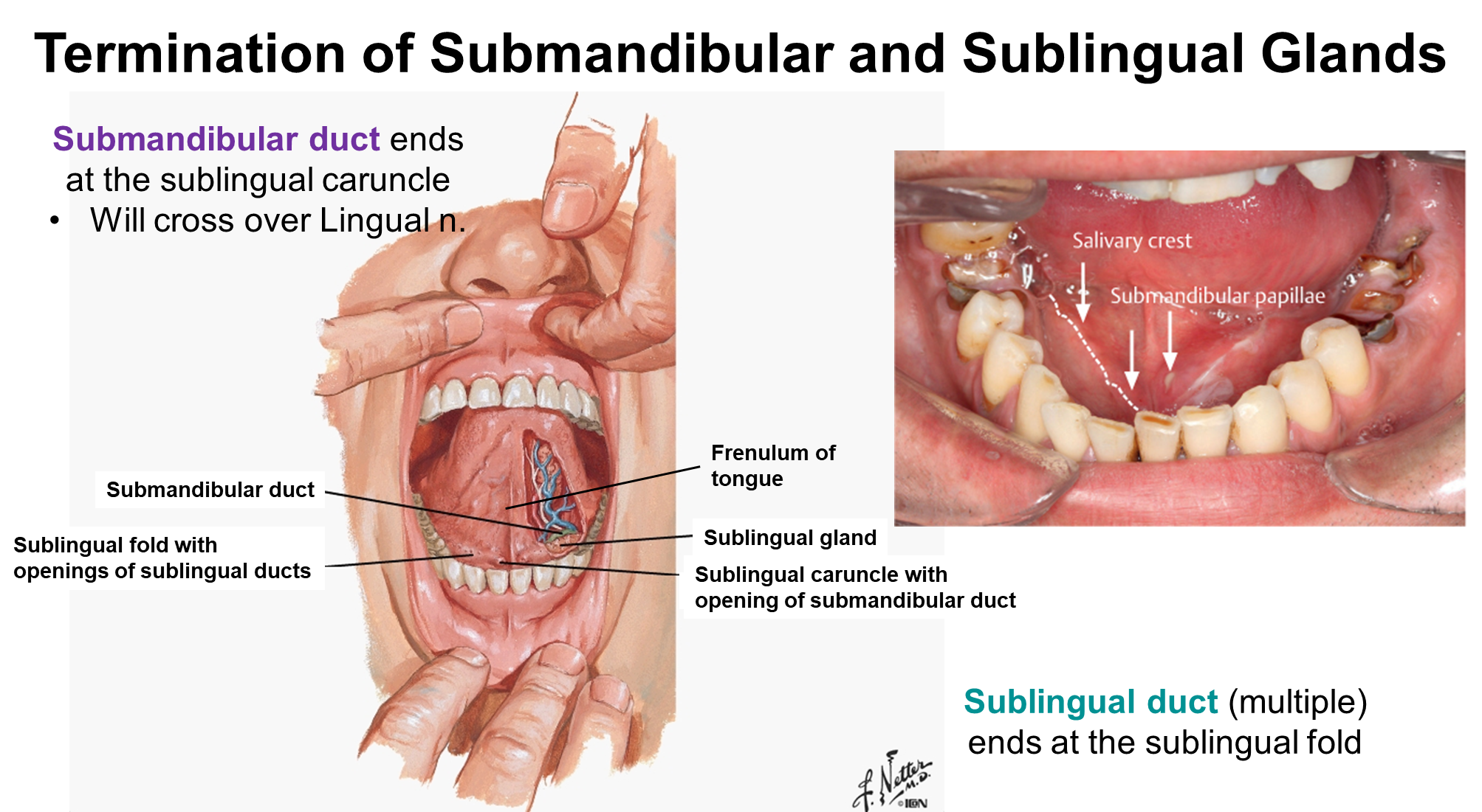
Two of the Major Salivary Glands
1) Sublingual Gland
→ lies against the internal surface of the anterior mandible
2) Submandibular Gland
→ responsible for creating most of the salivation when eating
→ found around the mylohyoid muscle and in the submandibular triangle of the neck
→ facial artery and vein pass through the gland
→ has a single duct that exits underneath your tongue at the sublingual caruncle
What are the Four Muscles of Mastication? (Origin, Insertion, Action, and Innervation)
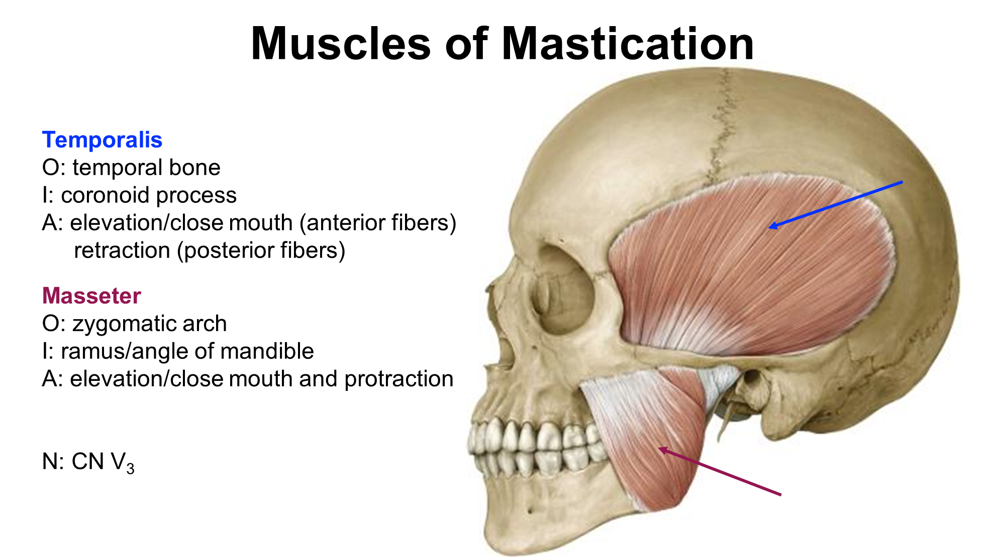
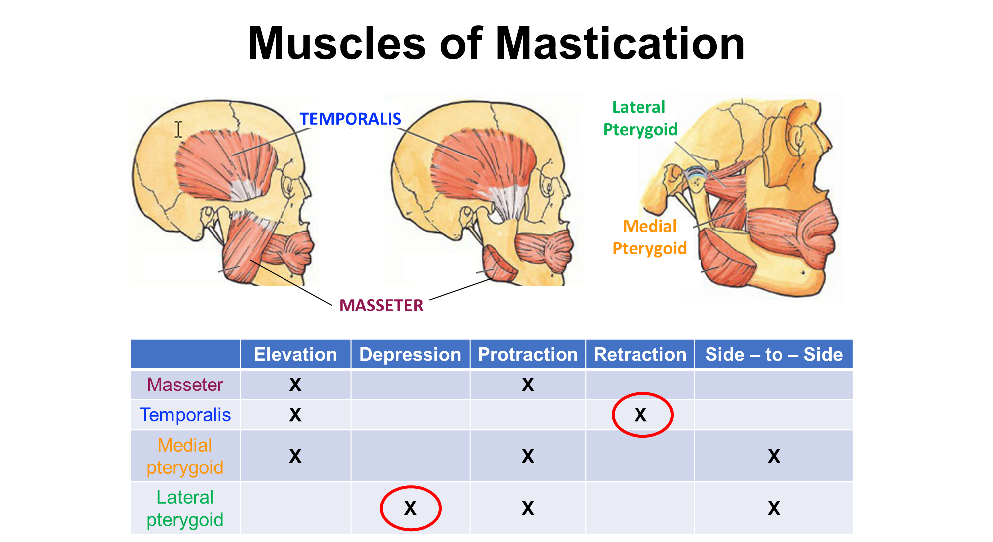
The Four muscles of mastication are:
1) Temporalis originating in the temporal bone and inserting into the coronoid process
-> works to elevate and close the mouth
-> involved in retraction
2) Masseter originating at the zygmoatic arch and inserting into the ramus angle of the mandible
-> elevates and closes the mouth
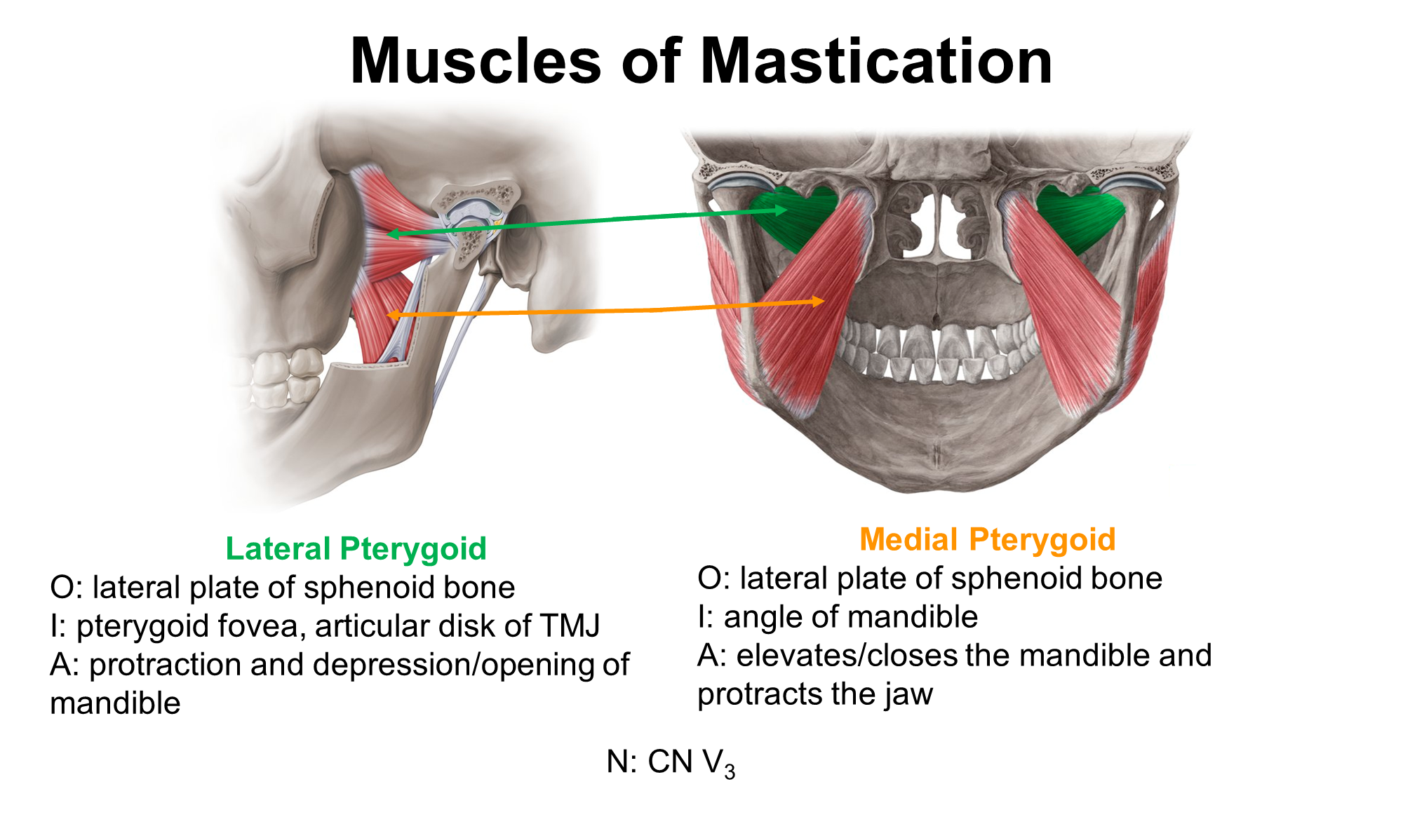
3) Lateral Pterygoid originating in the lateral plate of the sphenoid bone and inserting into the pterygoid fovea
-> protracts and depresses the mouth
4) Medial Pterygoid originating at the lateral plate of the sphenoid bone and inserts into the mandible
-> elevates and closes the jaw
What does a lesion of Cranial Nerve V3 cause?
Atrophy of the muscles of mastication leading to a sunken cheek
→ people that will open their mouth will have their jaw deviate towards the side of the lesion
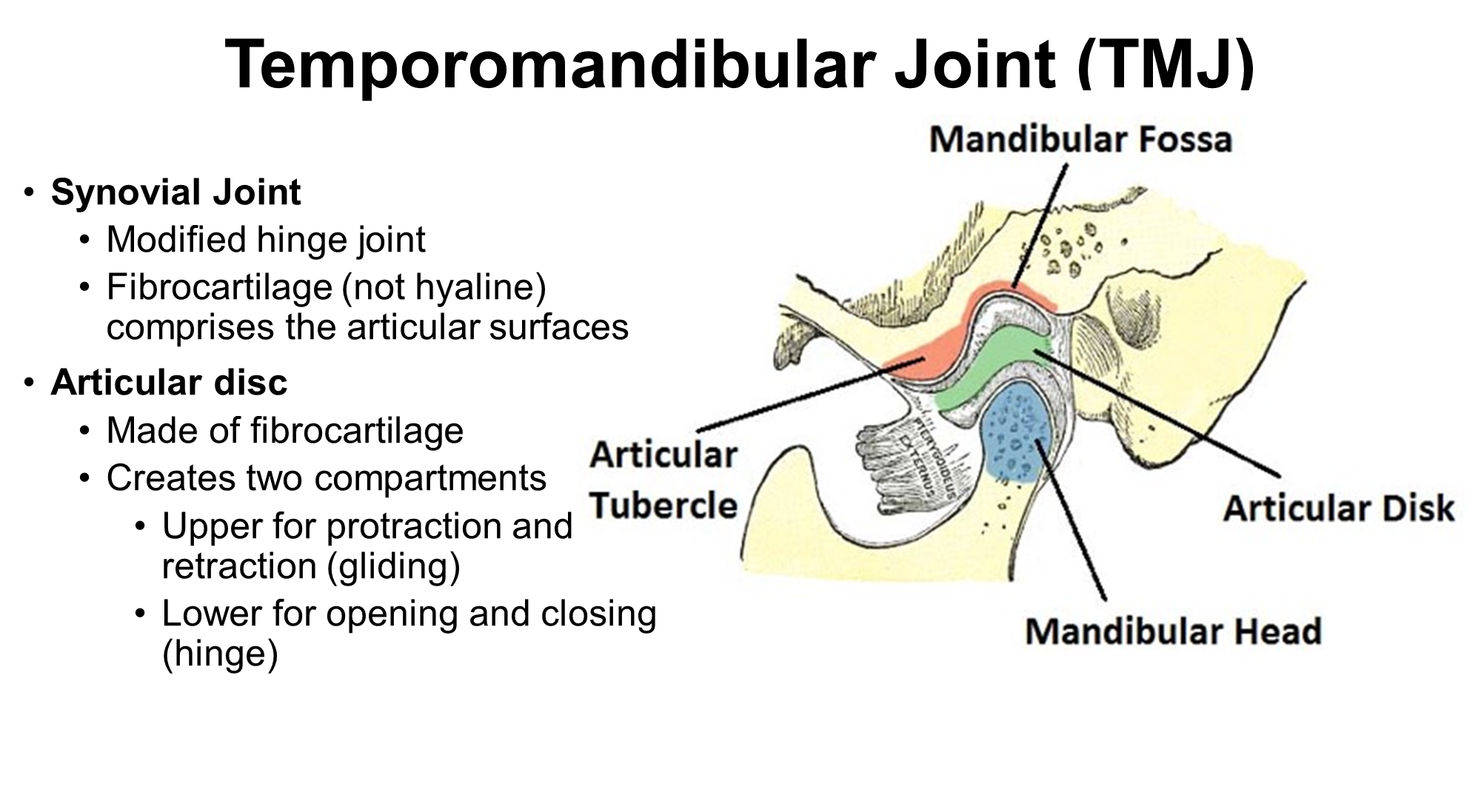
What is the Temporomandibular Joint?
What is the Trigeminal Nerve 2?
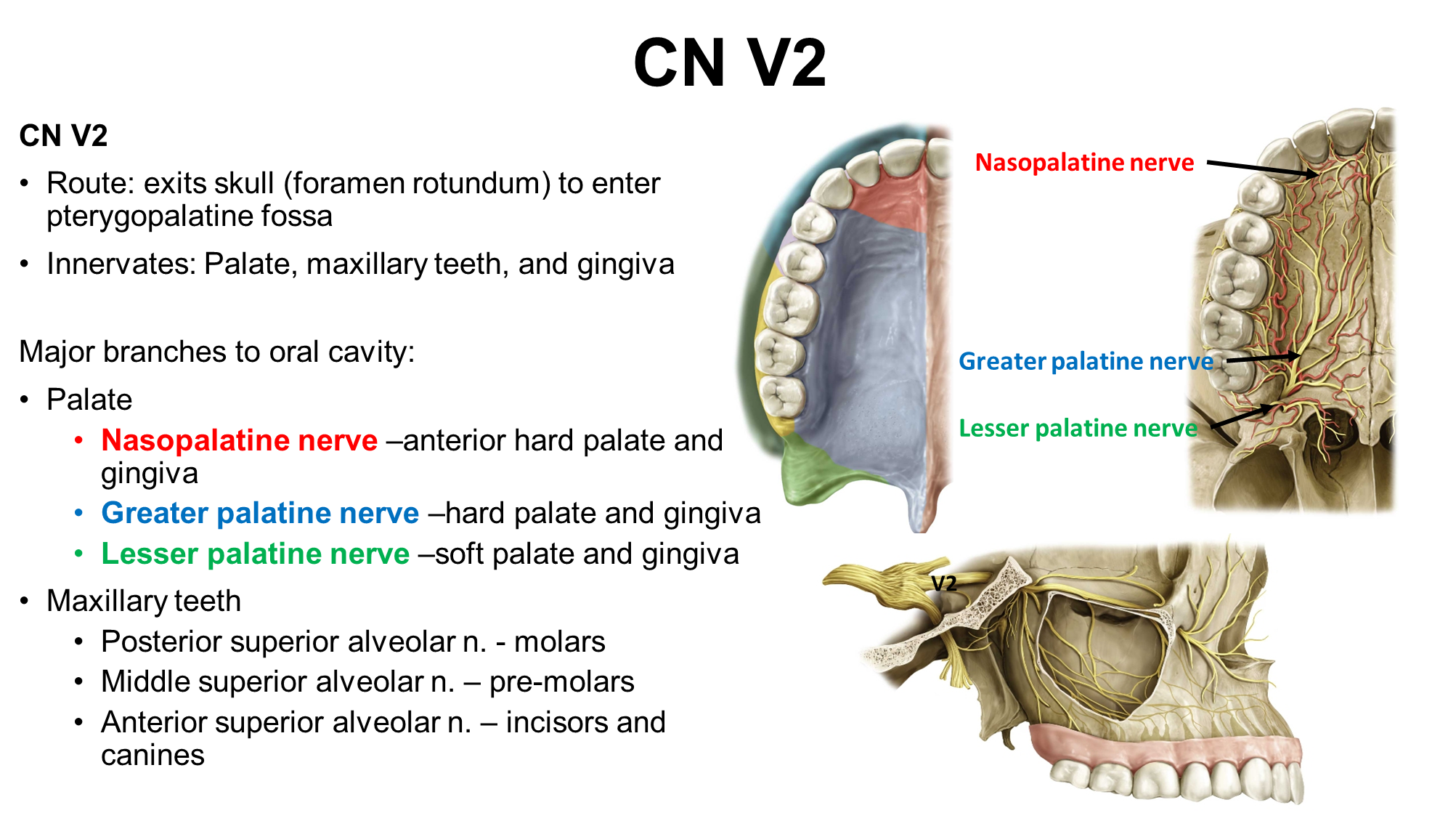
CN V2 or the Maxillary Branch of the Trigeminal Nerve
→ this nerve is responsible for innervation of the palate, maxillary teeth and gingiva. It has six major branches as seen above
What are the two branches of the trigeminal nerve 3 and how is it related to CN VII
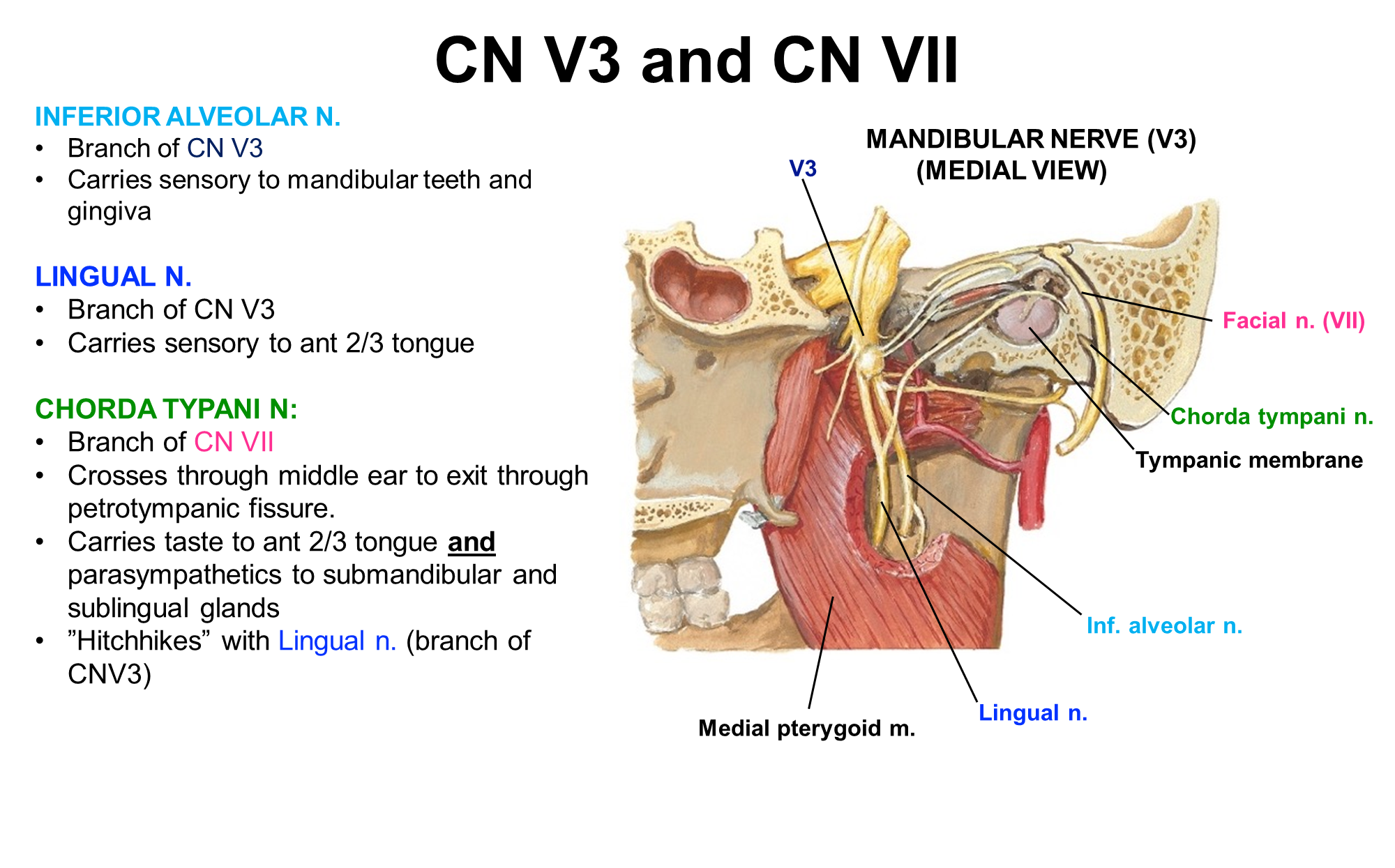
Trigeminal Nerve 3 is subdivided into two branches
1) Inferior Alveolar Nerve - carries sensory information from the mandibular teeth and gums
2) Lingual Branch - carries sensory information from the anterior 2/3rd of the tongue
These branches will hitchhike along Cranial Nerve VII or the Facial Nerve
→ these branches will ride the chorda tympani nerve which is a branch of CN VII
→ these nerves travel along the same route but exit independently from this nerve
What is the route for Cranial nerve IX through the skull?

GLOAPS
What is the neurovasculature of the mouth floor? (3)
What are the five CN VII Motor Fibers?
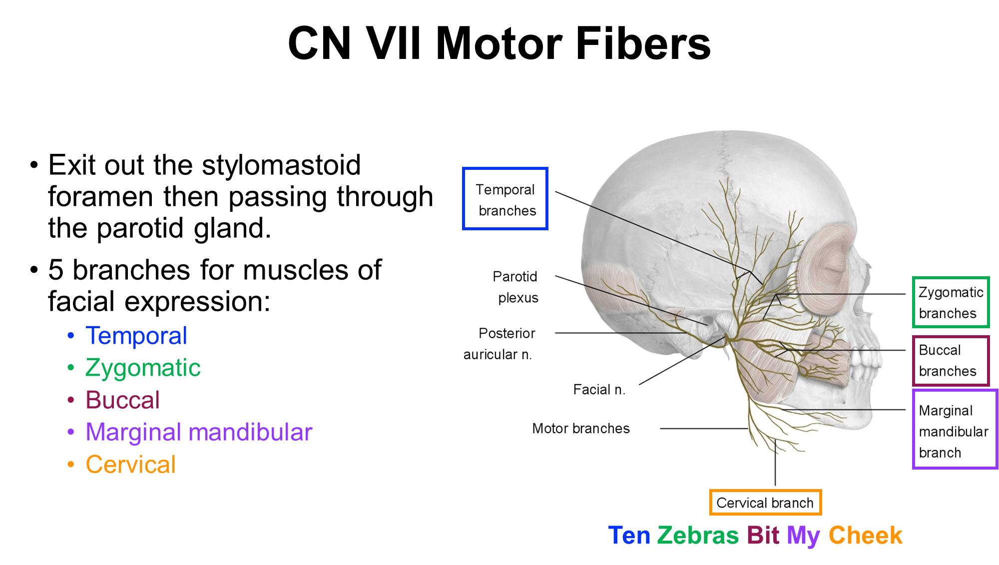
CN VII or the Facial Nerve has five branches that are used in facial expression. They pass through the parotid gland and can be remembered by Ten Zebras Bit My Cheek
→ Temporal
→ Zygomatic
→ Buccal
→ Marginal Mandibular
→ Cervical