MBB1 - Structure and Action of Neurons and the Anatomical Divisions of the Brain
1/56
There's no tags or description
Looks like no tags are added yet.
Name | Mastery | Learn | Test | Matching | Spaced |
|---|
No study sessions yet.
57 Terms
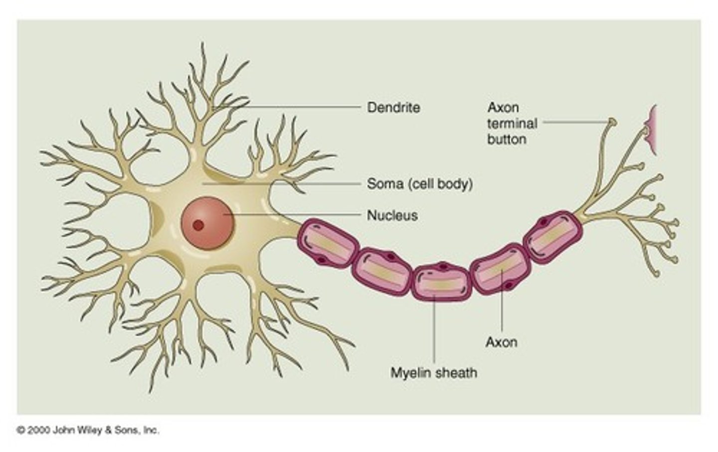
features of a typical neuron
soma (cell body), dendrites, myelin sheath, axon (inside myelin sheath), terminal buttons
action potential
rapid change in the membrane potential of the neuron caused by the movement of ions (when a neuron fires)
ions
charged molecules
membrane
edge of cell
membrane potential
difference in total charge between the inside and outside of a cell
what is the resting membrane potential
-70mV
polarise
extreme separation
Step 1 of an action potential
-sodium channels open
-sodium ions flow rapidly into the neuron through sodium channels
-influx of positive ions increases charge within the neuron, increasing the membrane potential in a process known as depolarisation
Step 2 of an action potential
-if there is sufficient sodium influx to depolarise the membrane potential beyond the threshold of excitation of -50mV, this triggers the opening of potassium cahnnels
-potassium flows out of the neuron while sodium is still entering, causing further depolarisation of the membrane potential
Step 3 of an action potential
-after approx. 1 millisecond, the actions potential reaches its peak and the sodium channels become refractory (close)
-the charge inside the neuron is more positive (+40mV) than the outside
-sodium channels will not be able to open again until the membrane potential returns to the resting potential
Step 4 of an action potential
-as the potassium channels are still open, potassium continues to exit the neuron
-this makes the charge within the neuron increasingly negative as the proportion of positive ions decreases
-hyperpolarisation means the charge is more negative than the resting rate during the refractory period
Step 5 of an action potential
-once the membrane potential returns to the resting rate of -70mV, the potassium channels close and the sodium channels reset so that another depolarisation can cause them to open again
-due to hyperpolarisation, a subsequent action will require a greater sodium influx immediately after the neuron has fired
what causes sodium channels to open
-change in the charge around the membrane (depolarisation), eg. when signals such as neurotransmitters are received
what does the sodium-potassium pump cause
a greater concentration of sodium ions outside the neuron, a greater concentration of potassium ions inside the neuron
rate law of action potentials
stronger stimulus = faster threshold for activation = more frequent action potentials
neuron firing as an 'all or none' event
will always go from -70mV to +40mV - can't have a bigger action potential and will always flow down the neuron in the same way - will never be faster or bigger
what can change in terms of action potentials
the gap between individual action potentials (frequency/rate)
what determines the strength of a neural signal
frequency of firing
rostral/anterior
towards the nose - front
caudal/posterior
toward the tail - back
dorsal/superior
toward the back - top
ventral (inferior)
toward the belly - bottom
lateral
away from the midline
medial
toward the midline
ipsilateral
on the same side of midline
contralateral
on the opposite side of midline
corpus callosum
thick section of the brain that consists of large bundles of axons that connect two hemispheres
homotopic connections
connect complimentary region of the other hemisphere
heterotopic
communicate to a different brain region
callosotomy
procedure to cut the corpus collosum to stop severe epileptic seizures - prevents communication between hemispheres
3 major divisions of the brain
forebrain, midbrain, hindbrain
subdivisions of the forebrain
telencephalon and diencephalon
what does the telencephalon consist of
the limbic system, basal ganglia and cerebral cortex
cerebral cortex
-the largest structure of the human brain - divided into two cerebral hemispheres
-inner 'white matter' is pale because of high proportion of axon fibres covered in fatty myelin layer
-outer 'grey matter' is where the neurons synapse and connect together
what is the basal ganglia responsible for
coordinating involuntary movement, particular aspects that are highly automatised or involuntary (eg. walking)
-different to cerebellum because basal ganglia refer to highly automatised movements
what happens in Parkinson's patients
the basal ganglia are dysfunctional, leading to weakness, tremors, limb rigidity, poor balance and difficulty initiating movements
limbic system
-neural system including the hypothalamus, thalamus, hippocampus and amygdala
-thought of as the 'emotion circuit'
-first proposed by James Papez in 1937
what does the diencephalon consist of
thalamus and hypothalamus
thalamus
major relay station for sensory inputs to cerebral cortex - divided into several nuclei
-eg. when playing tennis, info (sight of ball) will be relayed to areas controlling hand movement
hypothalamus
controls autonomic nervous system and endocrine (hormone) system - regulates survival behaviours (fighting, feeding, fleeing, mating)
what does the midbrain consist of
mesencephalon
mesencephalon
-located within the brainstem at the topmost region, directly above the hindbrain
-connects pons and cerebellum with the forbrain
-important role in motor movement, particularly movement of the eye and in auditory/visual processing
-consists of tectum/tegmentum
hindbrain subdivisions
metencephalon and myelencephalon
metencephalon components
cerebellum and pons
cerebellum
-'little brain'
-helps coordination of movement - different to basal ganglia as it uses visual feedback to fine tune movement to carry out a particular task
-damage to it causes problems with walking and leads to jerky, poorly coordinated movements and problems retaining balance
pons
-lies on the ventral surface of the brainstem
-contains several nuclei important in regulating sleep and arousal
-also relays info from cerebral cortex to the cerebellum
myelencephalon
-medulla
-links hindbrain to the spinal cord and contains neurons important for autonomic function like respiration and heart rate
-damage makes it impossible to live
What are the lobes of the cerebral cortex?
frontal, parietal, occipital, temporal
primary visual cortex
the medial and lateral region of the posterior occipital lobe whose primary input is from the retina.
where is peripheral vision processed?
anterior occipital lobe that extends into the calcarine fissure.
What did Hubel and Wiesel find in 1981?
Neurons show orientation selectivity
parietal lobe
attention and spatial awareness on the dorsal surface- the "where" pathway.
temporal lobe
auditory processing and complex visual processing on the ventral surface- "what"
primary auditory cortex
- superior part of the temporal cortex and a section within the Sylvian fissure.
- receives auditory info. from the cochlea (tonotopic map)
primary somatosensory cortex
- area of the parietal lobe where messages from the sense receptors are registered (somatotopic)
- posterior to the central sulcus
primary motor cortex
- the section of the frontal lobe responsible for voluntary movement
- located on the precentral gyrus
frontal lobe
associated with reasoning, planning, parts of speech, movement, emotions, and problem solving
- Railway man Phineas Gage (1848) - damage to frontal lobe, had severe personality changes