A&P Practical 1
1/107
Earn XP
Description and Tags
Georgia Southern Anatomy 1 Practical 1 Terms
Name | Mastery | Learn | Test | Matching | Spaced | Call with Kai |
|---|
No analytics yet
Send a link to your students to track their progress
108 Terms
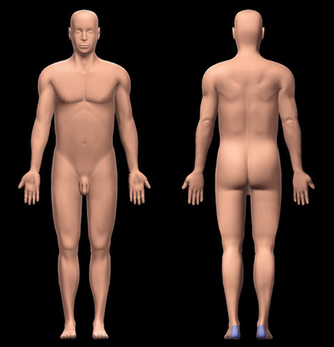
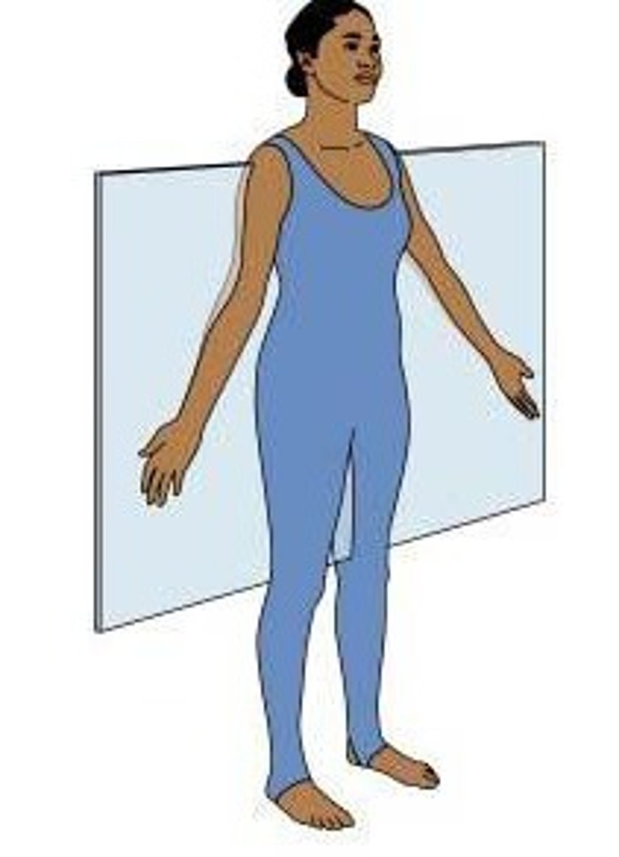
anatomical position
erect, feet forward, arms at side with palms facing forward, head facing forward
-all positions are switched to be applied as the conduit would be
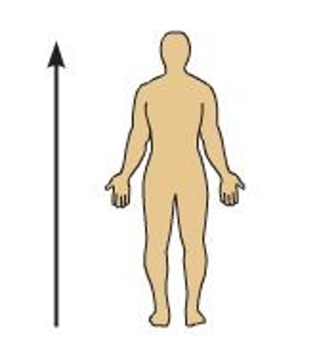
Superior (cranial)
above, higher than
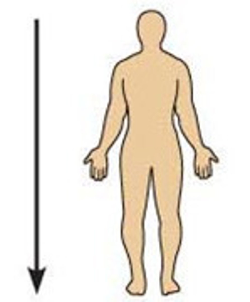
inferior (caudal)
below, lower than
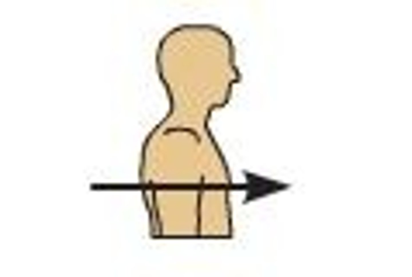
anterior (ventral)
front, forward
posterior (dorsal)
back, behind
remedial (medial)
near, close to body, in a limb
lateral
away from the middle, nearest to the outside
proximal
near, close to body, in a limb
distal
far away, farthest from body
superficial
on the surface, exterior
deep
inner layer, away from surface
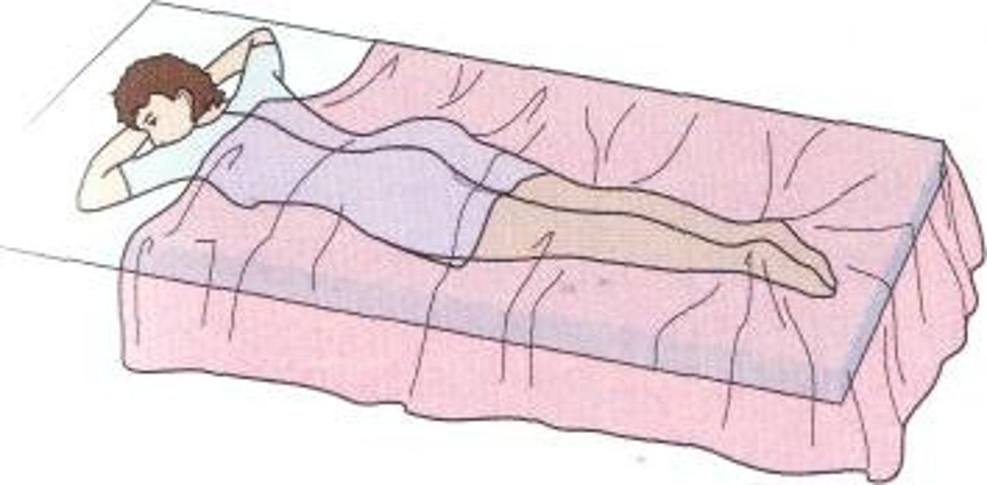
prone
lying down
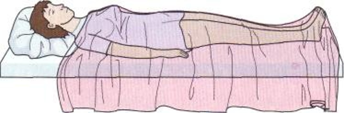
supine
facing up
rostral
toward the forehead or nose
caudal
toward the tail
oral
pertaining to the mouth
buccal
pertaining to the cheek
orbital
pertaining to the eye socket
cervical
7 vertebrae closest to the head

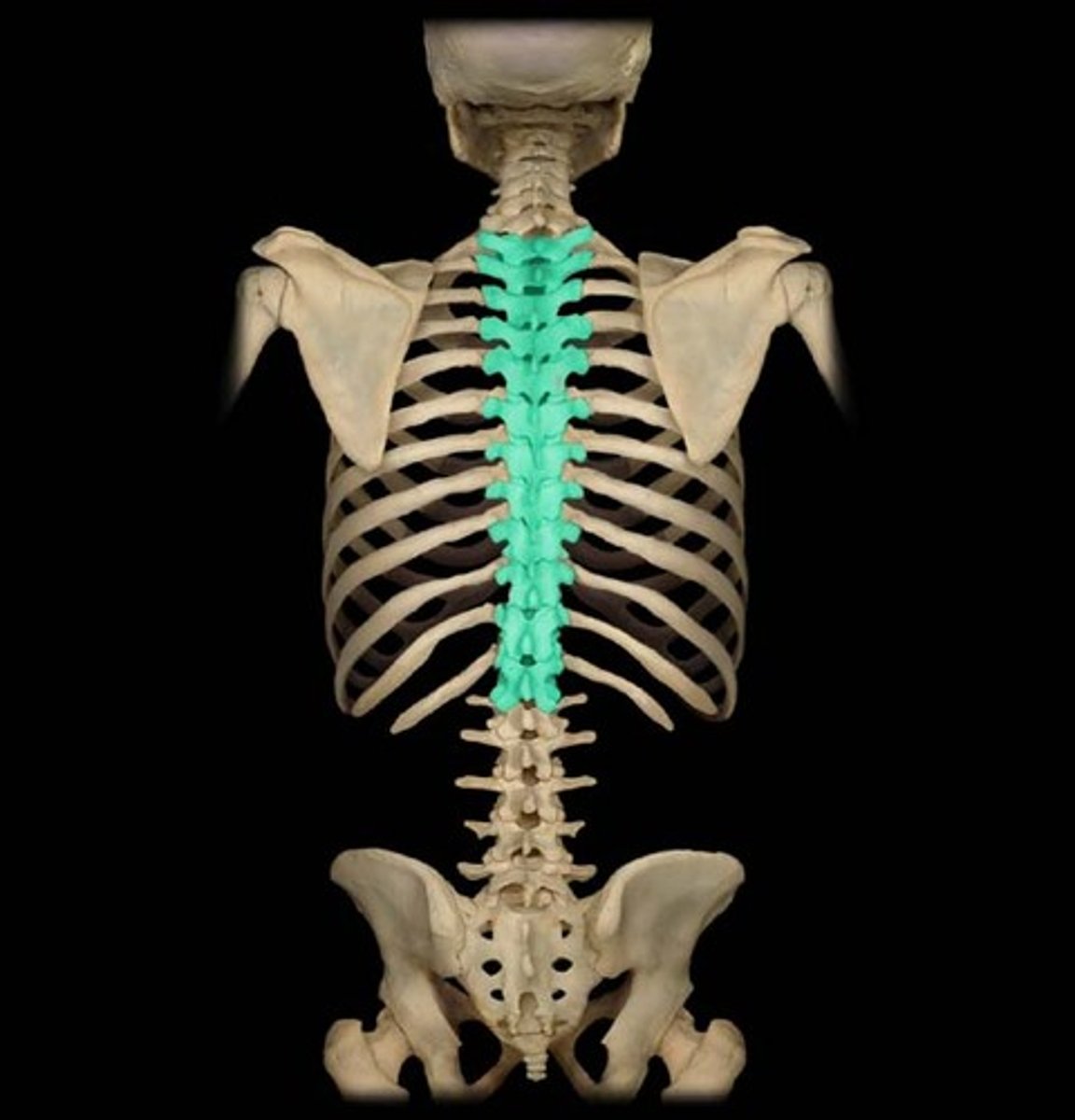
thoracic
12 vertebrae connected to the ribs
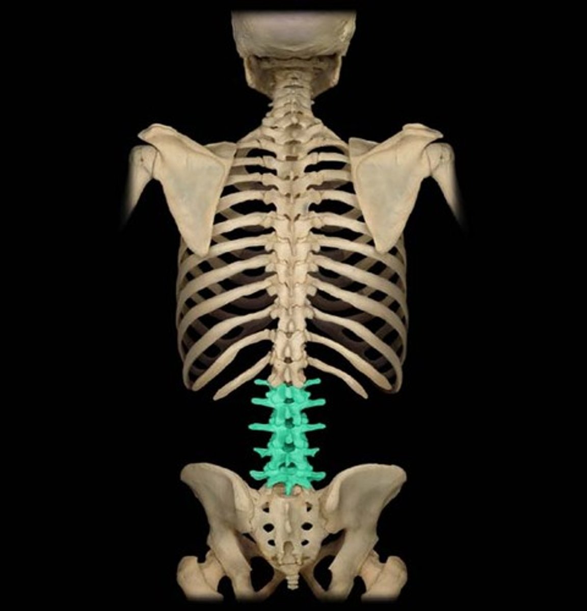
lumbar
5 vertebrae in the lower back
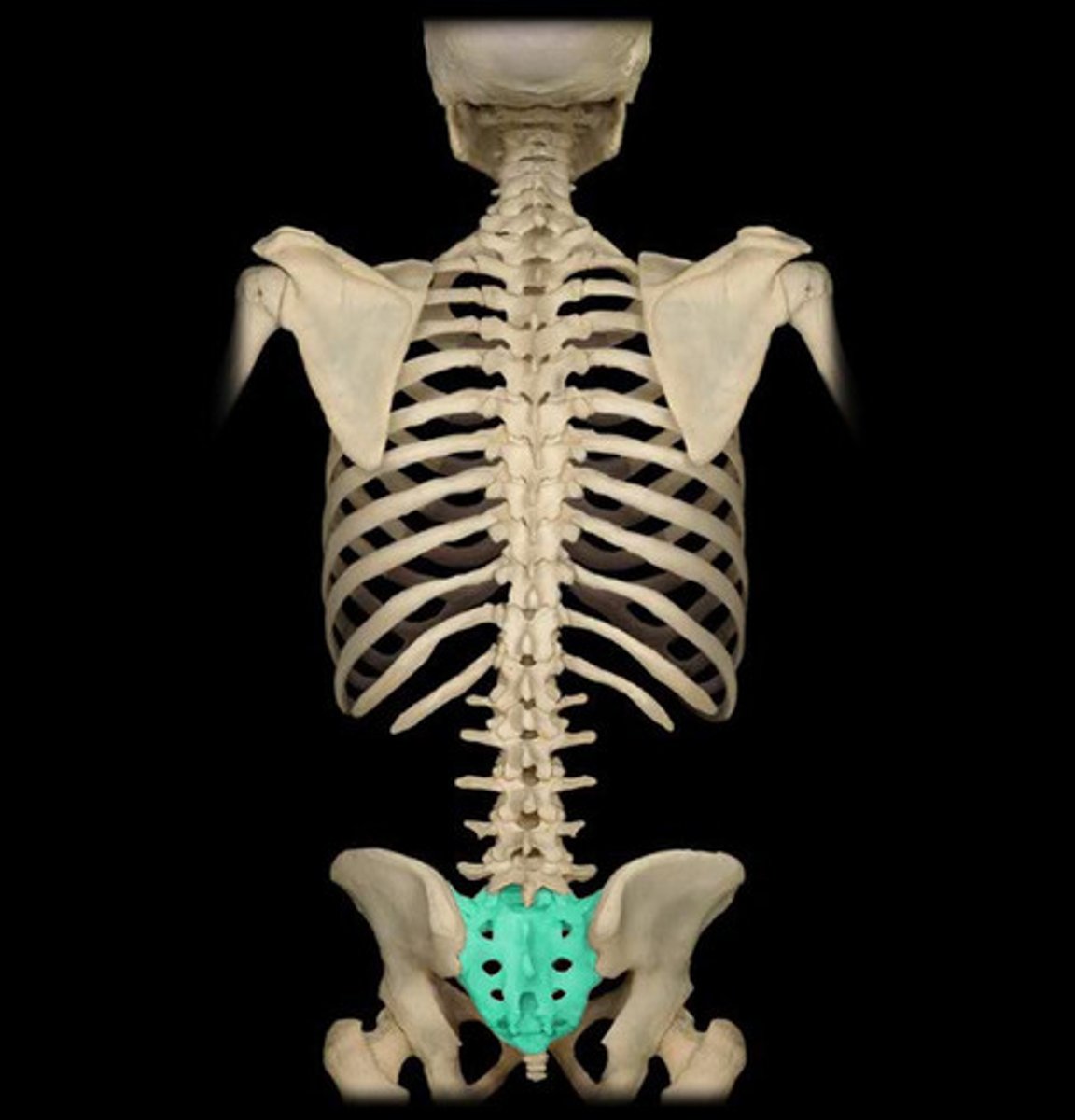
sacral
5 fused vertebrae connected to the pelvis
antecubital
anterior to the elbow
umbilical
pertaining to the belly button
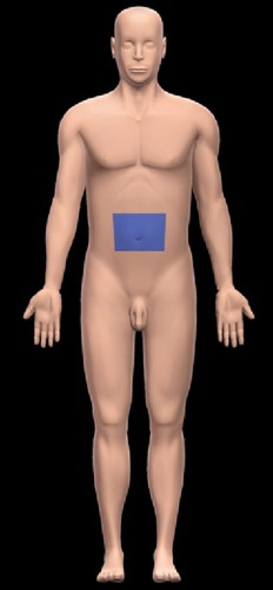
abdominal
below the chest and above the pelvis
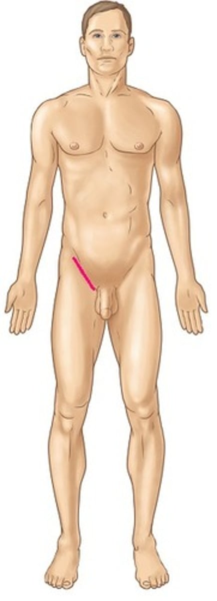
inguinal
pertaining to the groin
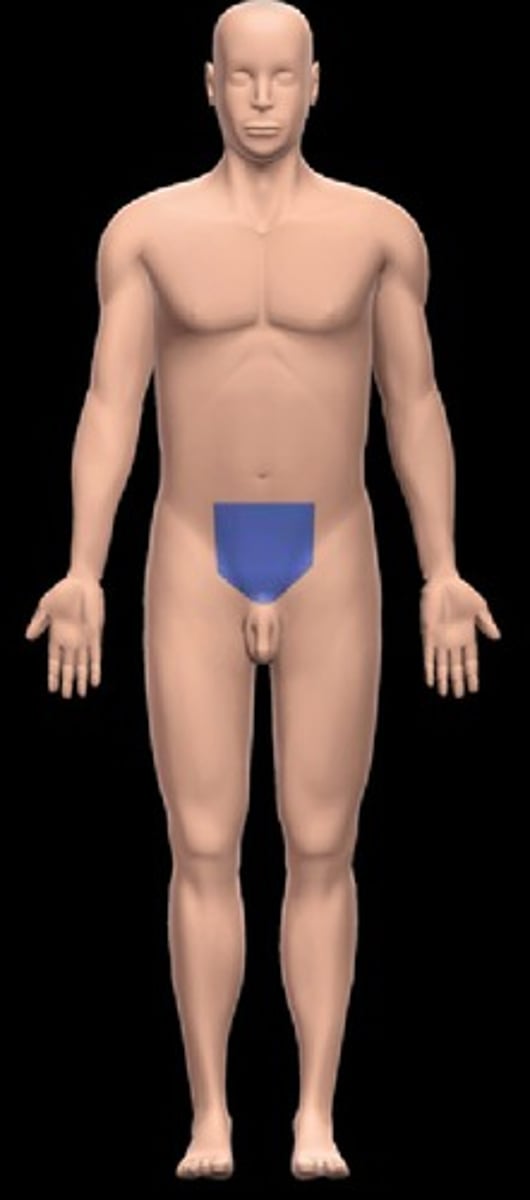
pubic
near pubic bone
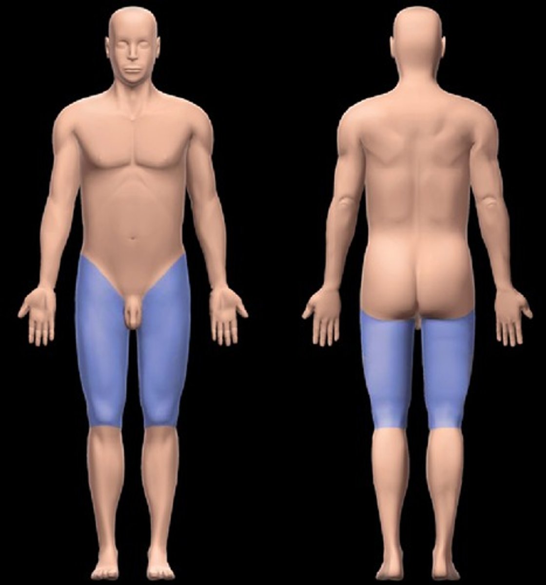
femoral
pertaining to the thigh
patellar
knee cap
digital
fingers and toes
occiptial
back of the skull
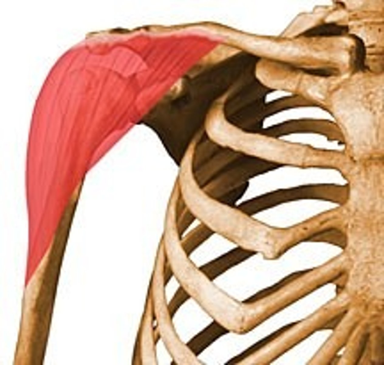
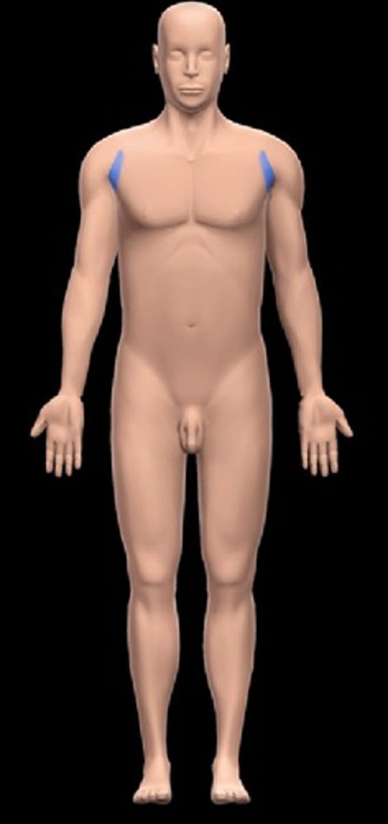
deltoid
pertaining to the shoulder
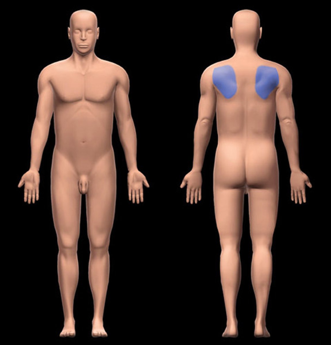
scapular
shoulder blade
axillary
armpit
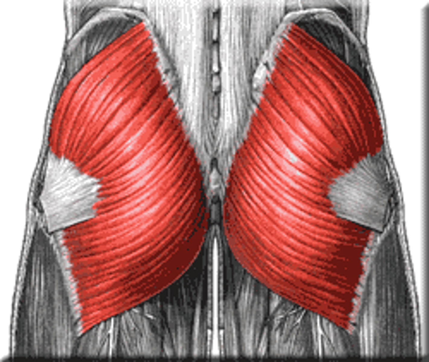
gluteal
the buttocks
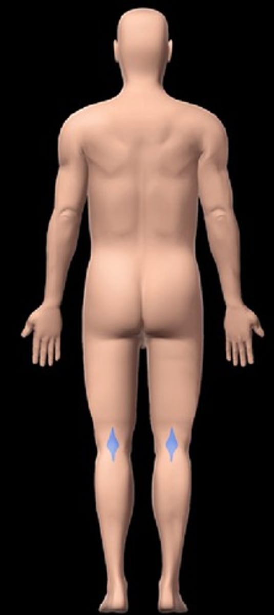
popliteal
posterior knee
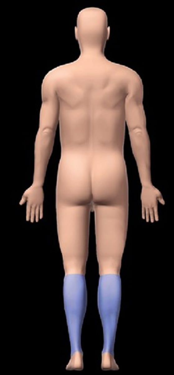
sural
calf
calcaneal
heel of foot
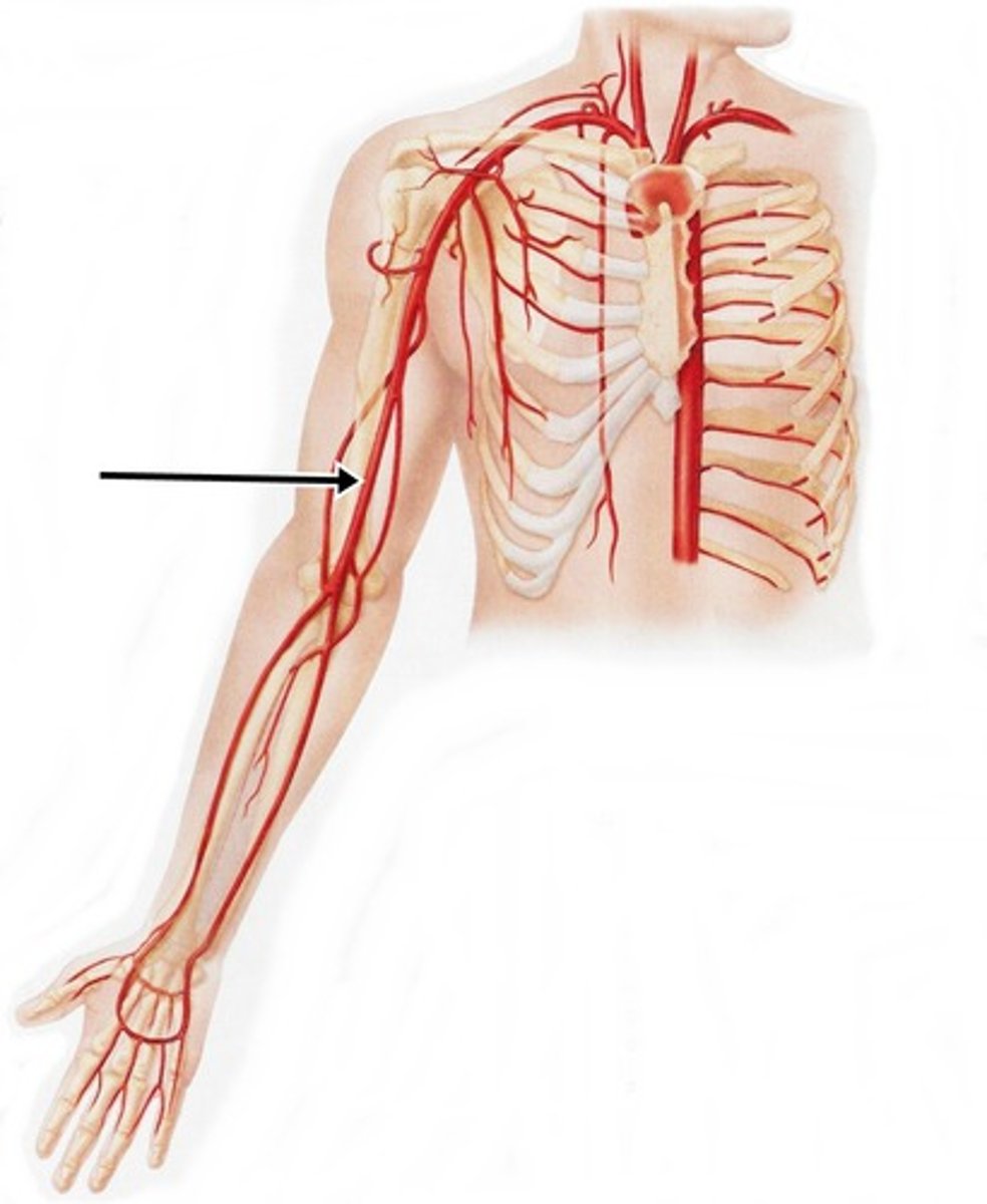
brachial
pertaining to the arm
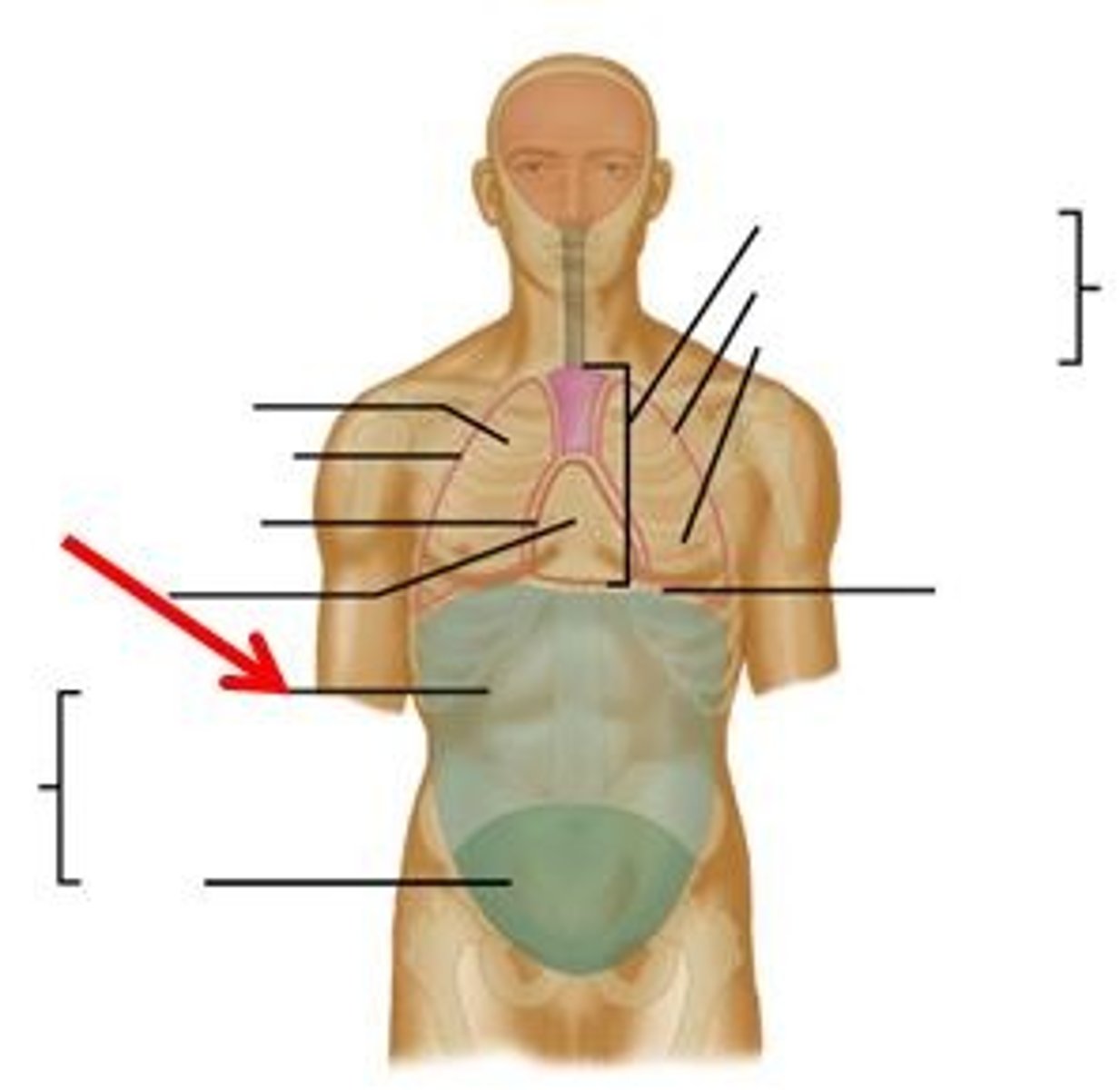
Regions of the stomach
right hypochondriac, epigastric, left hypochondriac, right lumbar, umbilical, left lumbar, right iliac, hypogastric, left iliac
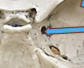
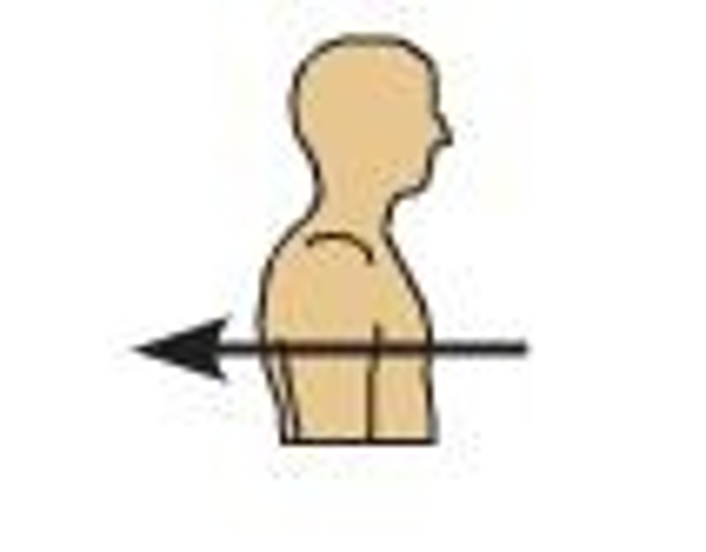
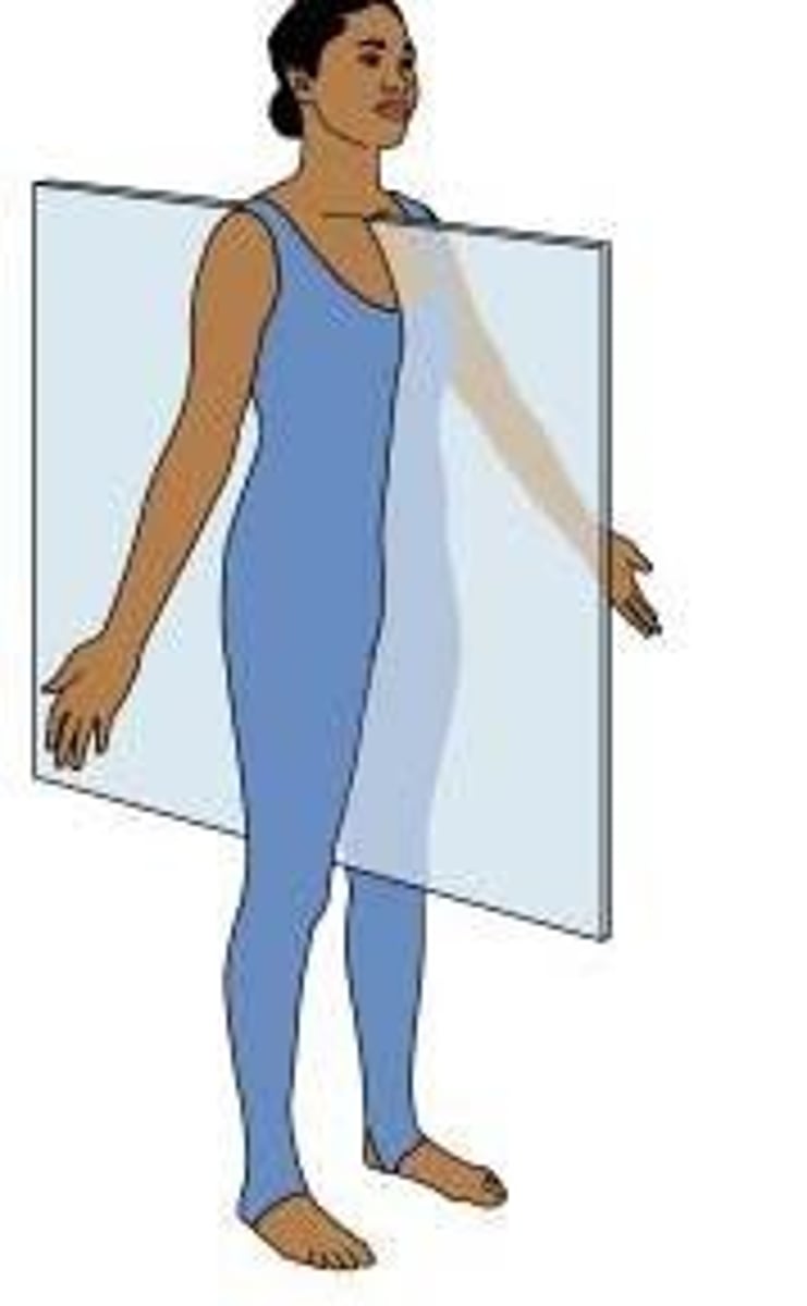
sagittal plane
splits body into left and right
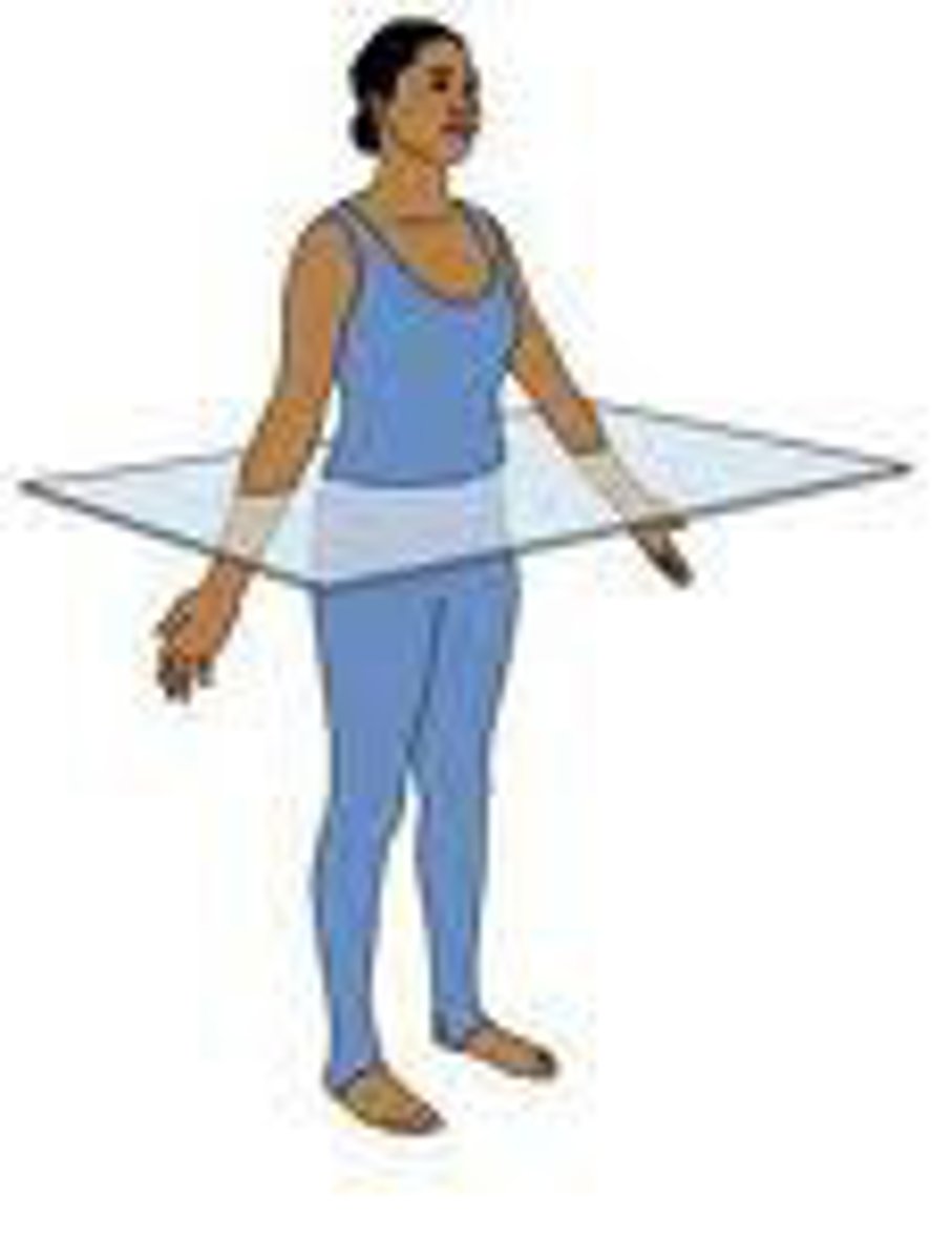
Transverse plane
horizontal division of the body into upper and lower portions
frontal plane
Divides the body into front and back portions.
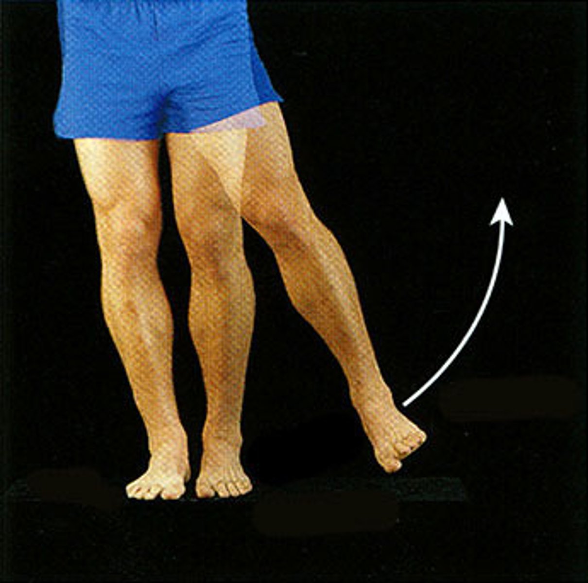
extension
increases the angle of a joint

Flexion
Decreases the angle of a joint
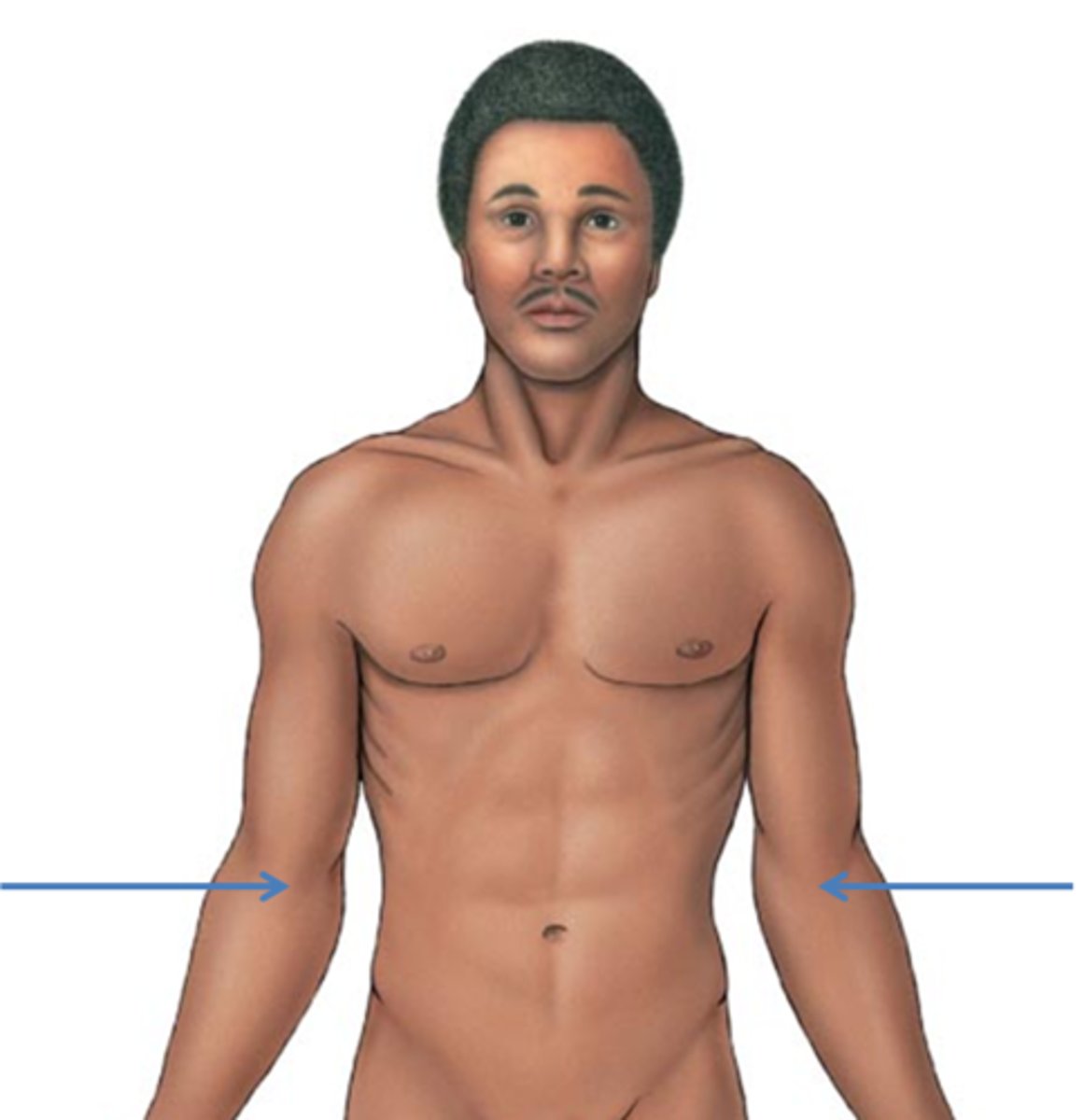
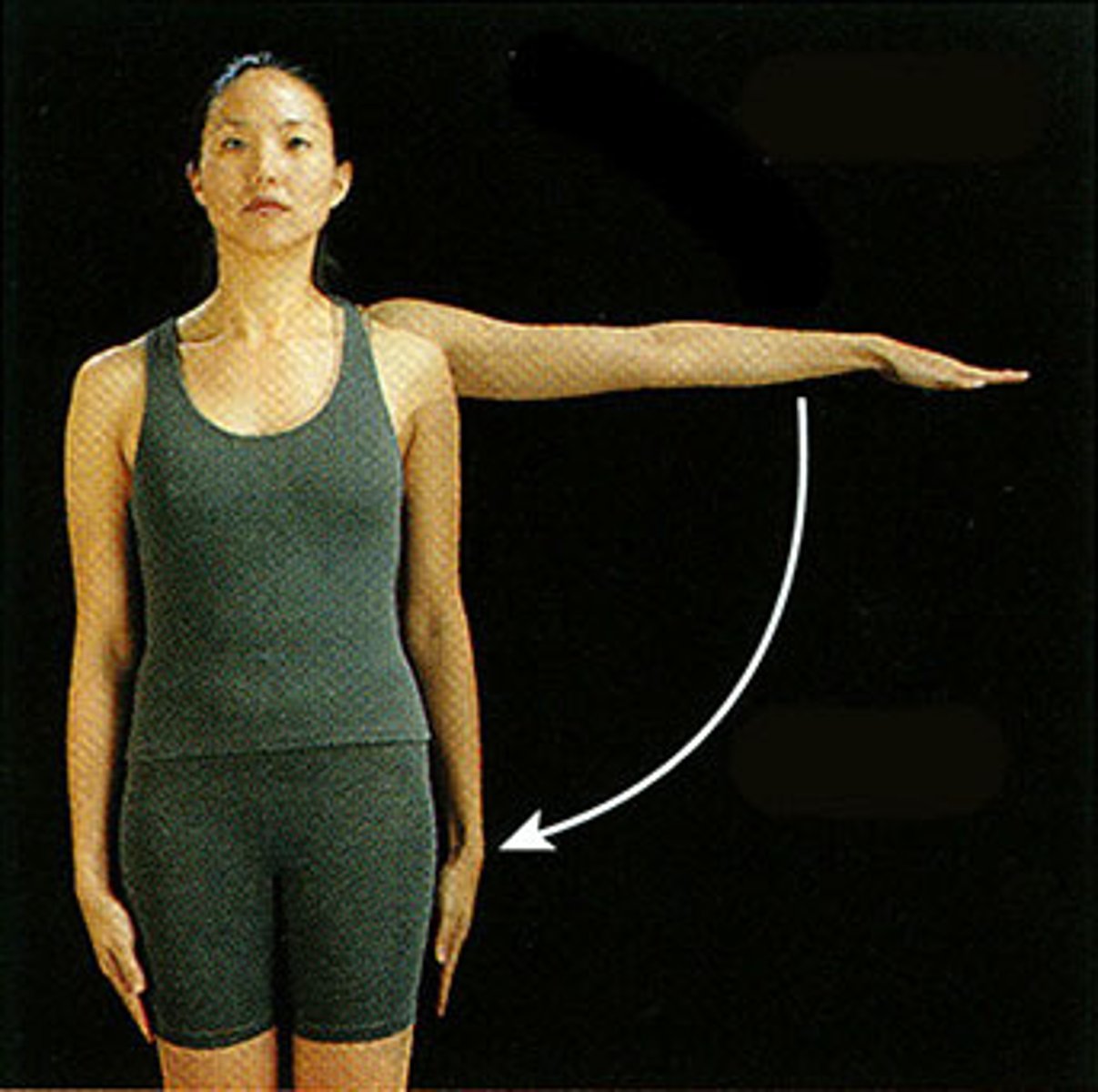
abduction
movement away from the midline
Adduction
Movement toward the midline of the body
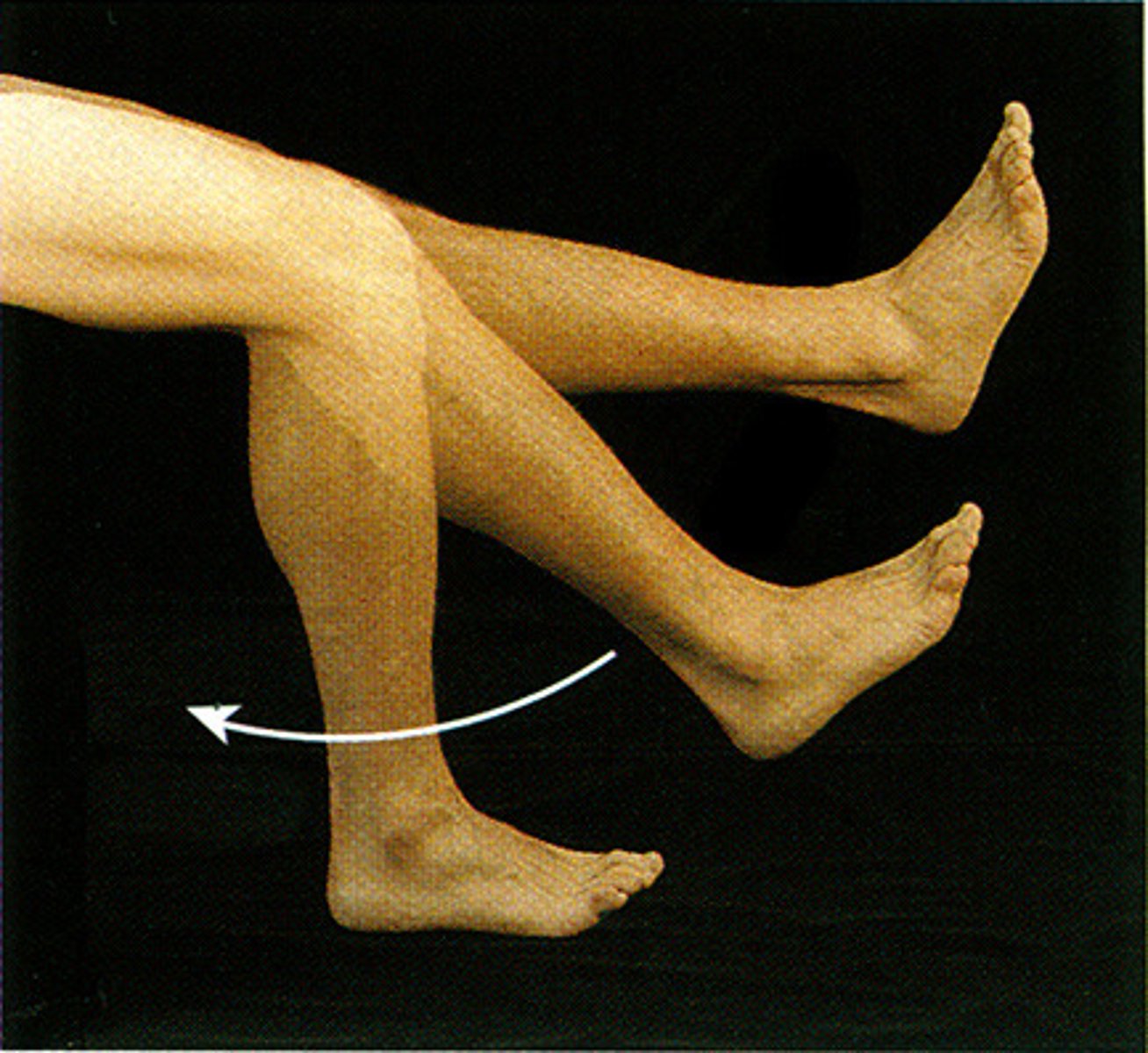
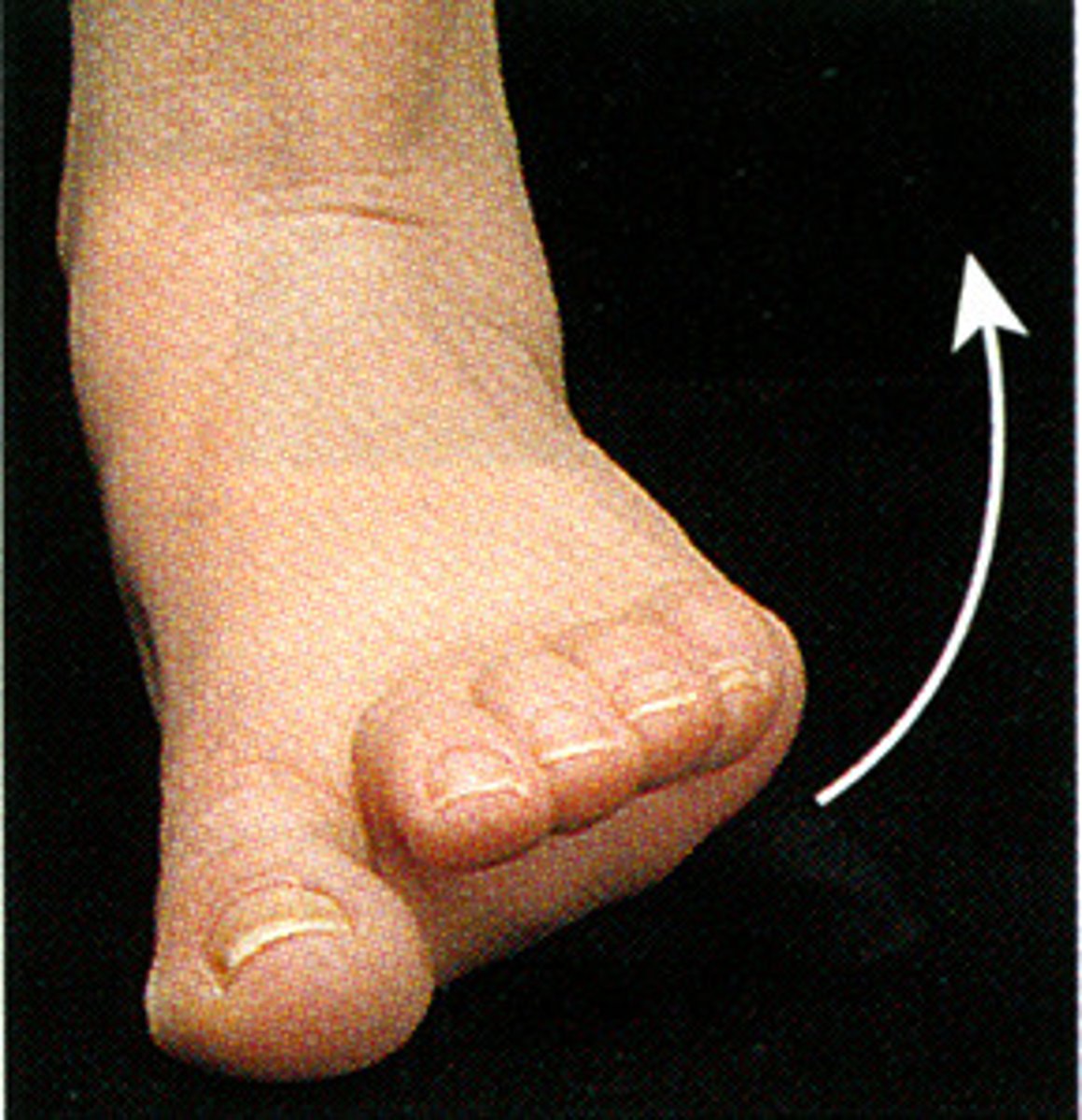
Inversion
turning inward
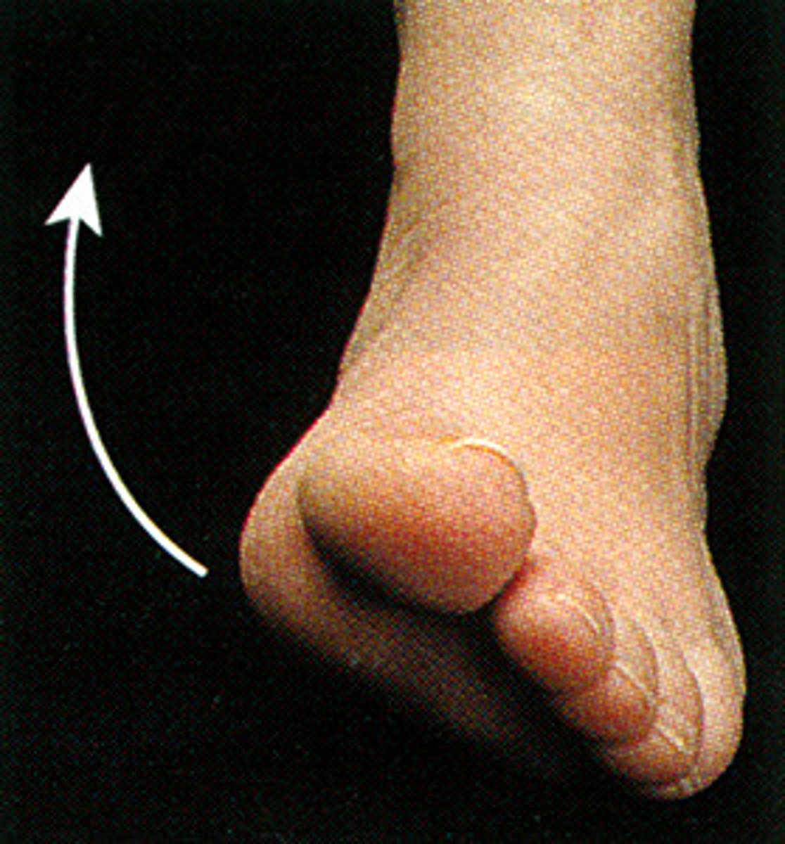
Eversion
turning outward
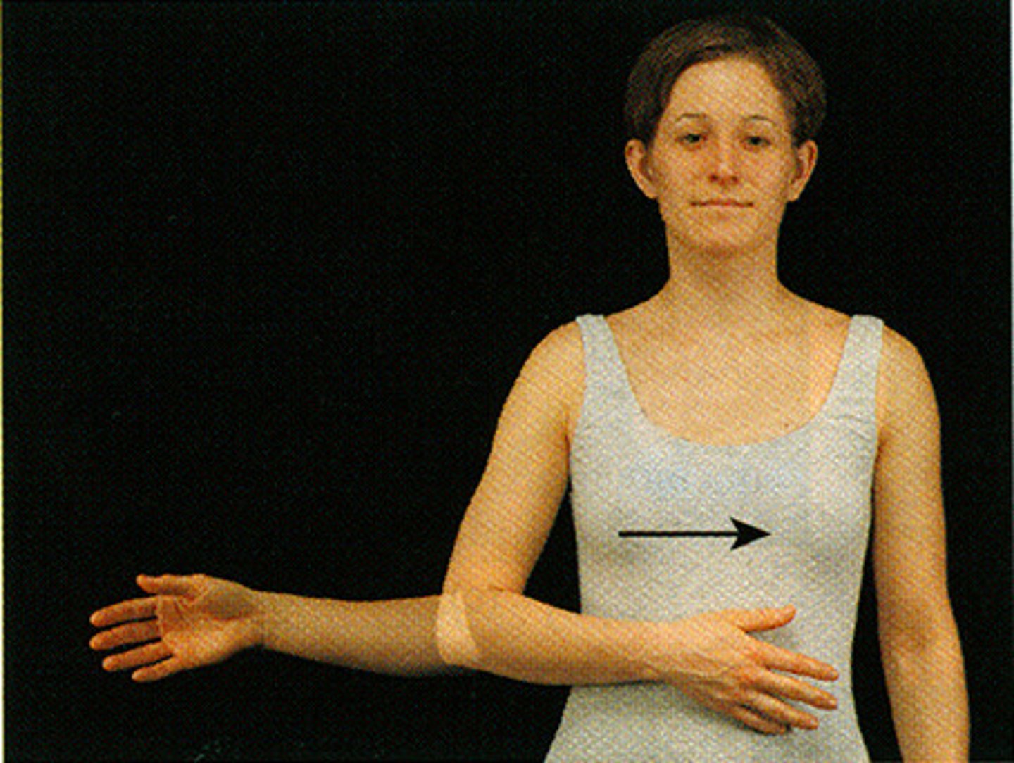
Supination
movement that turns the palm up
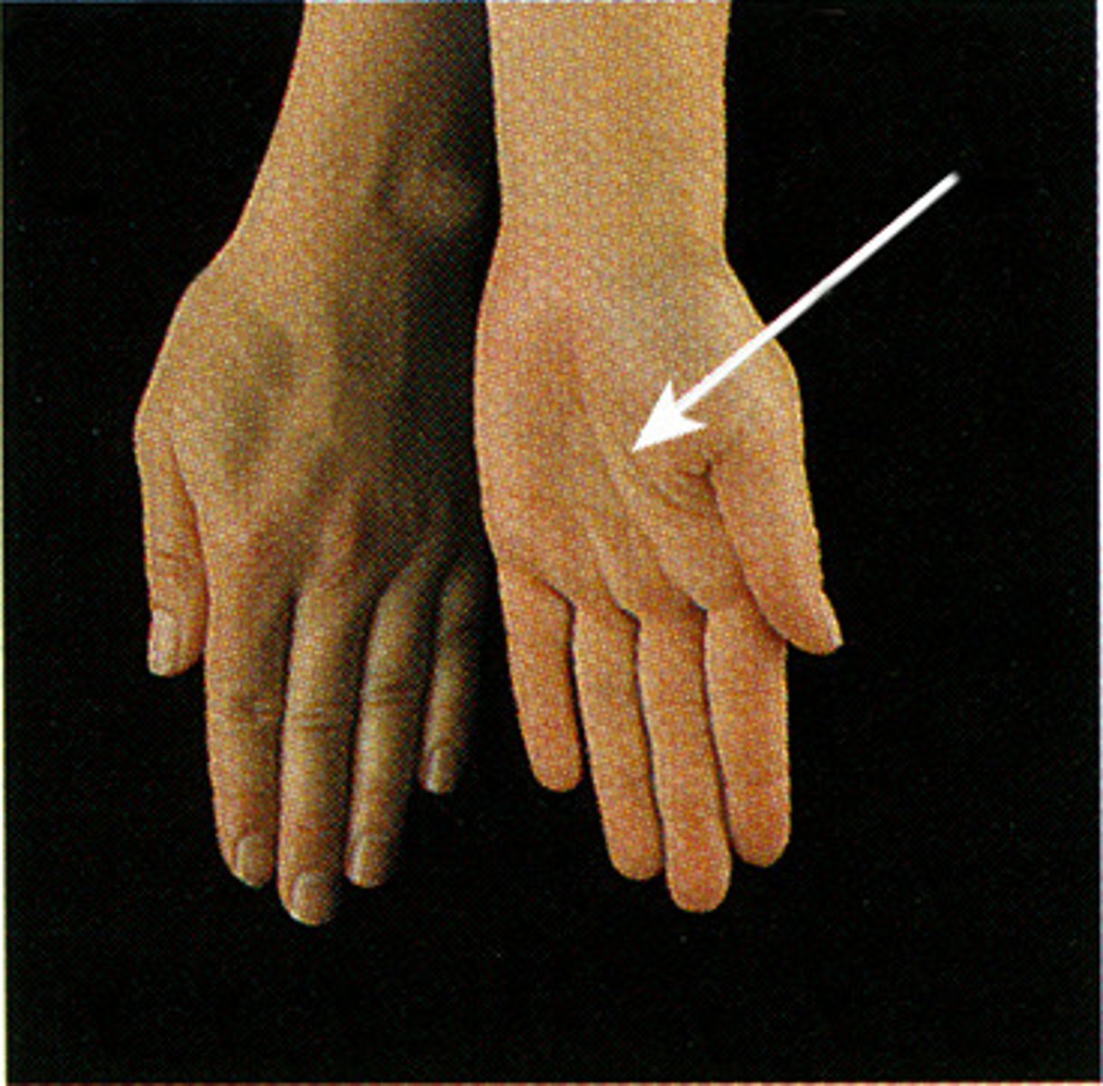
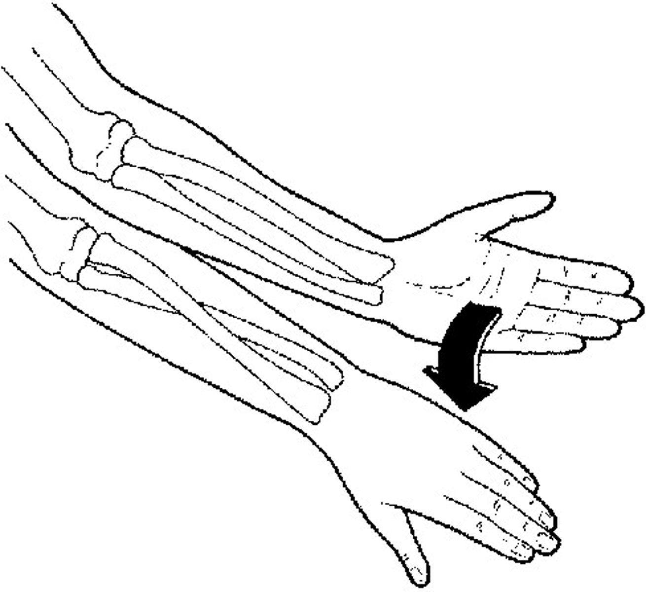
Pronation
turning the palm downward
neutral
Not favoring either side
medial rotation
inward (medial) movement of a body segment in the transverse plane
lateral rotation
outward (lateral) movement of a body segment in the transverse plane

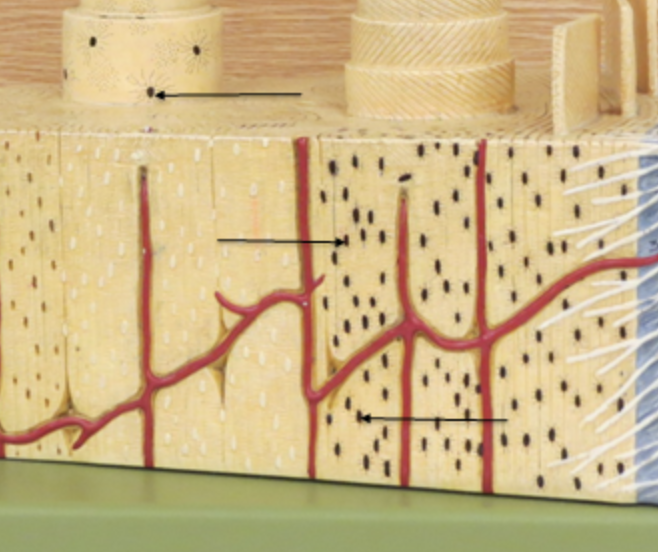
Endosteum
membranous lining of the hollow cavity of the bone
endosteum
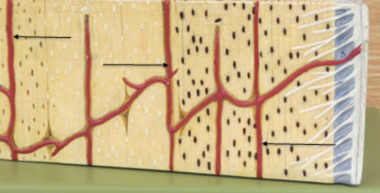
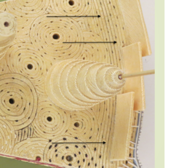
periosteum
surrounding area, outside area
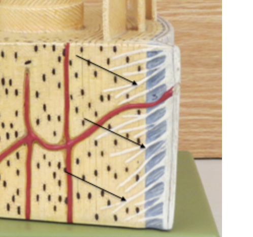
sharpeys fibers
spikes on the side
Volkmanns canal
horizontal canal
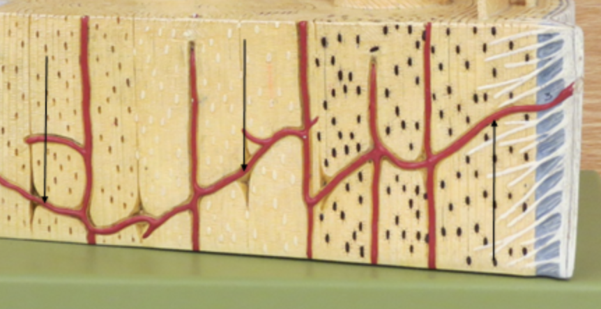
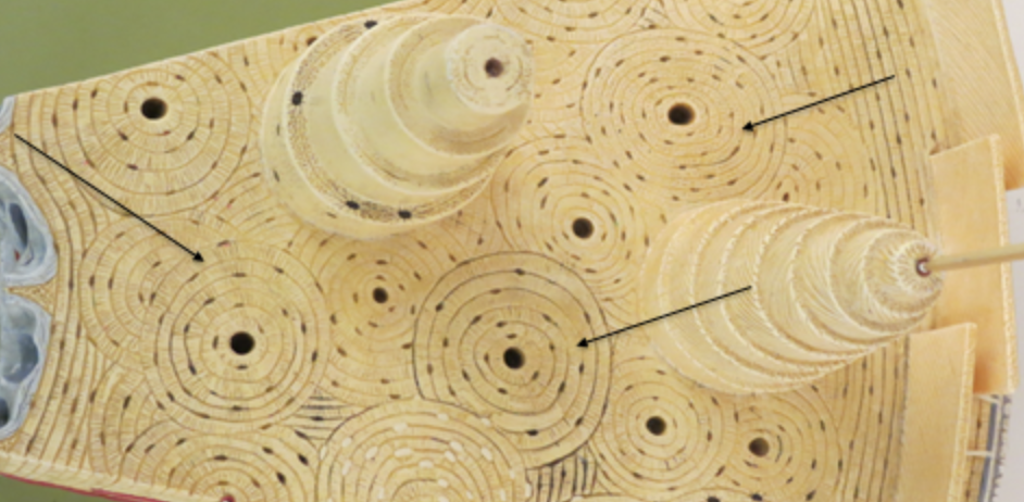
Haversian Canal
vertical canal
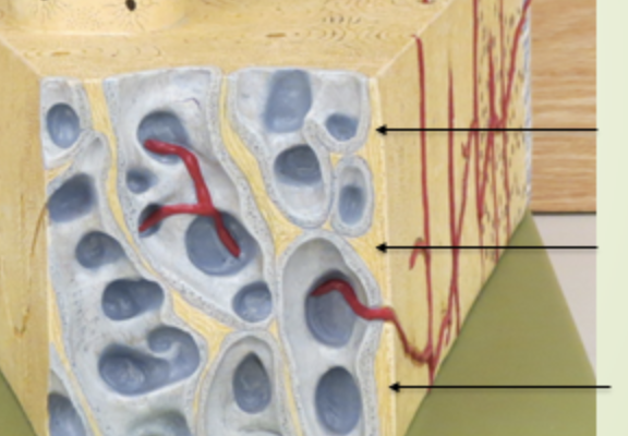
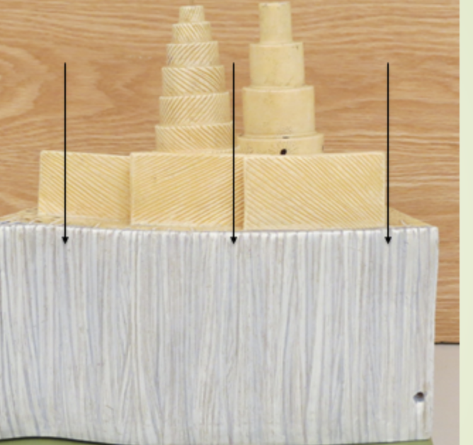
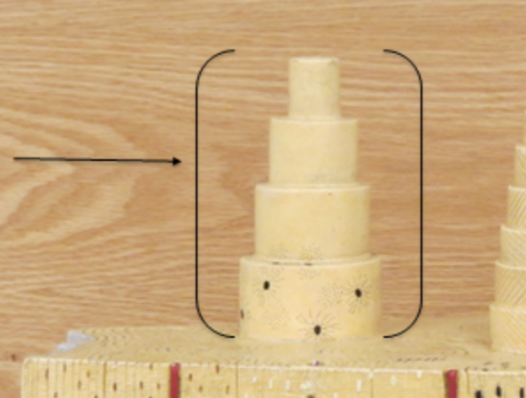
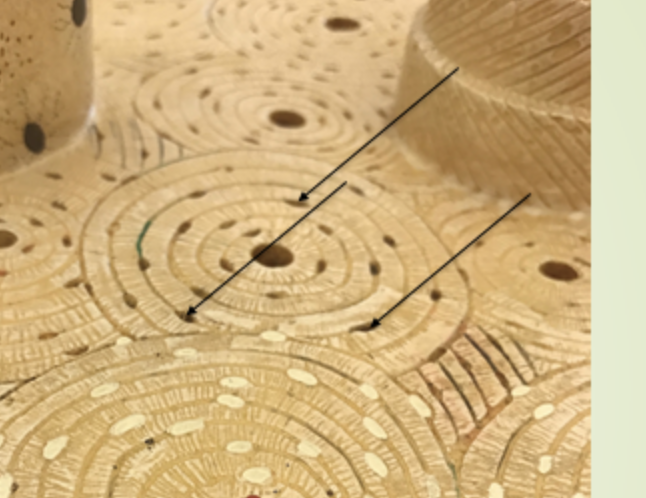
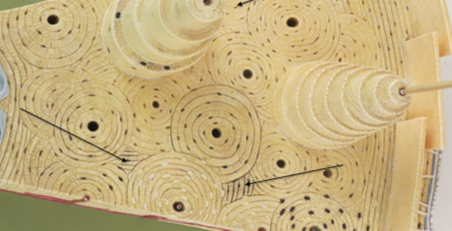
osteon
pillars
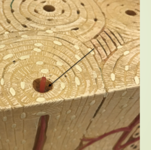
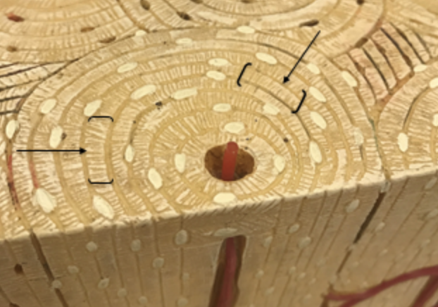
central canal
canal of blood vessels, what hole the blood moves through
osteocytes
bone cells
lacuna
where the osteocytes (bone cells) are located
canaliculi
parts of the rings around the canals , communicates with the osteocytes
interstitial lamellae
irregularly shaped and fill in the spaces between osteons
concentric lamellae
cylindrical rings of lamellae which are rich in collagen.
circumferential lamellae
one of the layers of bone that underlie the periosteum
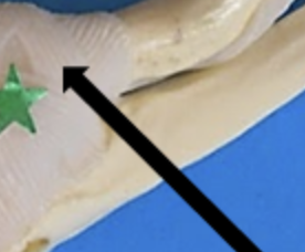
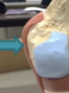
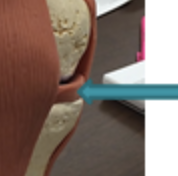
acromioclavicular ligament
on top of shoulder joint
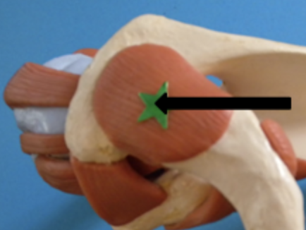
Coracoacromial ligament
middle of shoulder joint
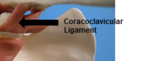
Coracoclavicular ligament
under shoulder joint
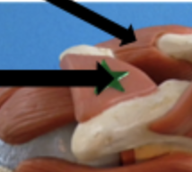
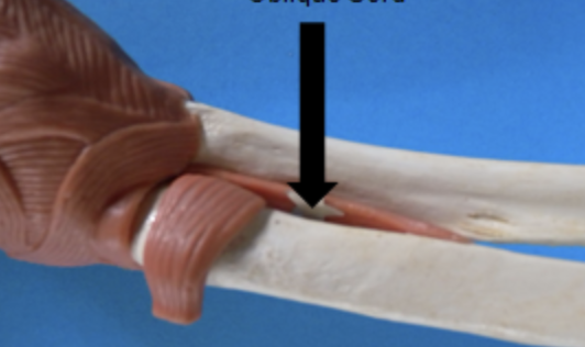
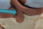
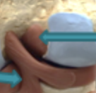
oblique cord
middle of elbow joint
ulnar collateral ligament
above funny bone in elbow joint
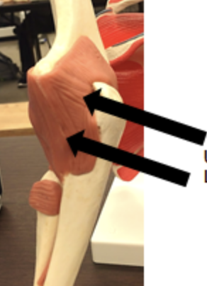
radial collateral joint
very inside (medial) elbow joint
annular ligament
posterior cruciate ligament
anterior cruciate ligament
tibial collateral ligament
fibular collateral ligament
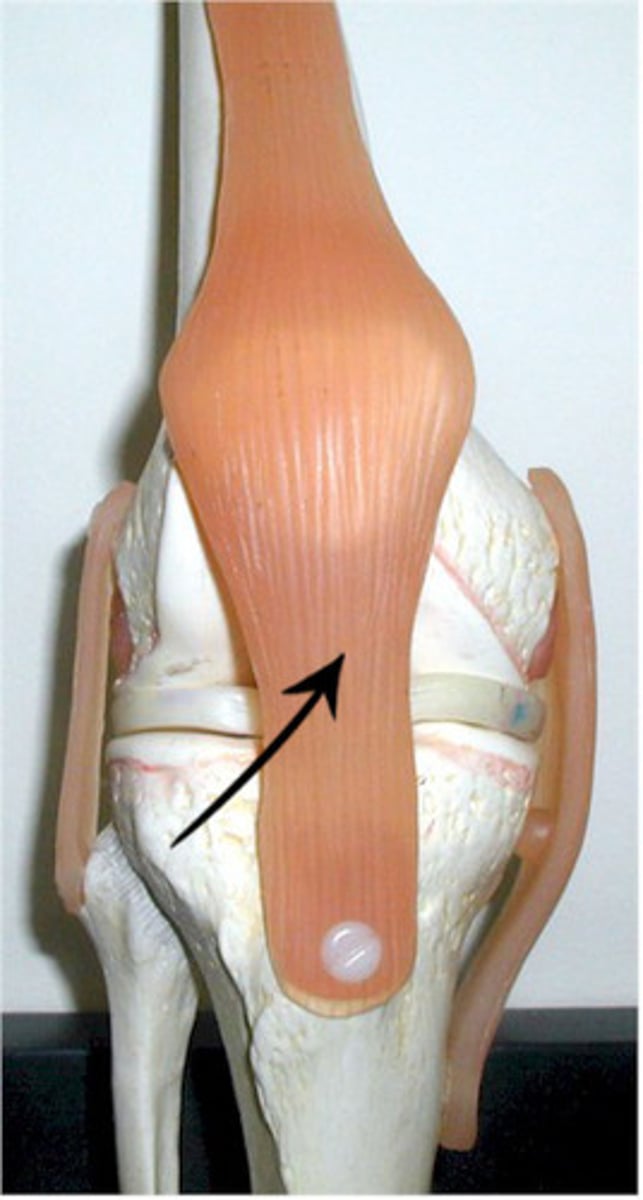
patellar tendon
patellar ligament
lateral Meniscus
medial meniscus
frontal bone
forehead
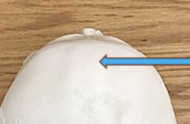
left parietal bone
right parietal bone
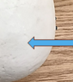
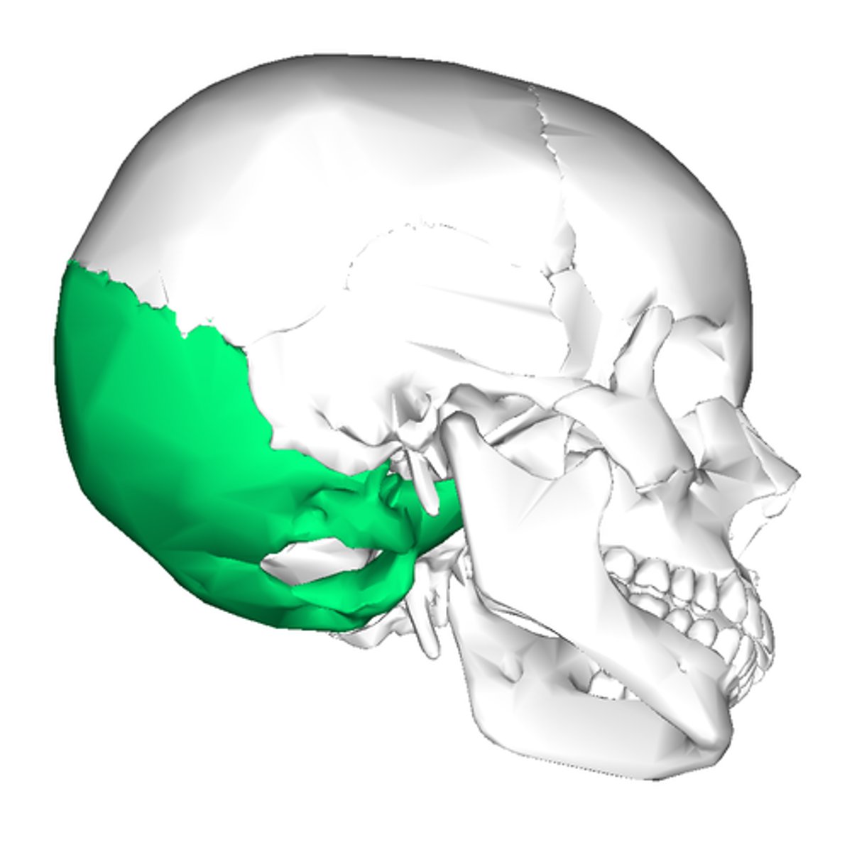
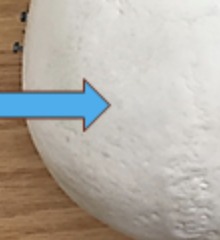
occipital bone

nuchal line
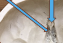
crista Galli
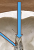
cribriform plate
lesser wing of sphenoid
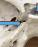
foramen lacerum
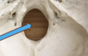
foramen magnum
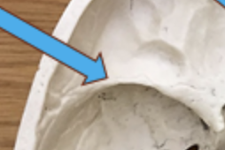
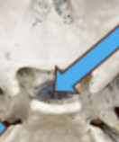
sella turcica
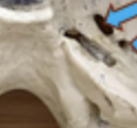
foramen ovale
foramen spinosum
ethmoid bone
greater wing of sphenoid
optic canal
foramen rotundum