CNS Supporting Cells
1/74
There's no tags or description
Looks like no tags are added yet.
Name | Mastery | Learn | Test | Matching | Spaced |
|---|
No study sessions yet.
75 Terms
Glial cells surround
Neurons, axons, and synapses
Glial cells provide
structural, metabolic and functional support in the CNS and PNS
Glia =
“glue”
Glial cells can be categorized as
macroglia and microglia
Macroglia are derived from
ectoderm (the neural tube (CNS glia) or the neural crest cells (PNS glia))
five major types of glial cells located in the CNS:
astrocytes, oligodendrocytes, microglia, ependymal cells, and pericytes
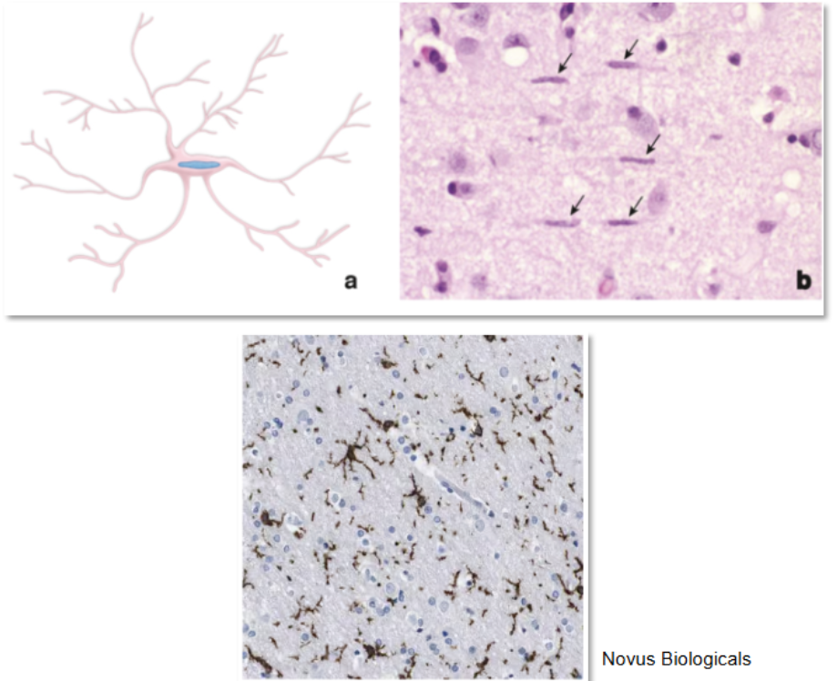
Astrocytes
are morphologically heterogeneous cells that provide physical and metabolic support for neurons of the CNS
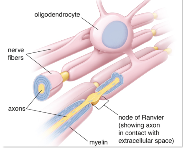
Oligodendrocytes
are small cells that are active in the formation and maintenance of myelin in the CNS
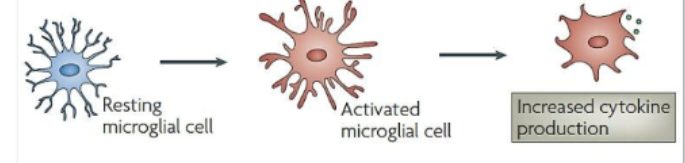
Microglia
are resident immune cells with small, dark, elongated nuclei that possess phagocytotic properties
Ependymal Cells
columnar cells that line the ventricles of the brain and the central canal of the spinal cord
Pericytes
surround capillary endothelial cells and help maintain the blood-brain barrier (BBB)
Astrocytes
Provide
structural and metabolic support in the nervous system
Appearance
star like “astro” + cell “cyte”
Most ___ glia in the CNS
abundant
Type I protoplasmic astrocytes
support neuronal cell bodies and dendrites in the gray matter
Type II fibrous astrocytes
support axons and their myelinating cells (oligodendrocytes) in the white matter
Important Astrocyte Activities
Repair of neuronal injury
• Regulation of the internal fluid environment of the CNS
• Clearance of neurotransmitters from synaptic cleft
• Metabolic exchange between the vascular system and the neurons of the
nervous system.
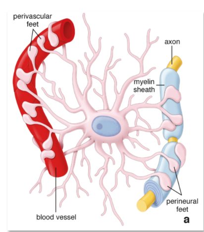
• Astrocyte end feet assist with blood brain barrier maintenance
Astrocytes form structures called
end feet on capillaries, a component of the BBB
Transporter porteins on the end feet allow astrocytes to take up important molecules such as
glucose, lactate, amino acids, and other metabolites from the blood
These transporter proteins then
release them into the extracellular fluid around
Through this mechanisms astrocytes provide key nutrients to
nearby neurons, axons, and oligodendrocytes
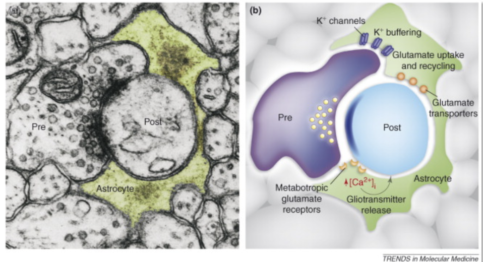
Astrocytes also express
ion channels and ion transporters, which enable them to regulate the ionic concentration of the extracellular fluid
In response to neuronal activity, neurons and astrocytes
release vasodilators and vasoconstrictors that affect capillary
blood flow
By releasing Vasomodulators
neurons and astrocytes may regulate vascular endothelial cells directly and/or through another type of glial cell, the pericytes.
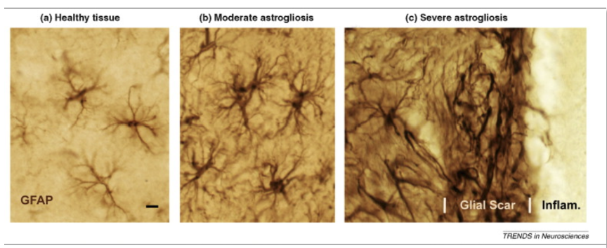
Following injury to the CNS astrocytes respond by
transforming into reactive glia, releasing immune modulatory factors, and forming glial scars
These glial scares can replace
regions of lost neurons but impede the regeneration of axons
Astrocytes are important mediators of synaptic transmission, forming the
tripartite synapse
Astrocytes help regulate
synaptic glutamate levels at glutamateric synapses
Glutamine is imported into the
presynaptic glutamatergic neuron and converted into glutamate by glutaminase
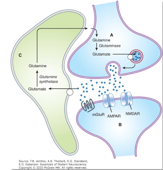
The glutamate is then transported in
vesicles by the vesicular glutamate transporter
Upon release into the synapse
glutamate can bind to and activate AMPA and NMDA types of ionotropic glutamate receptors (AMPAR, NMDAR) on the postsynaptic neuron
Synaptic transmission is terminated by
transport of the synaptic glutamate into a neighboring astrocyte or into the presynaptic and postsynaptic neurons, which is not shown by a glutamate transporter
It is converted into
glutamine by glutamine synthetase and transported back into the extracellular fluid where it can be taken up by the presynaptic axon
By regulating the levels of glutamate
the magnitude and duration of the postsynaptic responses can be modulated
Astrocyte Images
Oligodendrocytes
Produce and maintain the myelin sheath in the CNS, which aids in insulating neurons to maximize action potential speed (electrical signaling).
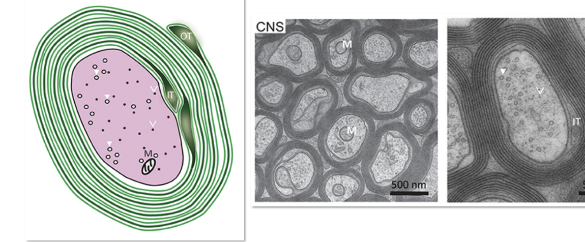
Myelin Sheath in the CNS is formed by
concentric layers of oligodendrocyte plasma membrane
The multiple processes of a single oligodendrocytes may myelinate
one axon or several nearby axons
The nucleus containing region of the oligodendrocyte may be at some
distance from the axons it myelinates.
Myelin specific proteins
proteolipid protein (PLP)
myelin oligodendrocyte glycoprotein (MOG)
oligodendrocyte myelin glycoprotein (OMgp)
With its high lipid content (80% lipid and 20% protein) the myelin membranes form an
insulating sheath that ensures fast and efficient conduction of action potentials by salutatory conduction
In the CNS, each oligodendrocyte can form __ segment of myelin for up to ___ adjacent axons
1; 50
An individual axon (especially the longer ones) is myelinated by
multiple oligodendrocytes along its length
If oligodendrocytes become damaged or die
oligodendrocyte precursor cells in the subventricular zone can differentiate and replace oligodendrocytes to remyelinate axons.
Microglia
innate immune cells in the brain
In regions of injury and disease
microglia become actively phagocytotic (reactive microglial cells)
What % of the total cells iin the brain are resident immune cells in the CNS?
15-20%
Microglia are likely derived from the
embryonic yolk sac and are distinguished by their small cell bodies and short processes
Microglia exhibit both
phagocytic and antigen presenting functions that defend the CNS from infection by bacteria, viruses, and fungi
Microglia are involved in maintaining
overall brain health as they constantly scavenge the CNS for extracellular protein aggregates called plaques and damaged neurons and synapses
As phagocytic cells microglia have also been implicated in
dendritic spine removal underlying spine plasticity
They remove
bacteria, injured cells, and the debris of cells that undergo apoptosis
they also mediate neuroimmune reactions
such as those occurring in chronic pain conditions
When activated they become
round shaped
Can be activated to
inflammatory or anti inflammatory
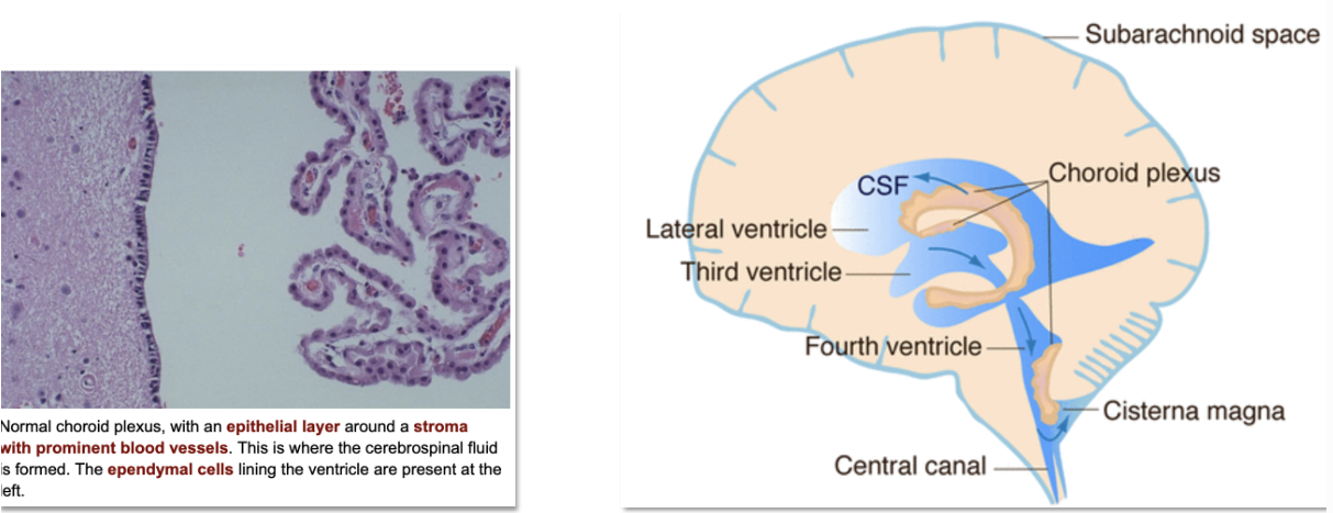
Ependymal Cells
Line the ventricles and central canal and form the choroid plexus
Forming a epithelial layer ependymal cells produce and circulate
CSF for the brain and spinal cord
Within the 4 brain ventricles
a population of specialized ependymal cells and capillaries together form a structure called the choroid plexus, which produces the majority of CSF
CSF is formed as
plasma is filtered from the blood through the ependymal cells
Choroid plexus ependymal cells actively
transport sodium, chloride, and bicarbonate ions, with the accompanying water, into the ventricles
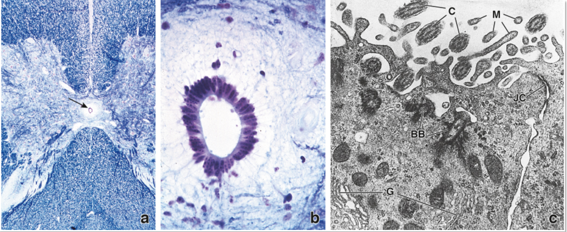
Tight junctions formed between
ependymal cells establish the blood–CSF barrier, and cilia on their apical surface facilitate the circulation of CSF
The other part of the blood CSF barrier is formed by the
tight junctions between the cells in the arachnoid membrane
Ventricles are lined with
ependymal cells
Every ventricle as well as the central canal in the
spinal cord
Blood Brain Barrier is formed by
specialized vascular endothelial cells in CNS capillaries
The BBB forms
tight junctions that prevent the diffusion of water-soluble molecules, and movement of immune and pathogenic cells from the blood into the CNS
The endothelial cells are covered by
pericytes a type of contractile glial cell (smooth muscle type cell),
Pericytes
help regulate the tight junctions and also control the diameter of the capillary and modulate blood flow.
Surrounding the pericytes are
astrocytic end feet that function to transport essential nutrients from the blood into CNS tissue and release factors that affect contraction of pericytes
Pericytes and the BBB
Pericytes are
contractile cells that surround capillary endothelial cells and help form and maintain the tight junctions that ensure the BBB.
As contractile cells
pericytes can regulate contraction and relaxation that control capillary blood flow
Arterioles
which are larger blood vessels lined with
vascular smooth muscle, have also been implicated as
cells that respond to neuronal activity–dependent
regulation of brain blood flow
fMRI
functional neuroimaging technique that detects changes
in blood flow as an indirect measure of neuronal activity.