THE EYE
0.0(0)
Card Sorting
1/105
Earn XP
Description and Tags
Last updated 7:03 AM on 11/21/22
Name | Mastery | Learn | Test | Matching | Spaced | Call with Kai |
|---|
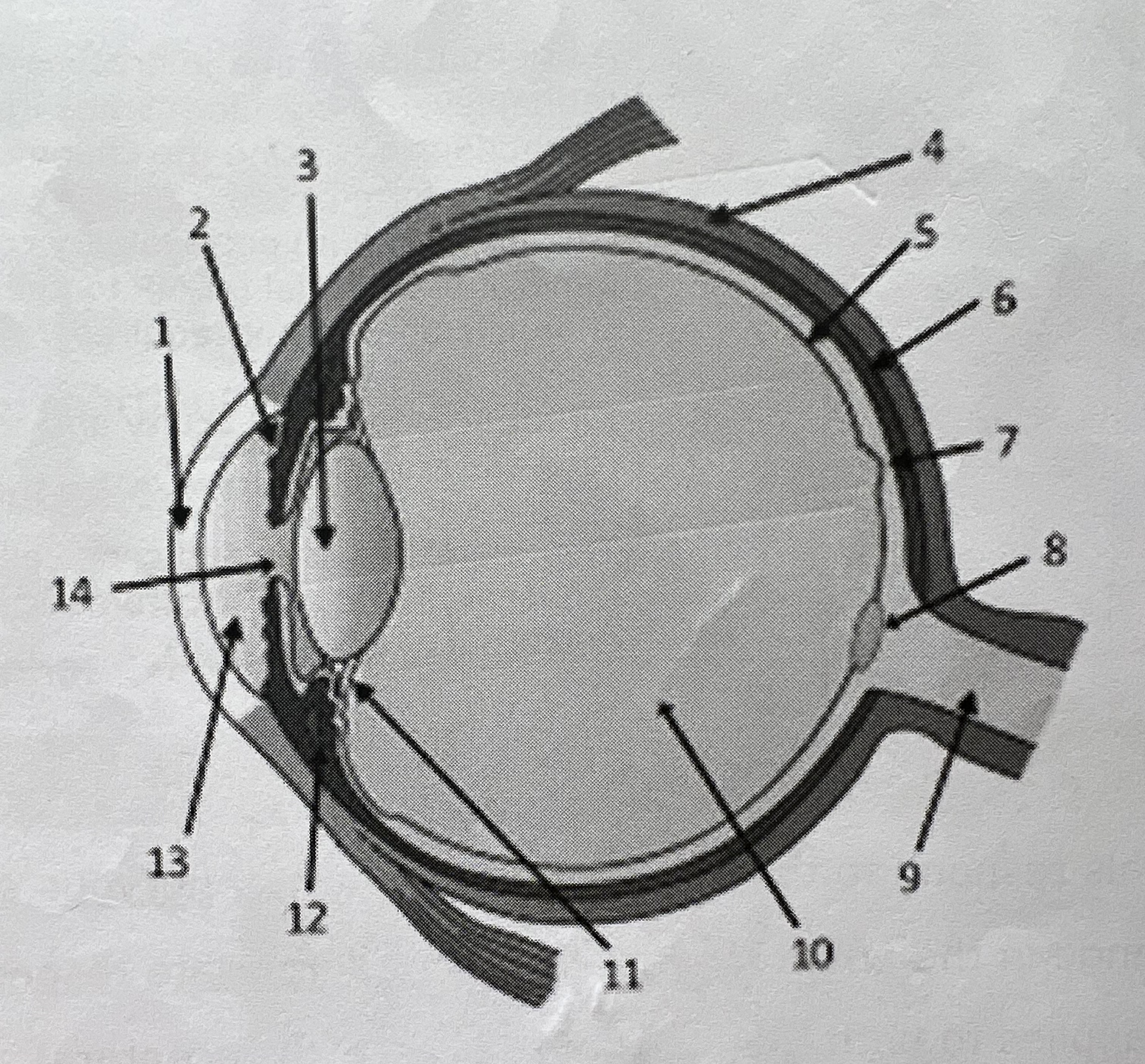
No analytics yet
Send a link to your students to track their progress
106 Terms
1
New cards
a internal or external environmental change
what stimulates a response ?
2
New cards
A chemical or physical change in the internal or external environment detected by receptors which evoke a response
What is a stimulus ?
3
New cards
they perceive a specific form of energy (heat, force, kinetic) and then convert the stimulus energy into a chemical energy called a nervous impulse
What do receptor cells do
4
New cards
converts stimulus energy into chemical energy in the form of an impulse
What is a transduction?
5
New cards
biological transducers
what are receptors called
6
New cards
fill in
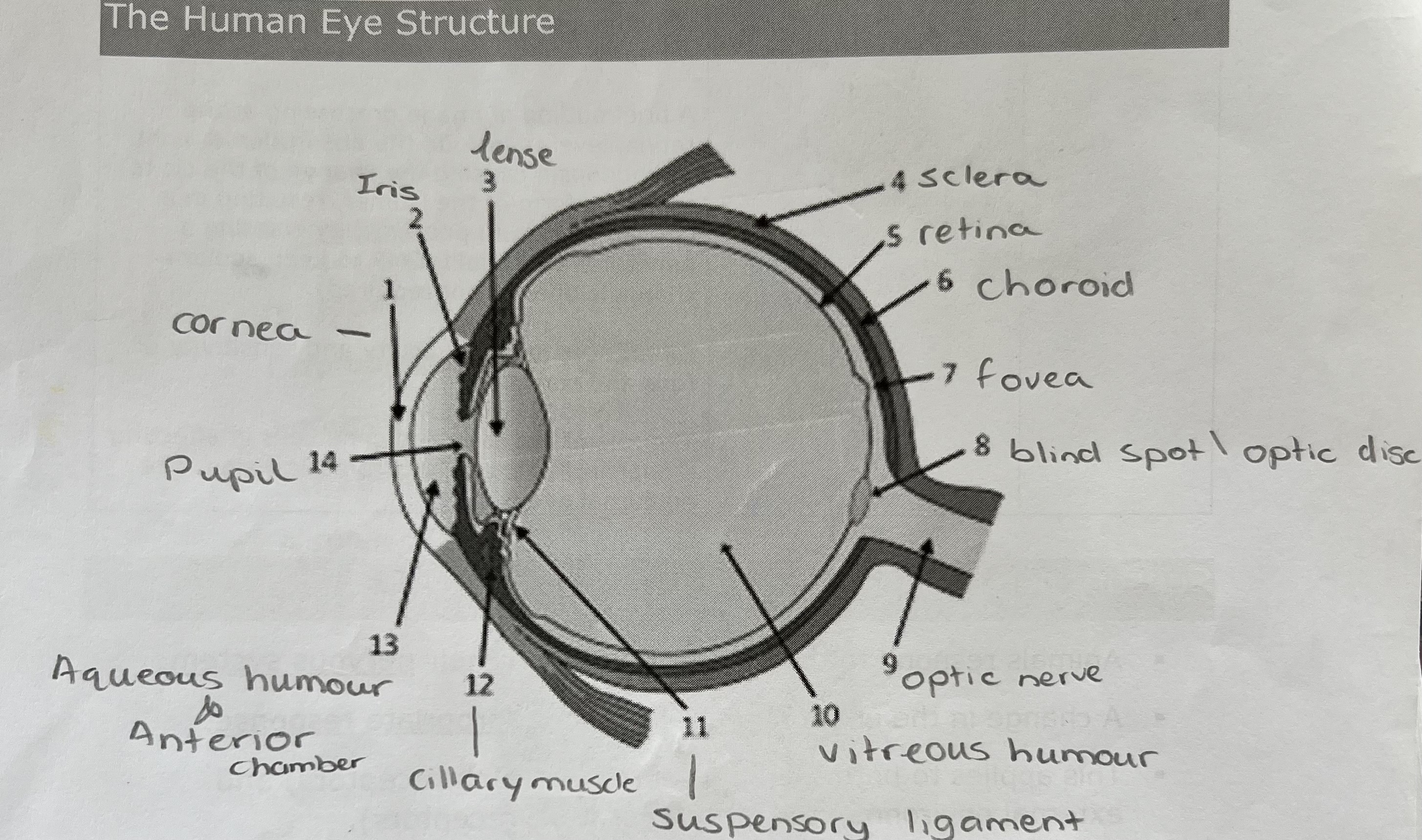
7
New cards
Orbits in the skull (sockets with tiny hole at the end
where is the eye located?
8
New cards
there are no bones in the eye rather it is supported via hydrostatic pressure created by the vitreous humour
how are the eyes supported
9
New cards
—> Sclera & cornea
—> choroid, ciliary body, lens & iris
—> the retina
—> choroid, ciliary body, lens & iris
—> the retina
outer to inner most tissue layers of the eye
10
New cards
they are external to the eye
Where are the ancillary structures located?
11
New cards
eyebrows & eyelashes — protect from foreign objects, perspiration and sunlight
eyelid — retractable shade (baths eye by blinking)
Conjunctiva — transparent, mucous membrane (continuous with sclera and eyelids. Lubricates the eye surface by secreting mucilage. It secrets lysozyme (bacterial enzyme) and contains blood vessels.
eyelid — retractable shade (baths eye by blinking)
Conjunctiva — transparent, mucous membrane (continuous with sclera and eyelids. Lubricates the eye surface by secreting mucilage. It secrets lysozyme (bacterial enzyme) and contains blood vessels.
what structures make up the ancillary structures and what are their functions?
12
New cards
Lacrimal glands
Where are tear produced?
13
New cards
above both eyes
where is the lacrimal gland located?
14
New cards
water, salts, mucilage, oils (lipids), antibodies & lysozymes
what do tears consist of
15
New cards
Lacrimal canaliculi, Lacrimal sac (surplus tears flow into nasal cavity)
where do tears drain?
16
New cards
The outermost layer of the eye, which is tough and opaque with a yellowish-white surface due to collagen fibres
What is the sclera ?
17
New cards
To protect the eye and support by maintaining shape
What is the function of the sclera ?
18
New cards
it is a transparent dome covering the iris
What is the cornea?
19
New cards
To refract light onto the retina, this is possible due to its curved surface. This function is aided by the aqueous humour found just behind the cornea as it increases the refracted index.
what is the cornea’s function?
20
New cards
a circular muscular diaphragm, made of smooth muscle.
What is the Iris?
21
New cards
it closes and opens the pupil controlling the amount of light which enters
what is the function of the iris?
22
New cards
the pigment over the iris (the amount of melanin)
what determines the pigment of the iris?
23
New cards
an opening in the iris, it looks black due to it absorbing light.
What is the pupil?
24
New cards
when intense light reflects off of the choroid blood vessels inside.
what is the red pupil effect ?
25
New cards
iris contracts (circular muscle contracts) therefore the pupil contracts and allows less light in
Closing of pupil:
26
New cards
longitudinal muscle contracts, the pupil dilates, allowing more light in.
opening of pupil:
27
New cards
The contraction and dilation of the pupil is a reflex, meaning that it is an involuntary movement in response to a stimulus
what is a reflex, give an example :
28
New cards
the autonomic nervous system
What part of the NS controls the pupil ?
29
New cards
—> It is rich in blood vessels to supply the retina
—> contains pigment cells which absorb light and prevent reflection of light (eye appears black)
—> contains pigment cells which absorb light and prevent reflection of light (eye appears black)
Function of the choroid layer>
30
New cards
ciliary body: marks the point where the sclera and cornea join, contains muscles and blood vessels as well as secretes aq humour.
ciliary muscle: smooth muscle, circular and longitudinal which bring about lens accommodation
ciliary ligaments: attaches ciliary body to lense
ciliary muscle: smooth muscle, circular and longitudinal which bring about lens accommodation
ciliary ligaments: attaches ciliary body to lense
what make up the accommodation structures? and what are their functions?
31
New cards
The ability to alter its shape and shift focus (forward backward) to form image on retina.
What is lens accommodation ?
32
New cards
A plastic, transparent biconvex structure that focuses light via refraction.
What is the lens?
33
New cards
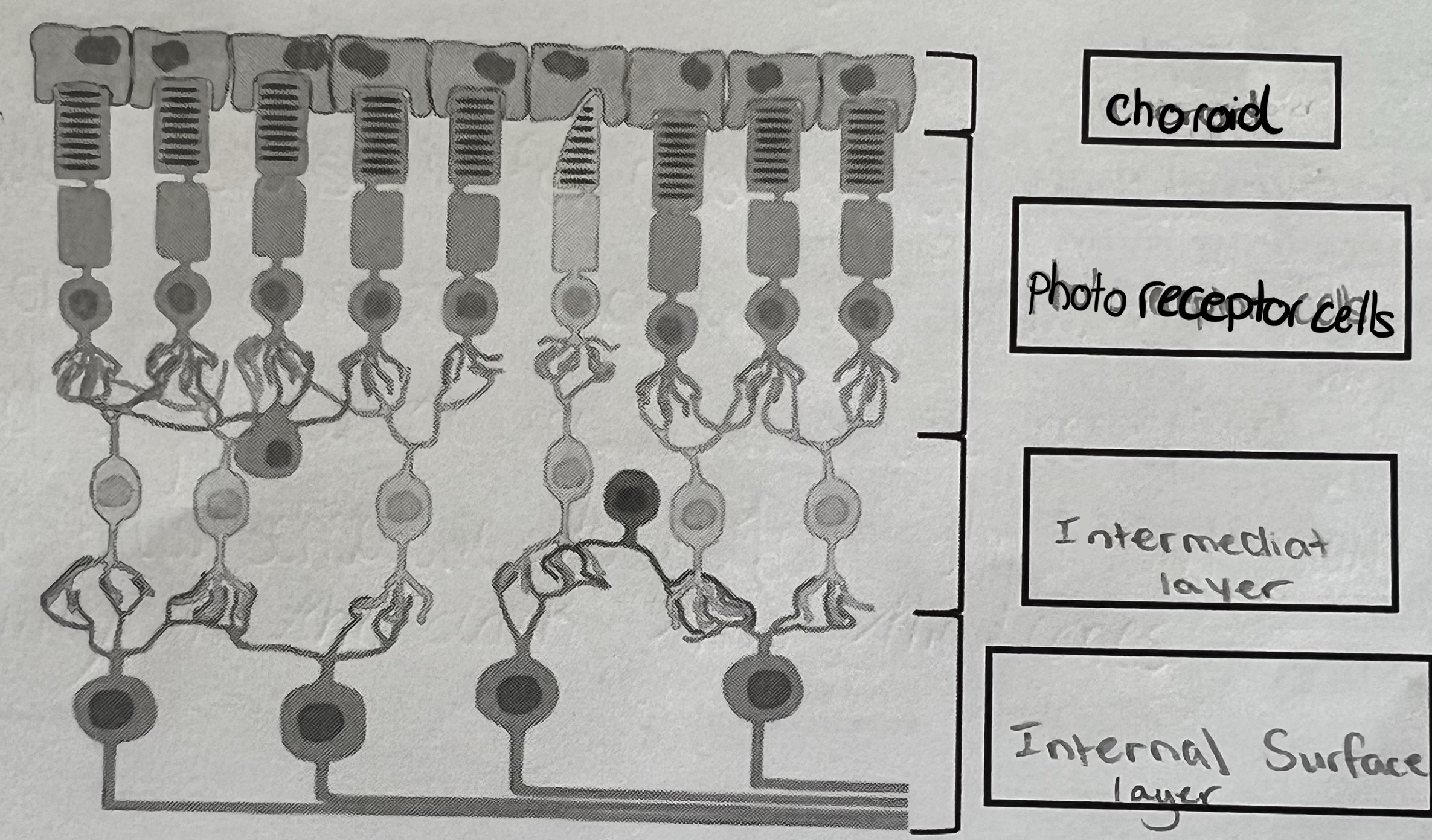
composed on sensory tissue with receptor cells, cell bodies and axons of neurones
What is the retina?
34
New cards
Rods: work best in low intensity light (convergence)
Cones: work best in high intensity light (visual acuity)
Cones: work best in high intensity light (visual acuity)
what are the two types of receptor cells ?
35
New cards
The optic disc which is the blind spot
Where do all neurones lead to ?
36
New cards
The most sensitive spot which contains the most cones, it is the point where most light focuses (sharpest vision)
What is the Fovea ?
37
New cards
a clear salt solution that refracts light (secreted by the ciliary body and drains into the blood vessels (via the cabal of schlemm)
What is Aqueous humour ?
38
New cards
Glaucoma
What happens if the canal of schlemm block up?
39
New cards
it is a clear semi- solid gel that refracts light further and supports the eyeball via hydrostatic pressure.
what is vitreous humour?
40
New cards
41
New cards
Photoreceptor layers: contains the rods and cones, particularly embedded in choroid to stop light travelling further.
intermediate layer: contains bio polar neurones and synapses connecting the photoreceptors layer and internal layer.
internal layer: includes ganglion cells and dendrites & axons of the optic nerve
intermediate layer: contains bio polar neurones and synapses connecting the photoreceptors layer and internal layer.
internal layer: includes ganglion cells and dendrites & axons of the optic nerve
what are the 3 tissues layers the retina consists of ?
42
New cards
—> outer segment : photosensitive region (flattened membranous vesicles containing pigment)
converts light energy into a generator potential (graded potential (may trigger an action potential)
—> A constriction: cytoplasm constricts due to pinch in outer membrane.
inner and outer segment connected via two cilia.
—> Inner segments: NUCLEUS (metabolically active part of the cell. MITOCHONDRIA (produces energy). POLYSOMES (protein synthesis — pigments and membranous vesicles)
—> Synaptic region: receptor synaptic connection with bipolar neurone.
what are the 4 main parts of cones and rods ?
43
New cards
a change in the membrane potential that is proportional to the size of the stimulus.
Graded potential
44
New cards
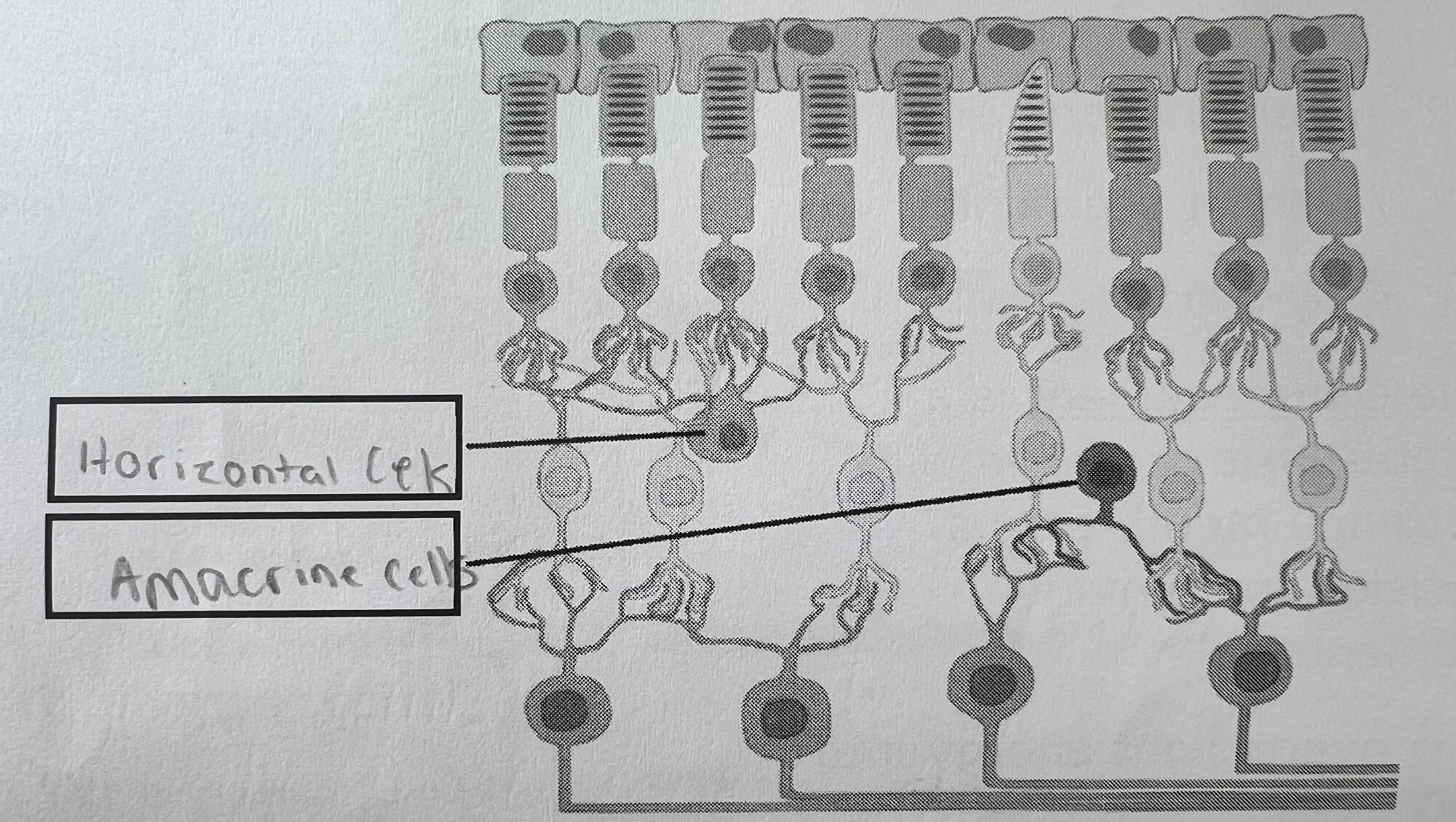
connect rods and integrate + regulate impulses
Horizontal cells
45
New cards
horizontally connected across several bipolar neurones (link both rods and cones). Allow for some visual processing before the impulse leaves retina
Amacrine cells
46
New cards
visual pigment — rods
rhodopsin
47
New cards
rod-shaped light sensitive cells which lies on the most peripheral parts of the retina in the vertebrate eye
rods
48
New cards
rod
outer segment is cylindrical
49
New cards
rod
synaptic connection is a single knob
50
New cards
rod
single type of cell
51
New cards
rod
monochromatic vision
52
New cards
several rods are interconnected to one horizontal cell
convergence
53
New cards
rod
used for low vision light conditions
54
New cards
rod
sensitive to scattered light
55
New cards
rod
highly light sensitive
56
New cards
rod
low visual acuity
57
New cards
rod
absent in fovea
58
New cards
rod
slower response to light
59
New cards
rod
about 12 million
60
New cards
rods
contain more pigments
61
New cards
rods
stacks of membrane - enclosed discs are not attached directly
62
New cards
rods
insufficiency of rhodopsin causes night blindness (black spots)
63
New cards
cones
trichromatic
64
New cards
cones
not sensitive to scattered light
65
New cards
cones
not highly sensitive to light
66
New cards
cones
around 6 million
67
New cards
cones
concentrated in fovea
68
New cards
cones
discs directly attached to outer membrane
69
New cards
cones
insufficiency of iodopsin causes colourblindness
70
New cards
cones
faster light response
71
New cards
cones
contains fewer pigments
72
New cards
cones
useful in bright light conditions
73
New cards
cones
each is connected individually
74
New cards
cones
3 types if cells (RBG)
75
New cards
cones
synaptic connection is branched
76
New cards
cones
outer segment is conical
77
New cards
cones
Iodopsin is the visual pigment
78
New cards
a type of photoreceptor in the retina which is responsible for colour vision in daylight.
cone
79
New cards
when they are directly stared at as there is a concentration of cones in the fovea.
when do objects look sharpest and why ?
80
New cards
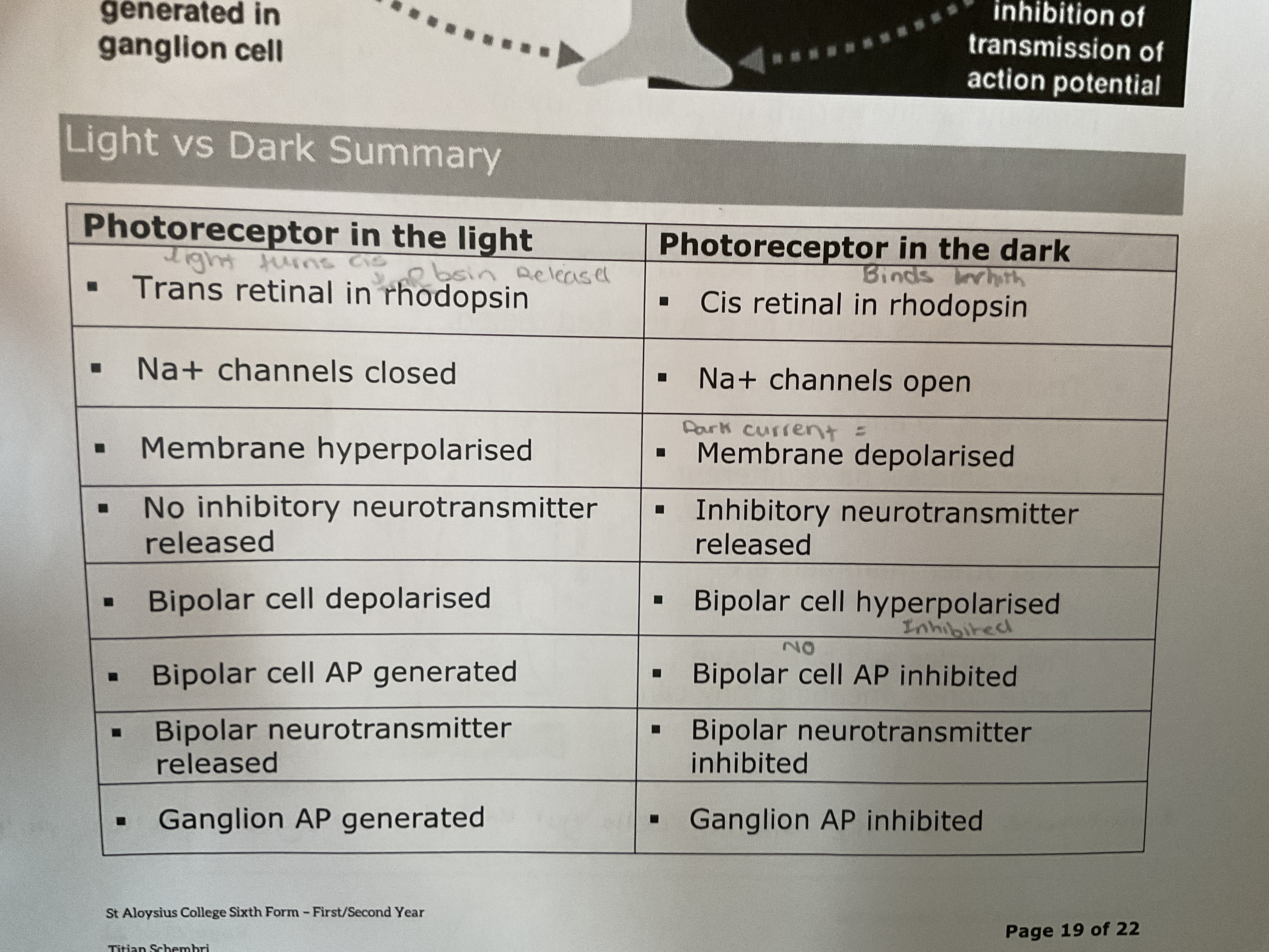
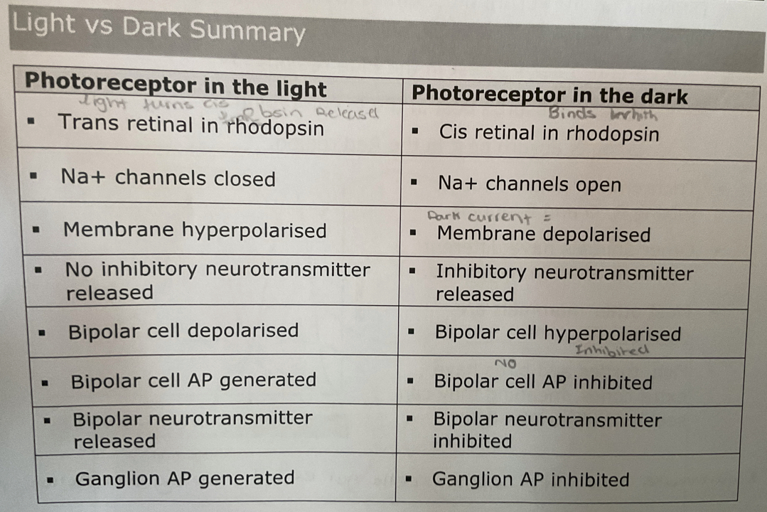
they work in inverse sequence compared to general sensory stimuli.
how do light energy transductions work ?
81
New cards
rods and cones work similarly:
—> absence of light = produce inhibitory neurotransmitter (glutamate)
—> glutamate — hyperpolarises the bipolar neurones (therefore wont excite ganglion cells)
—> presence of light — inhibitory neurotransmitter is inhibited
—> bipolar neurones exit ganglion cells and generate impulse leading to the brain.
mechanism of sensory transduction
82
New cards
opsin (protein + light absorbing group)
isomers of retinal (💡all-trans retinal 🌚 11-cris retinal)
isomers of retinal (💡all-trans retinal 🌚 11-cris retinal)
what does rhodopsin consist of?
83
New cards
11-cis retinal + light photon = all-trans retinal
opsin breaks off from all-trans retinal
—> molecule losses sensitivity (BLEACHING)
opsin breaks off from all-trans retinal
—> molecule losses sensitivity (BLEACHING)
Rhodopsin In light
84
New cards
bleaching happens quickly in bright light — dark spots appear and disappear when rhodopsin is recharged.
(11- cis retinal spontaneous turn to all trans retinal and opsin rebonds to it as they are recharged)
(11- cis retinal spontaneous turn to all trans retinal and opsin rebonds to it as they are recharged)
explain bleaching
85
New cards
when opsin is broken off and changes shape it triggers this effect.
what triggers the Na+ channels to close in the outer segment
86
New cards
Na+ channel in the outer segment are open in the dark and so Na+ ions flow from inner segment ——> outer segment.
opsin present :
Cyclic Guanosine Monophosphate —> Guanosine Monophosphate
Cyclic Guanosine Monophosphate —> keep Na+ channels open
opsin present :
Cyclic Guanosine Monophosphate —> Guanosine Monophosphate
Cyclic Guanosine Monophosphate —> keep Na+ channels open
Dark current:
87
New cards
light Na+ channels close therefore hyperpolarisation
Guanosine monophosphate closes channels
Guanosine monophosphate closes channels
hyperpolarisation of photoreceptors
88
New cards
hyperpolarised photoreceptors, which depolarises bipolar neurones and generate an Action Potential leading to a retinal ganglion cell Action potential sending an impulse to the brain
what stops the release of glutamate?
89
New cards
1 photon inhibits over 1m Na+
what triggers hyperpolarisation of photoreceptors
90
New cards
91
New cards
having 3 types of cones each with a different opsin molecule (different opsin = absorb different ranges of visible spectrum).
It involves mixing RBG signals
It involves mixing RBG signals
Trichromatic vision
92
New cards
blue region
S-cone
93
New cards
green
M-cone
94
New cards
red
L-cones
95
New cards
dichromatic
most mammals are ____chromatic
96
New cards
fish, turtles, birds
have extra cones so they see ultraviolet
97
New cards
deficiency of one or more cones, linked to X-chromosome (sex linked)
colourblindness
98
New cards
red-green
most common colourblindness
99
New cards
> only rods (or few cones)
> pupils stretch far wider (allow more light in)
> Tapetum lucidum (reflects more light onto receptors)
> Disproportionately large eyes
> Reduced eye movement
> Rotational ability of the neck
> spherical lens and wide cornea —> increase refraction
> pupils stretch far wider (allow more light in)
> Tapetum lucidum (reflects more light onto receptors)
> Disproportionately large eyes
> Reduced eye movement
> Rotational ability of the neck
> spherical lens and wide cornea —> increase refraction
Adaptations of nocturnal Animals (7)
100
New cards
the ability to distinguish two ore more stimuli of equal intensity separately. SHARPNESS OF VISION
visual acuity