MSK Part 1: CH6
1/155
There's no tags or description
Looks like no tags are added yet.
Name | Mastery | Learn | Test | Matching | Spaced | Call with Kai |
|---|
No analytics yet
Send a link to your students to track their progress
156 Terms
Textbook first
yay
Calcifications in the soft tissues can indicate what?
-Tumor
-Myositis Ossificans
-Systemic disorders (scleroderma, hyperparathyroidism)
Look at this image and tell me what view and deformity
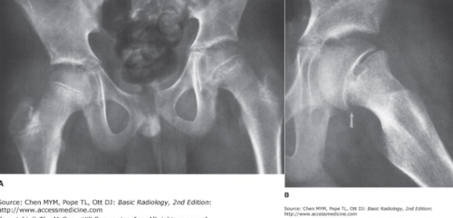
Analysis
Views: AP (left), frog leg lateral (right)
Analysis: Fracture through the physis of the left proximal femur. Lucent line demarcating the physis is slightly widened and alignment of the dges of the epiphysis and metaphysis is abnormal. Frog leg is more obvious. They have SCFE
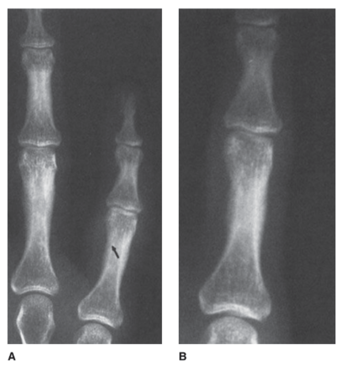
What is fractured here?
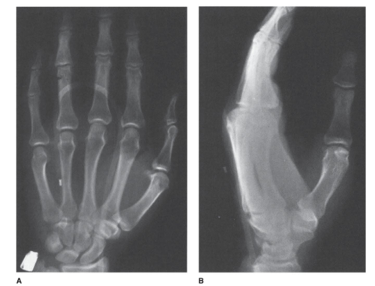
answer
View: AP and lateral radiographs
You cant see anything on the lateral, but on the AP you can see a phalangeal fracture of the 4th phalanx.
A radiograph of normal bone shows what
A smooth homeogenous cortex surrounding th emedullary space. The cortex will be thicker along the shaft of the long bones and thinner in the small bones at the ends of long bones (like carpal and tarsal bone).
Wheres the fracture in this one? 1st scan is 2.5 months earlier
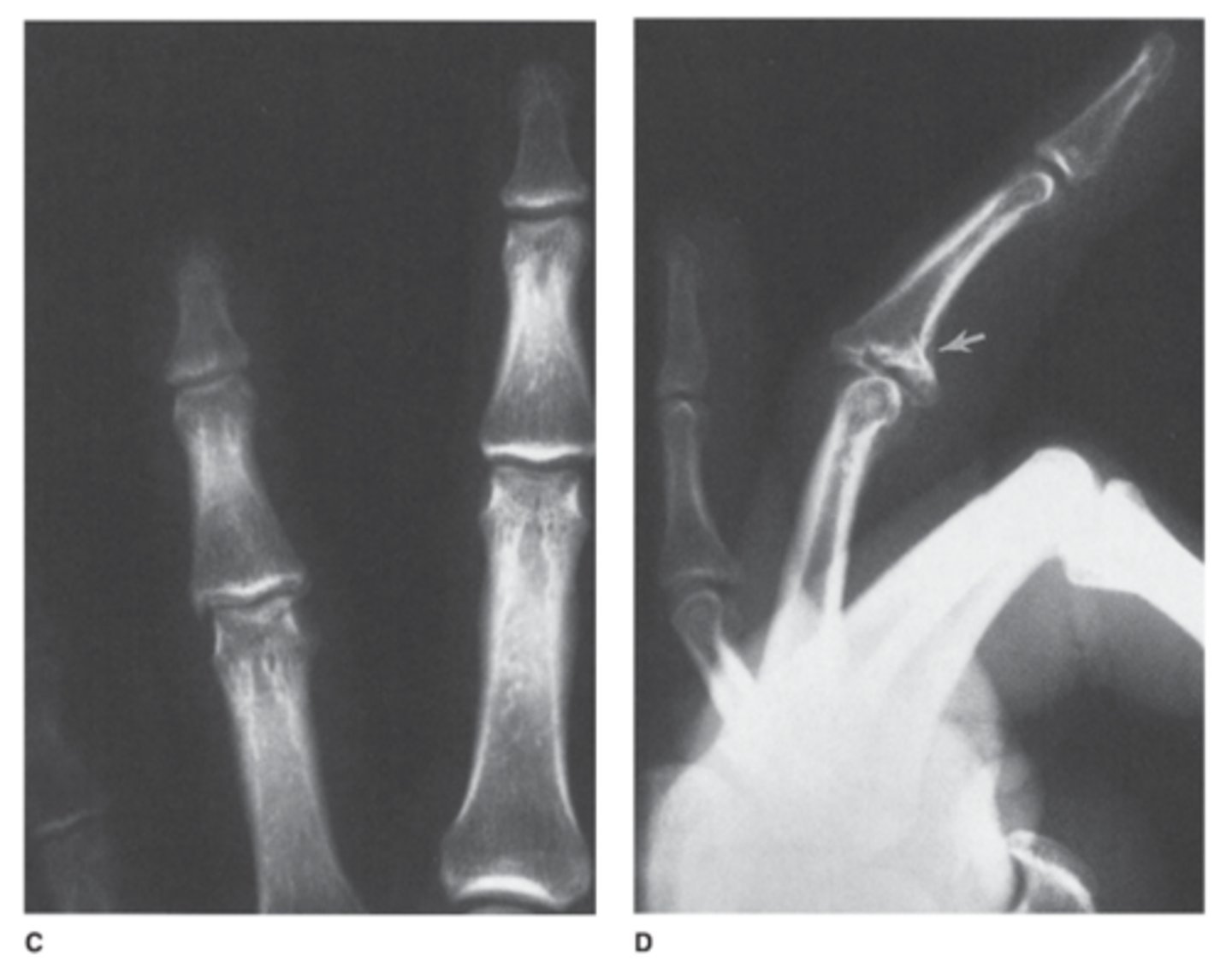
Answer
intra-articular fracture of the proximal aspect of the middle phalanx
Two uses for a CT in skeltal imaging
1) evaluating a fracture fragment position
2) evaluating bone tumors or tumor like diseases
Anything abnormal with this image of the metacarpals? 36 y/o woman
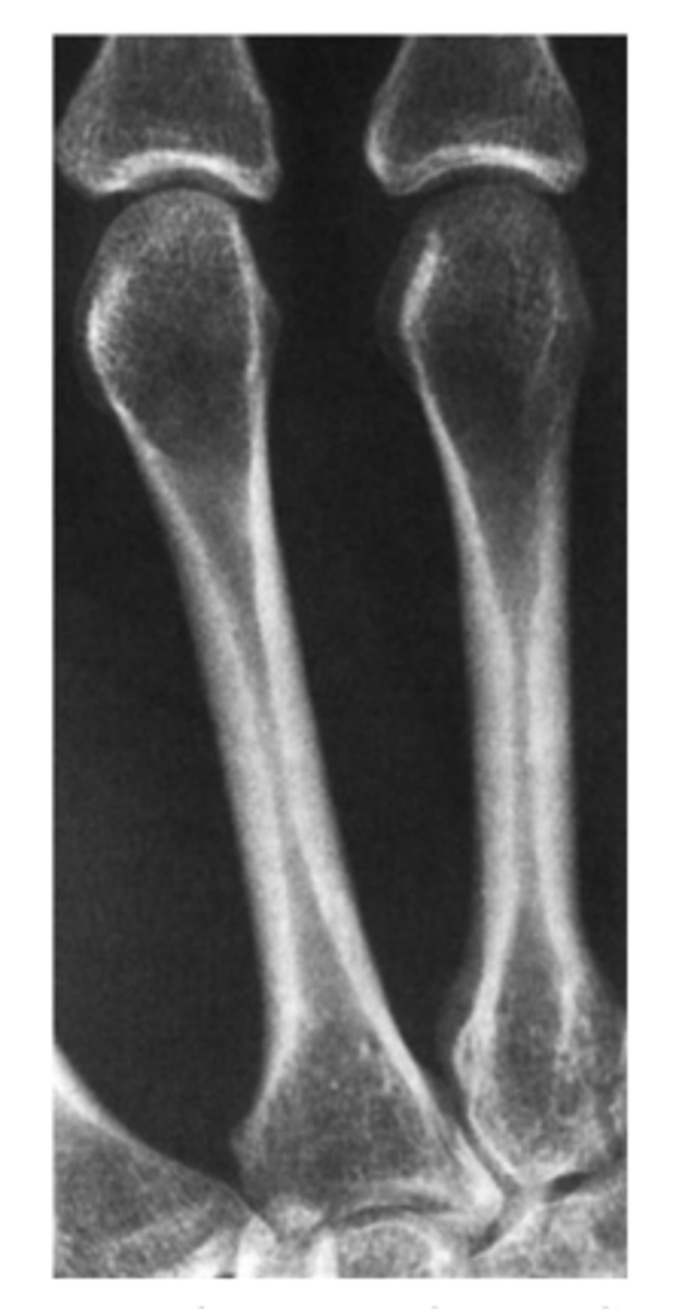
Answer
Normal metacarpals!
Note the cortex is thick and homogenous in the mid shaft of the metacarpal. it bones thinner as it approaches the ends of the bones.
MRI uses
-Great for soft tissue injury
-great for looking at neoplasm, marrow packing diseases, osteomyelitis, fractures that're occult, AVN
This is a 10 year old who 2.5 years earlier had soft tissue swelling due to cellulitis after a cat bite
-Note how the soft tissues are swollen in A.
-B is 2.5 earlier
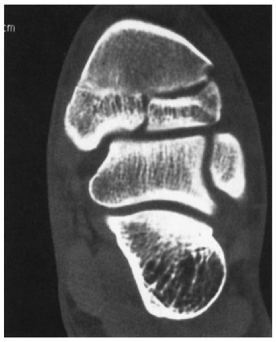
Where is the fracture here? This is the x-ray and CT
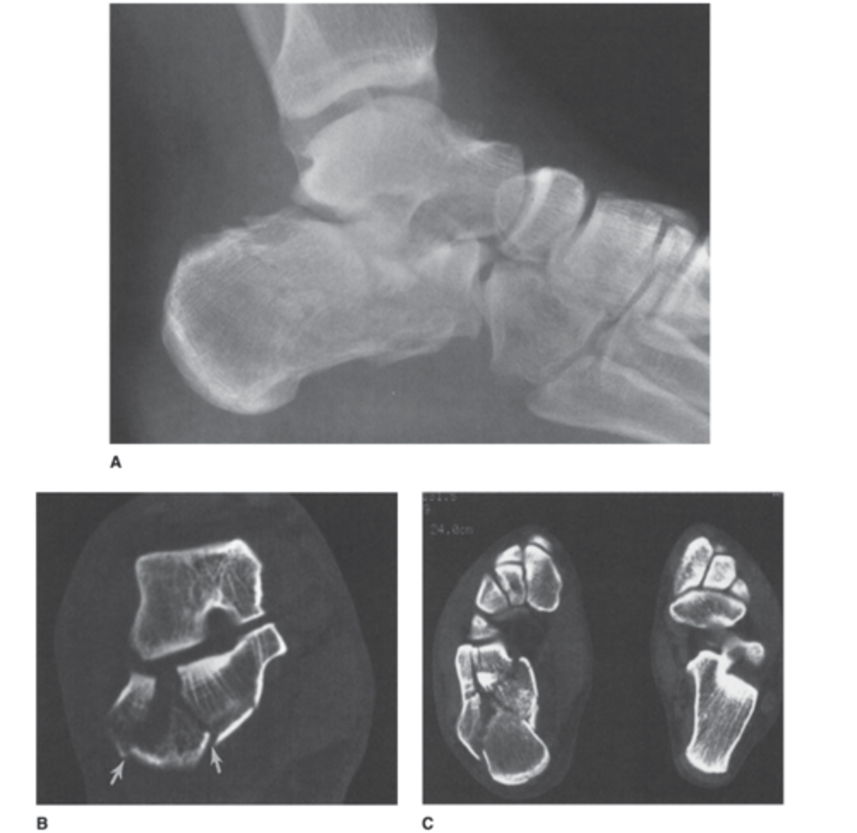
Answer
Calcaneal fracture. It is comminuted. We cant really see the degree to which it is involved on the x-ray.
CT is showing oriented fracture lines entering the posterior facet of the posterior and middle subtalar joints.
There is also a comminuted fracture of the calcenocuboid joint.
Moral of this: note how well the CT confirmed this for you
Two most common nuclear medicine imaging techniques for bone disease
-PET-CT
-Technetium bone scan
when a strongly suspected fracture is not identified on x-ray what may you do?
Repeat the x-ray in 7-10 days
What should you use second line when you think there is a bone tumor but you can't see it on x-ray
MRI
On the first day of your medical school rotation in orthopedic surgery, the resident and attending physician send you to the emergency department to see a 26-year-old man with a broken leg (AP and lateral views of the distal tibia and fibula).
You are supposed to look at the radiographs for
Case 6-1 (Figure 6-8) and call your colleagues in the operating room to describe the fracture. Which of the
following statements concerning the fracture would
you wish to make?
A. The distal tibial fragment is displaced 1 cm ante-
riorly.
B. There is no comminution of the tibial fracture.
C. There is slight valgus angulation of the distal tib-
ial fragment.
D. This is an open or compound fracture.
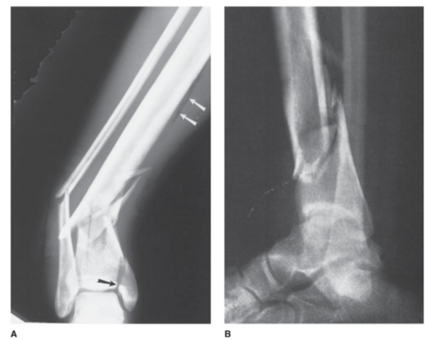
Answer
D.
comminuted fractures of the distal tibia and fibula with intra-articular extension of the tibial fracture (black arrow). The main distal fracture fragment is displaced posteriorly. As seen on the frontal view, the distal fragments are angulated so
they are pointing medially.
Is this an open fracture?
Gas density (white arrows) indicates that air has penetrated into the soft tissues through a skin wound, so this is an open fracture. A fracture line extends to the tibial articular surface (black arrow)
Infant with low-grade fever. You obtain a chest radiograph to evaluate for pneumonia (frontal view of the chest).
You interpret the chest radiograph for Case 6-2
(Figure 6-9) and render the following opinion:
A. Normal chest radiograph
B. Round pneumonia
C. Healing rib fractures
D. Pneumothorax
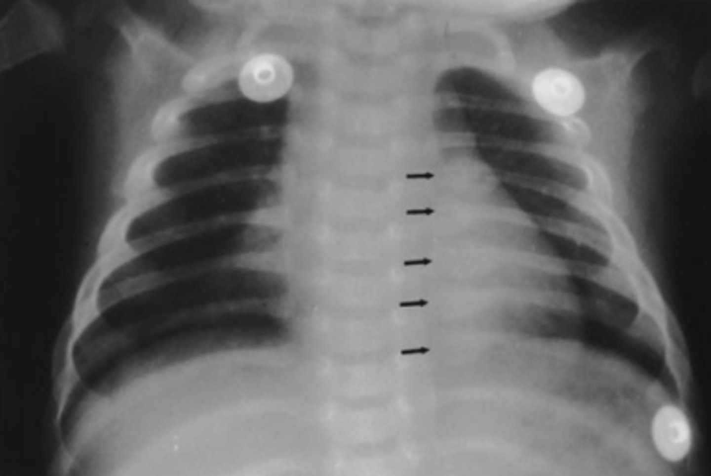
answer
C.
Unfortunately these are healing rib fractures in an infant (note the calluses). Suspected child abuse and an immediate CPS call.
While moonlighting in the emergency department of a small community hospital, you examine a 25-year-old man who fell on an outstretched
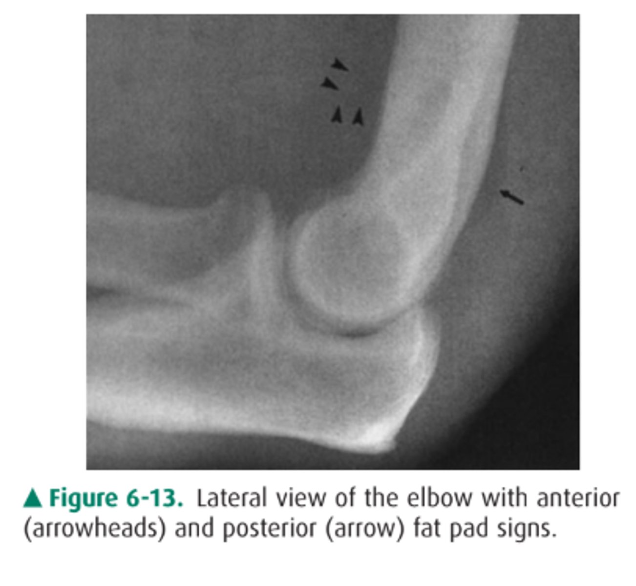
hand and now complains of elbow pain. You obtain an AP and lateral view of his elbow (lateral view of the elbow).
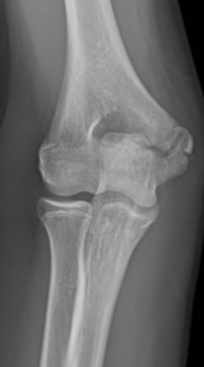
You first examine the lateral view of the elbow in Case
6-3 (Figure 6-10). You find
A. a lytic lesion in the distal humerus.
B. displacement of the fat pads of the elbow.
C. a fracture through the proximal ulna.
D. dislocation of the elbow.
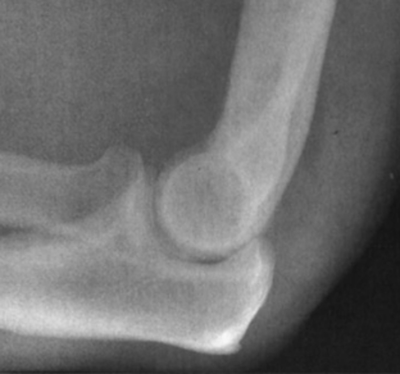
Answer
B.
You have the anterior sail sign and posterior fat pad sign.
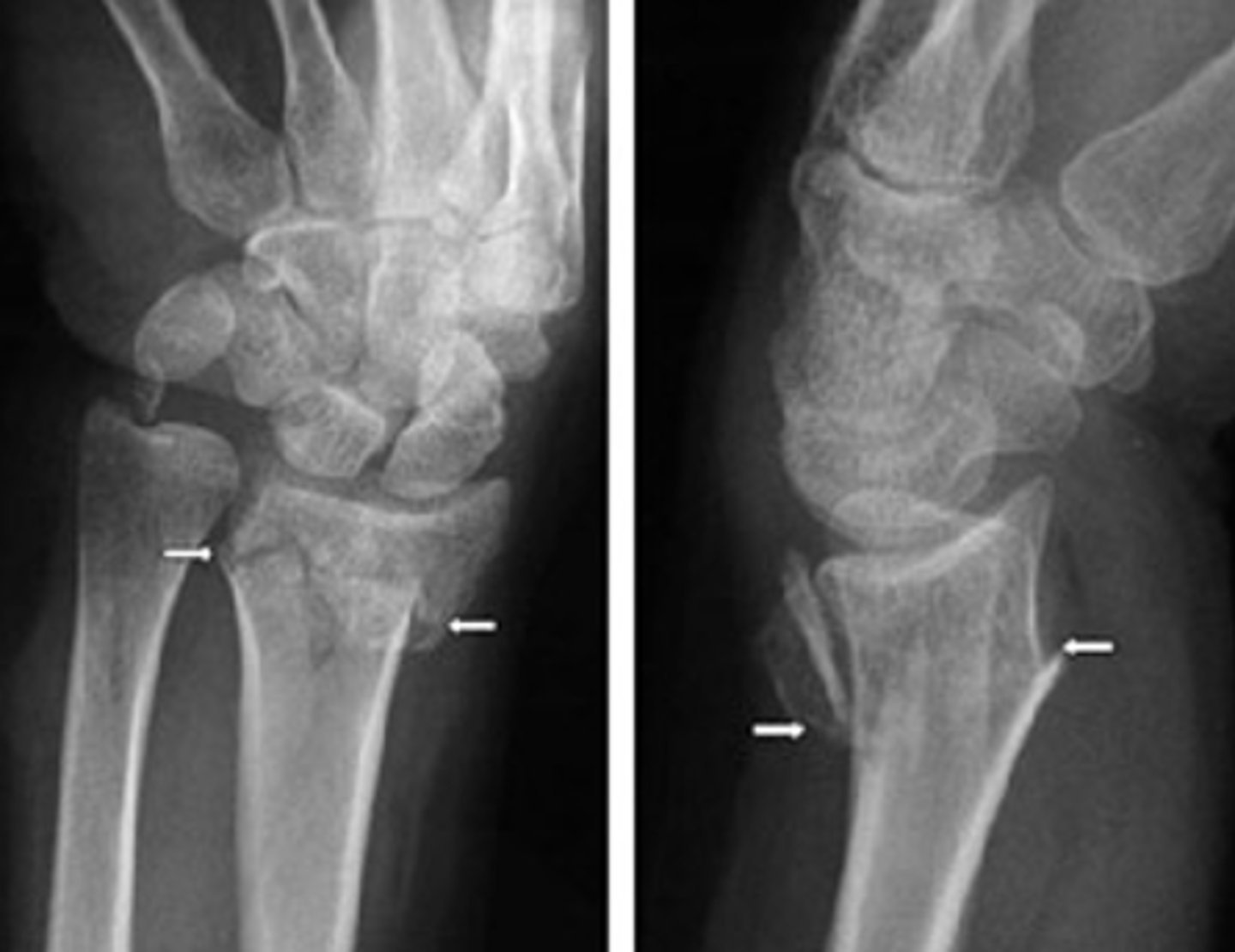
A week later you are once again moonlighting in the same small emergency department when a 29-year-old man is carried in complaining of ankle pain after a twisting injury. His ankle is swollen and ecchymotic, and he is tender to palpation along the medial malleolus. He has no other complaints.
You order frontal, lateral, and oblique views of the ankle (AP view of the ankle).
You examine the radiographs for Case 6-4. Only the
AP view is shown here (Figure 6-11). You tell the pa-
tient he has broken his ankle, but you want to get one
more study:
A. Entire tibia and fibula, to exclude more proximal
fractures
B. Ipsilateral foot, to exclude a fracture of the fifth
metatarsal
C. Contralateral ankle, for comparison purposes
D. CT, for precise evaluation of the alignment of the
fracture
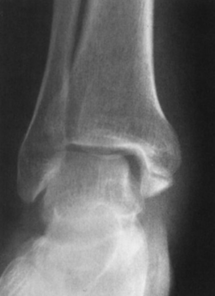
Answer
A.
Transverse fracture of the distal medial malleolus with widening of the medial aspect of the ankle joint. We see this with a proximal fibular fracture or maissoneuve. You should get these views to rule out involvement of the proximal fibula and syndesmosis injury.
A 15-year-old boy complains of ankle pain after a fall (AP and lateral views of the ankle).
What is the abnormality in Case 6-5 (Figure 6-12)?
A. Sprain of the lateral ligaments
B. Fracture of the distal fibula
C. Stress fracture of the talus
D. Triplane fracture of the distal tibia
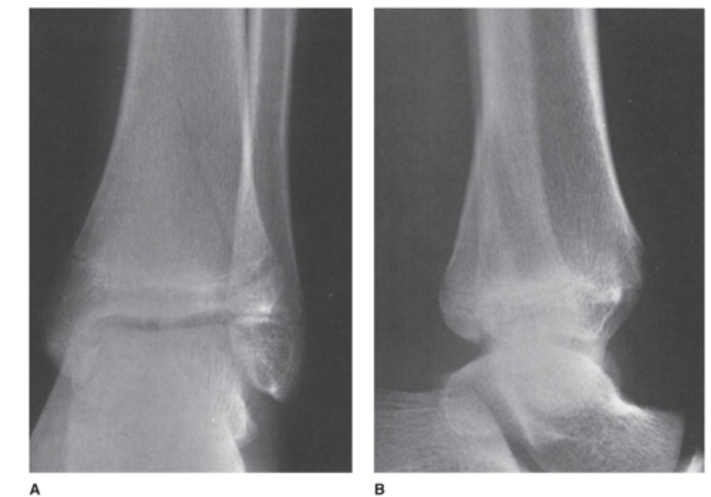
Answer
D.
Note how the lucencies rule vertically through the epiphysis and obliquely through the metaphysis. This is also called a Salter Harris IV fracture
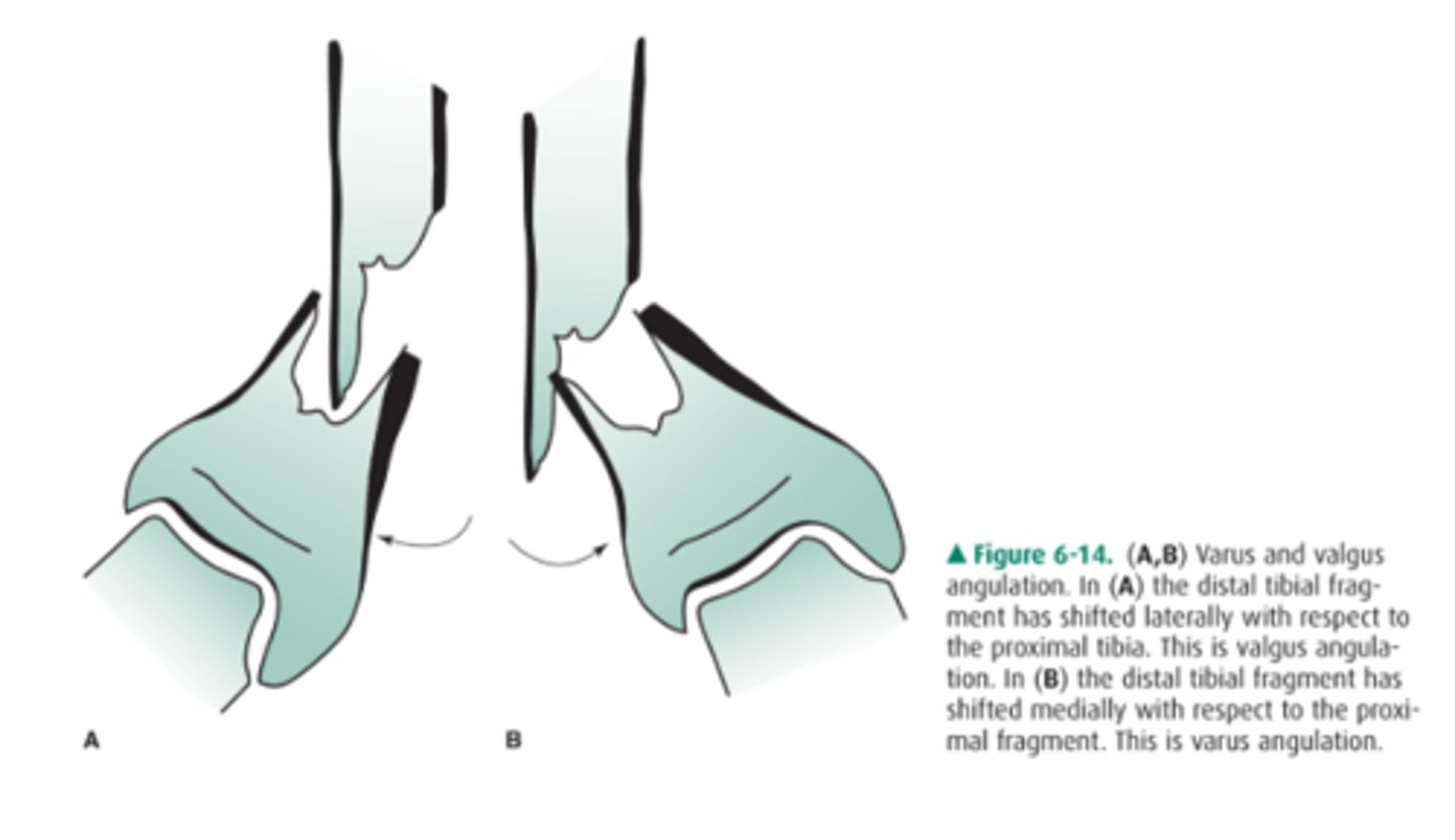
How do we describe aligment of fracture fragments?
You need to state displacement and angulation
Angulation
Use direction and degree:
EX: "30 degrees of varus angulation of the distal fragment"
What are the signs of child abuse seen here?
answer
Top is showing a metaphyseal corner and the bottom is showing a bucket handle fracture
What causes the anterior and posterior fat pads?
Displacement due to accumulation of blood or fluid in underlying tissues
Examples of what can cause the fat pad appearance
-Rheumatoid arthritis
-bleeding disorder
-Radial head fracture
Maissoneuve Fracture
∙Fibular neck fracture.
∙Proximal fibular fracture due to external rotation.
∙Energy of rotation transfers through the interosseous ligament and exits at proximal fibula.
∙Requires Syndesmotic fixation.
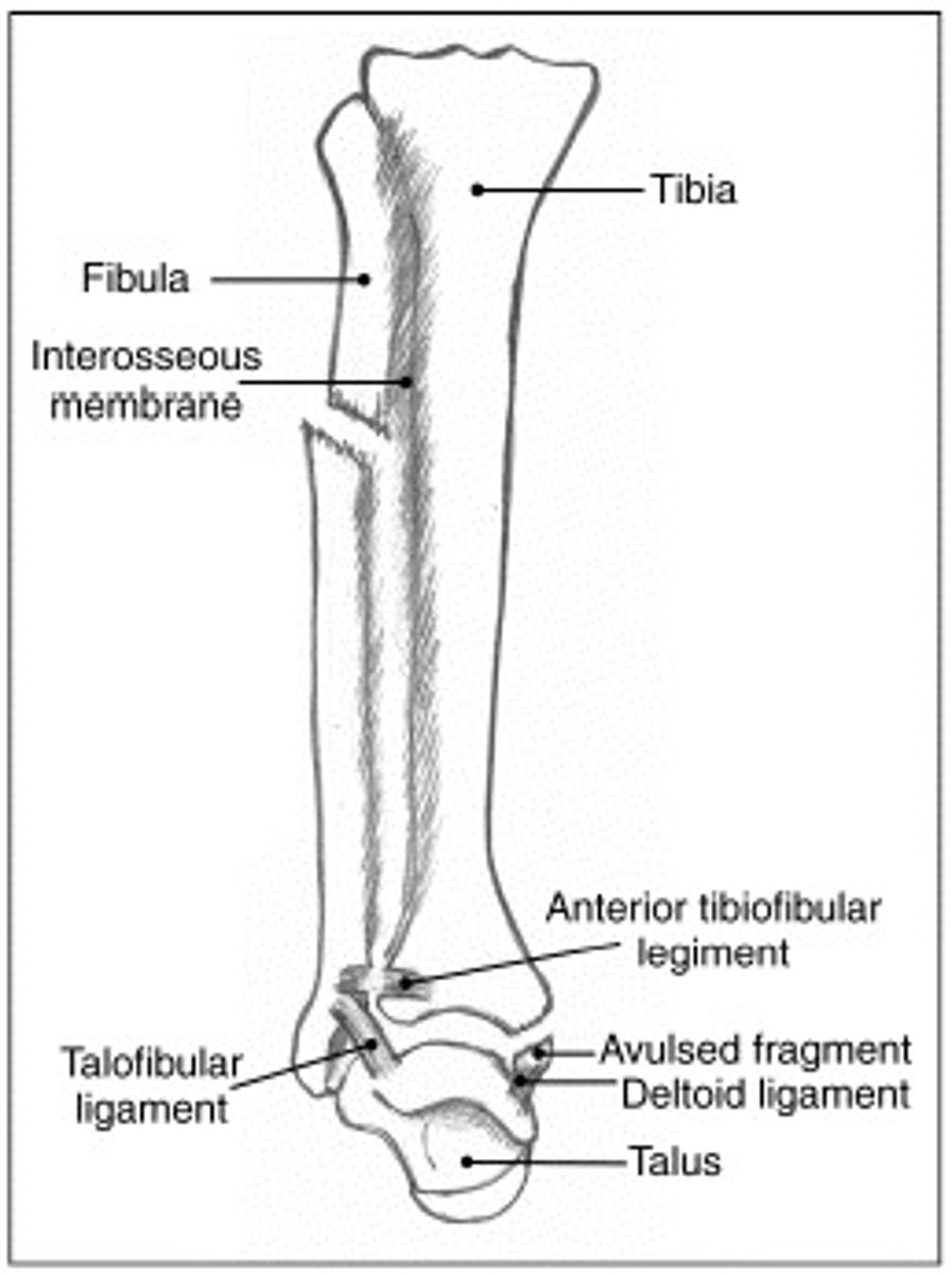
What views should you get to rule out a maissoneuve fracture?
-Fibula
-Tibia
-Ankle
Salter Harris IV fracture
-Triplane fracture
-Goes through all three the epiphyses, physes, and metaphyses
What is fractured here?
Answer
Proximal Fibula
What do we see transverse medial malleolar fractures with in terms of mechanism of injury?
Eversion ankle injuries. Then with lateral malleolar (more common) we see with inversion ankle injuries.
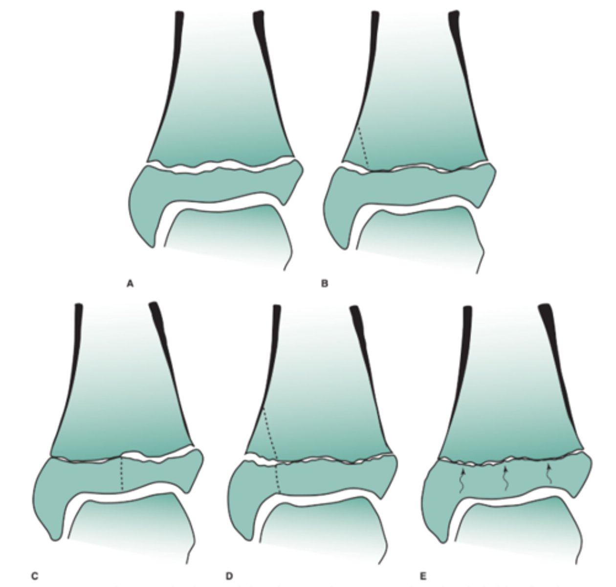
Salter Harris classification
SALTER-HARRIS - Physeal Injuries
(SALTR - Same, Above, Lower, Through, Really bad)
1 - fracture through physis
2 - fracture through physis into metaphysis
3 - intra-articular fracture through physis into epiphysis
4 - intra-articular fracture through epiphysis, physis, and metaphysis 5 - crush injury
What type of salter harris fracture is this?
Answer
Salter Harris III (through the epiphysis)
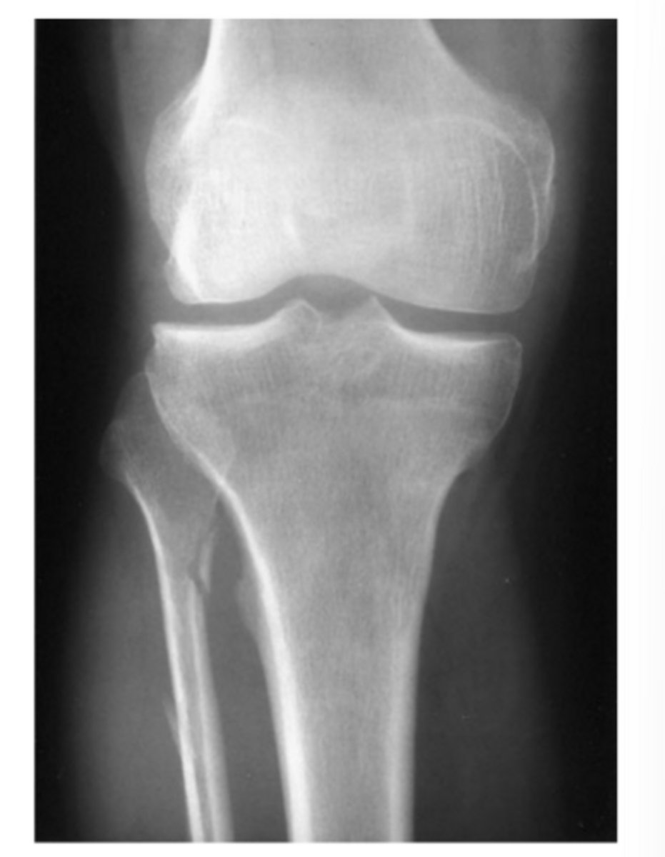
A 12-year-old girl comes to your pediatrics office complaining of 2 weeks of knee pain. There is no history of trauma. She is slightly swollen, tender, and erythematous over the proximal fibula. You obtain frontal
and lateral views of the tibia and fibula (AP and lateral views of the proximal tibia and fibula).
Based on the history, physical examination, and radi-
ographs for Case 6-6 (Figure 6-19), which of the following choices is the best working diagnosis?
A. A bone tumor, most likely benign
B. A bone tumor, most likely malignant
C. An infection of the bone
D. A stress fracture of the proximal fibula
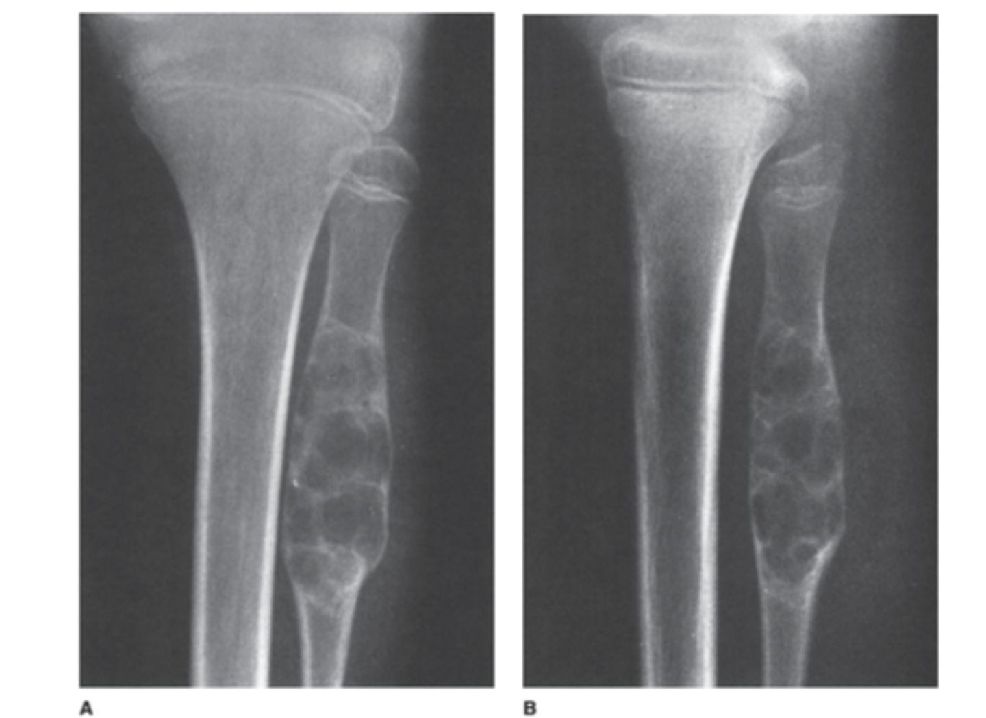
Answer
A.
This is a focal lytic lesion in the proximal fibulat metadiaphysis with an intact shell of new cortex and a well defined zone of transition betweenitself and adjacent normal bone. Its benign!
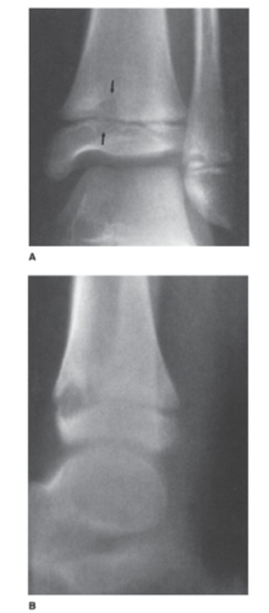
This 5-year-old girl has been limping off and on for 2 months. Her knee is warm and swollen (AP view of the knee).
What is the most likely diagnosis for Case 6-7
(Figure 6-20)?
A. Osteomyelitis
B. A malignant bone tumor
C. A Salter-Harris IV fracture
D. Langerhans cell histiocytosis
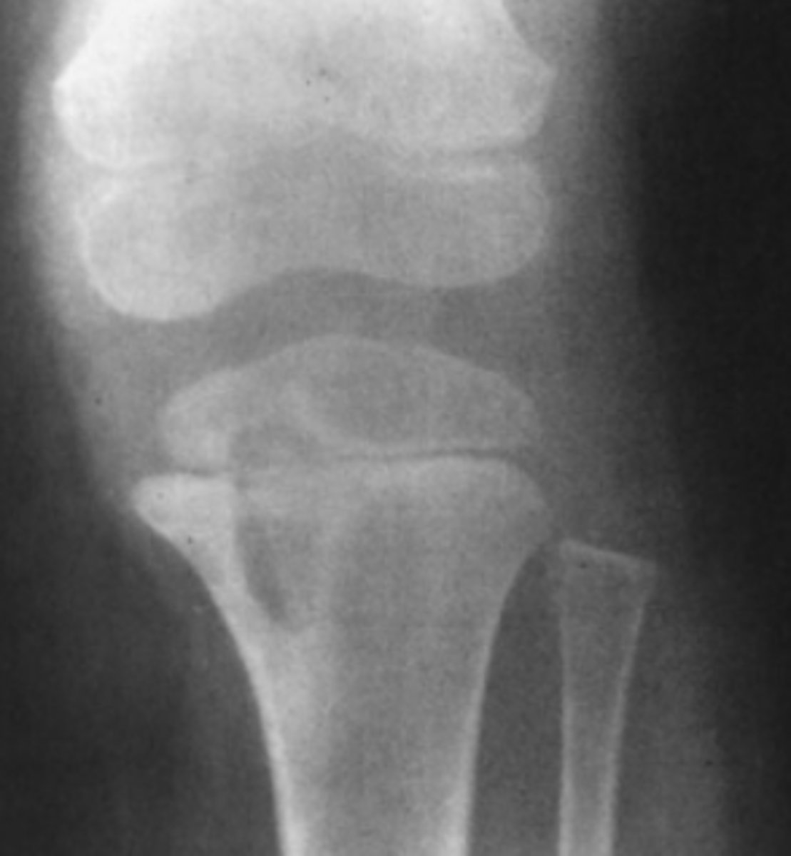
Answer
A.
Well defined lytic lesion in the proximal tibia with slcerotic edges. It is extending across the physis to involve portions of both the metaphysis and epiphysis.
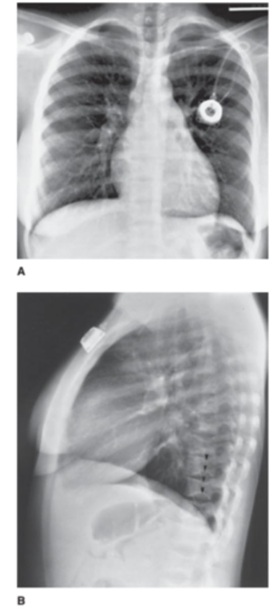
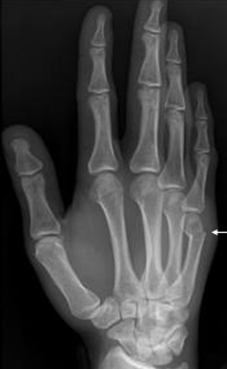
A 35-year-old man complains of a lump in the soft tissues of the right arm. He first noticed the lump 6 months ago after hurting his arm in a fall from a bicycle (AP view of the arm).
What should you do about the calcified lump in the
patient's arm in Case 6-8 (Figure 6-21)?
A. Needle biopsy
B. Open excisional biopsy
C. Reassure the patient
D. Bone scan
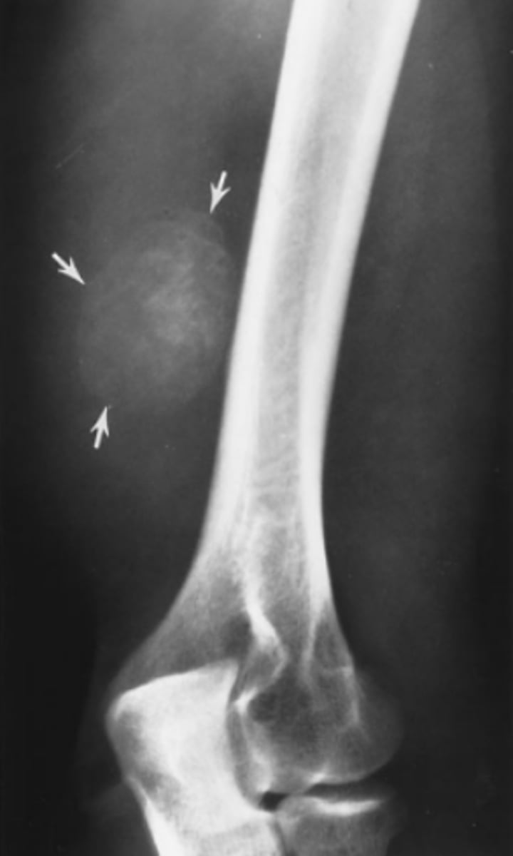
Answer
C.
Well defined ossified mass projecting into the musculature of the posterolateral arm. It has a thin but distinct cortex surrounding the trabeculae. It is myositis ossificans
6-9. A 10-year-old girl complains of a lump on the inside of her thigh near the knee. It has been there as long as she can remember but has been annoying her since she recently took up horseback riding (AP view of the distal femur).
What is the lump in Case 6-9 (Figure 6-22)?
A. An osteosarcoma
B. An osteochondroma
C. A normal variant
D. A soft tissue sarcoma
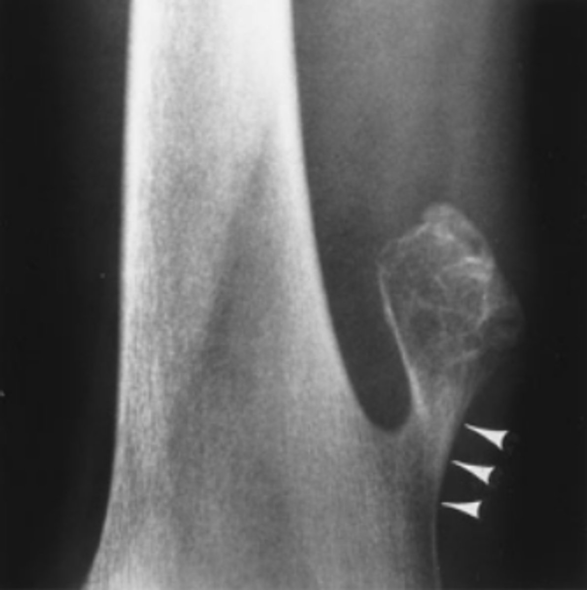
Answer
B.
Ossified mass topped by a cauliflower like thin shell of cortex. Note how its continuous with the cortex of the tumor the femur cortex. It blends together beautifully and it is growing away from the bone.
This is a osteochondroma
Is bone cortex pliable?
No. The cortex is not pliable and it wont stretch to accomodate a growing lesion. Instead it is slowly remodeled by resorption of endosteal bone and deposition of periosteal new bone. It is a timely process.
Characteristics of a malignant bone tumor
-Rapid growth rate
-Poorly defined borders
-Destroy cortex and the periosteum cant contain them with new mineralized bone. There may be gaps where it breaks through the cortex.
-You can see incomplete periosteal new bone mineralization (onion skin)
Organism that causes osteomyelitis
Staphylococcus and streptococcus
Myositis ossificians
calcium deposits resulting from repeated bruising
Myositis Ossificans vs sarcoma
Myositis Ossificans: Grows from the outside in
Sarcoma: Grows from inside out
Do you think this is potentially malignant?
Answer
-Yes its an osteosarcoma
-Note that the area is mottled lucent and sclerotic
-You cant see where the tumor begins and ends.
-Periosteum cant contain it.
-Smudged appearance
What does this person have?
Answer
Osteomyelitis.
Most common of all benign cartilaginous neoplasms
Osteochondroma
a 45-year-old woman with a history of breast cancer diagnosed 3 years previously. She underwent surgery and since that time has been free of disease. She has come for her routine follow-up appointment and complains only of vague, aching discomfort in her left hip. A chest x-ray shows no evidence of metastatic disease.
For Case 6-10 (Figure 6-28), which of the following
studies would be least useful today?
A. Chest CT
B. Left hip x-ray
C. Bone scan
D. Skeletal survey
E. Mammography
Answer
D.
It really is just the weakest out of all of those
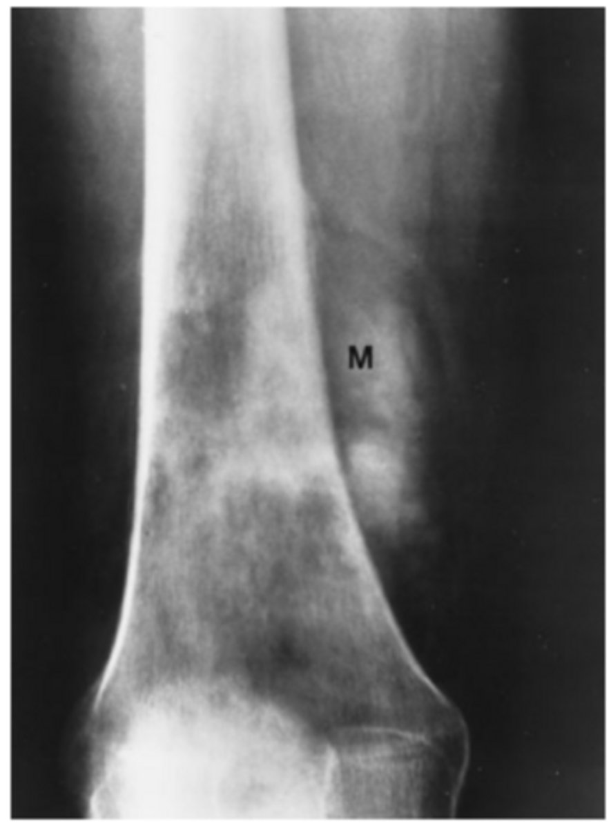
A 40-year-old man complains of knee pain and swelling of 3 weeks' duration. He has no other known disease. You order conventional radiographs of the knee.
For Case 6-11 (Figure 6-29), what is the next study
you should order?
A. Bone scan
B. MRI of the knee
C. Hand films
D. Chest radiograph
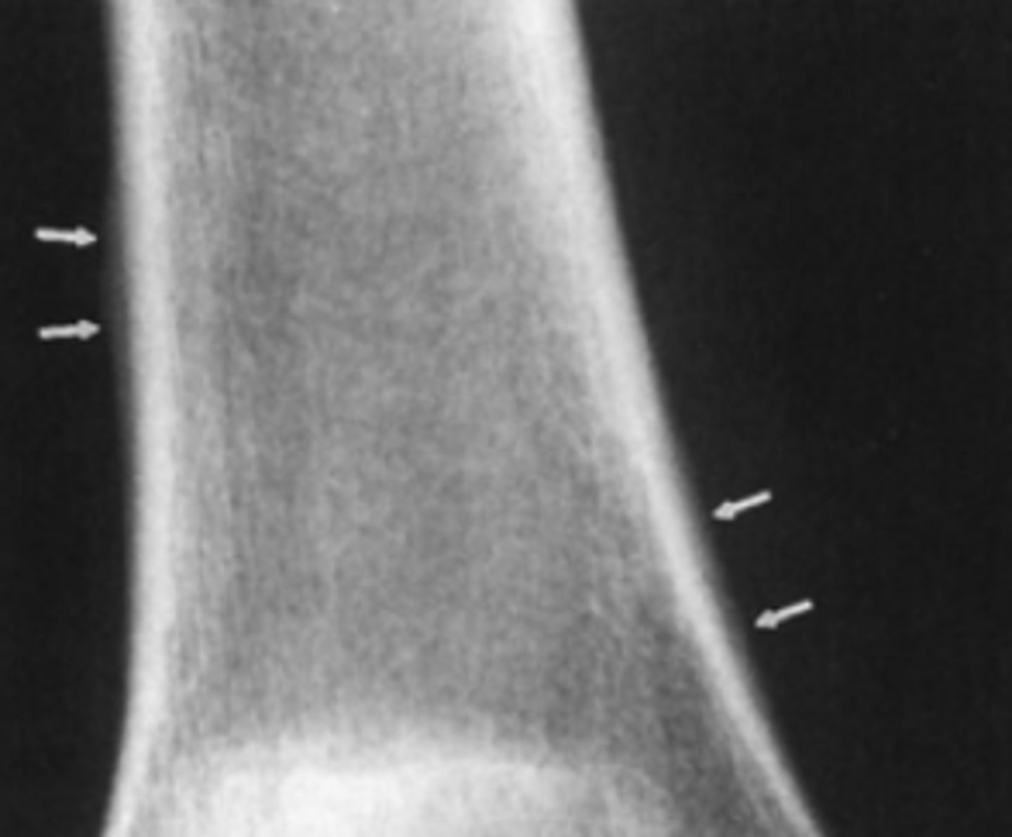
Answer
D.
A thin rim of calcium added to the bony contour of both sides of the right femoral metaphysis due to periosteal elevation. Similar findings are seen on the left femur and both tibiae
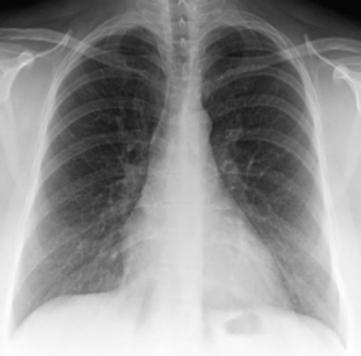
This chest radiograph was obtained to exclude pneumonia in a chronically ill 26-year-old woman (PA and lateral views of the chest).
For Case 6-12 (Figure 6-30), there is no evidence of
pneumonia, but there are several abnormalities that
are clues to the nature of this patient's chronic illness.
Which finding is such a clue?
A. The presence of a central venous line
B. Enlargement of the pulmonary artery segment of
the mediastinum
C. Depression in the endplates of numerous vertebrae
D. Asymmetry of the breast shadows
Answer
C.
Many vertebral bodies in the lateral view shaped like the letter H with central depressions in the superior and inferior end plates
What does this patient have in the previous question?
Sickle cell! When they clump together the sickled cells they can block blood vessels. She is having AVN due to this! The mottled appearance of the humeral heads is from AVN as seen in sickle cell anemia patients. Did you note as well her spleen is small? Splenic infarct is seen in these patients.
A 50-year-old man with multiple myeloma was treated successfully with chemotherapy and has been in remission for 2 years. Recent immunoelectrophoreses showed slight elevation of his paraproteins, which causes his oncologist to worry about progression of his myeloma.
For Case 6-13 (Figure 6-31), which of the following
imaging tests is most likely to help determine if this
patient has progressive myeloma?
A. PET-CT
B. Bone Scan
C. Chest x-ray
D. CT of the spine
Answer
A. PET CT is a good bet
Periosteal elevation as seen in 6-11
Nonspecific finding that occurs with local disorders such as fracture, bone tumors, osteomyelitis, and bone infarction.
Hypertrophic osteoarthropathy
Characterized by chronic proliferative periostitis of long bones , clubing of fingers and synovitis, Its asso with Squamou cell carcnimoa and Adenocarcinoma of the lung.
Important rule out with periosteal elevation
Now time for the slides
What is the mid shaft of the bone also known as?
Diaphyses
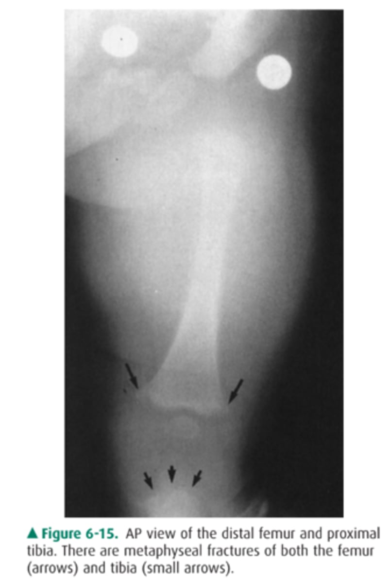
Based on the two age ranges what do each of these individuals have respectively?
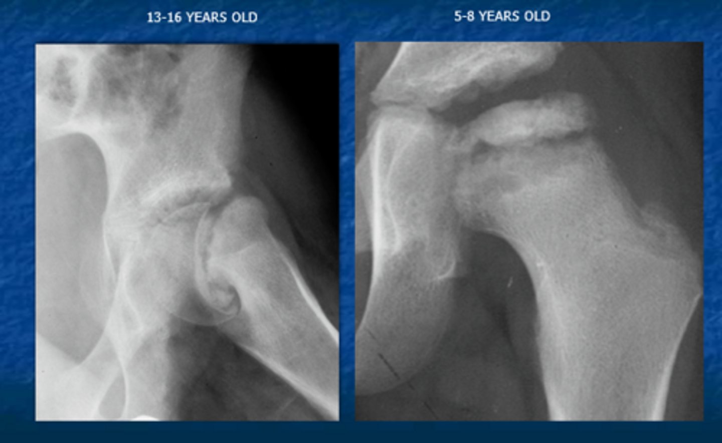
Answer
13-16 y/o: SCFE
5-8 y/o: Leggs Cathe Perthes
Leggs Cathe Perthes
Congenital AVN of the hip commonly in children around 4-8 y/o
What does this person have?
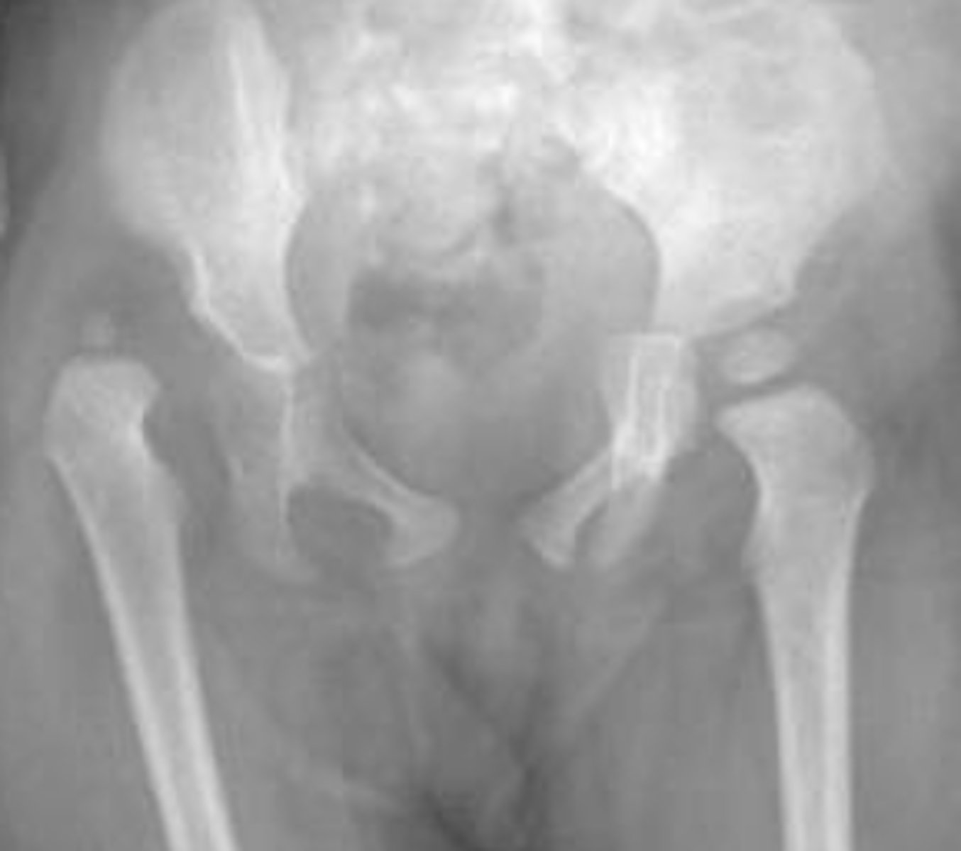
answer
Developmental dysplasia of the hip
Dislocation vs subluxation
Dislocation=no contact with the joint at all
Subluxation=partial contact with the joint
How long does it take for a callus to heal?
7 days
This is a 7 month old in tears
What do you want to do?
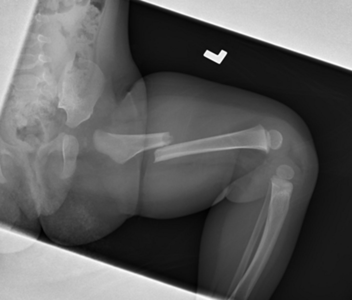
answer
Call CPS femur fx
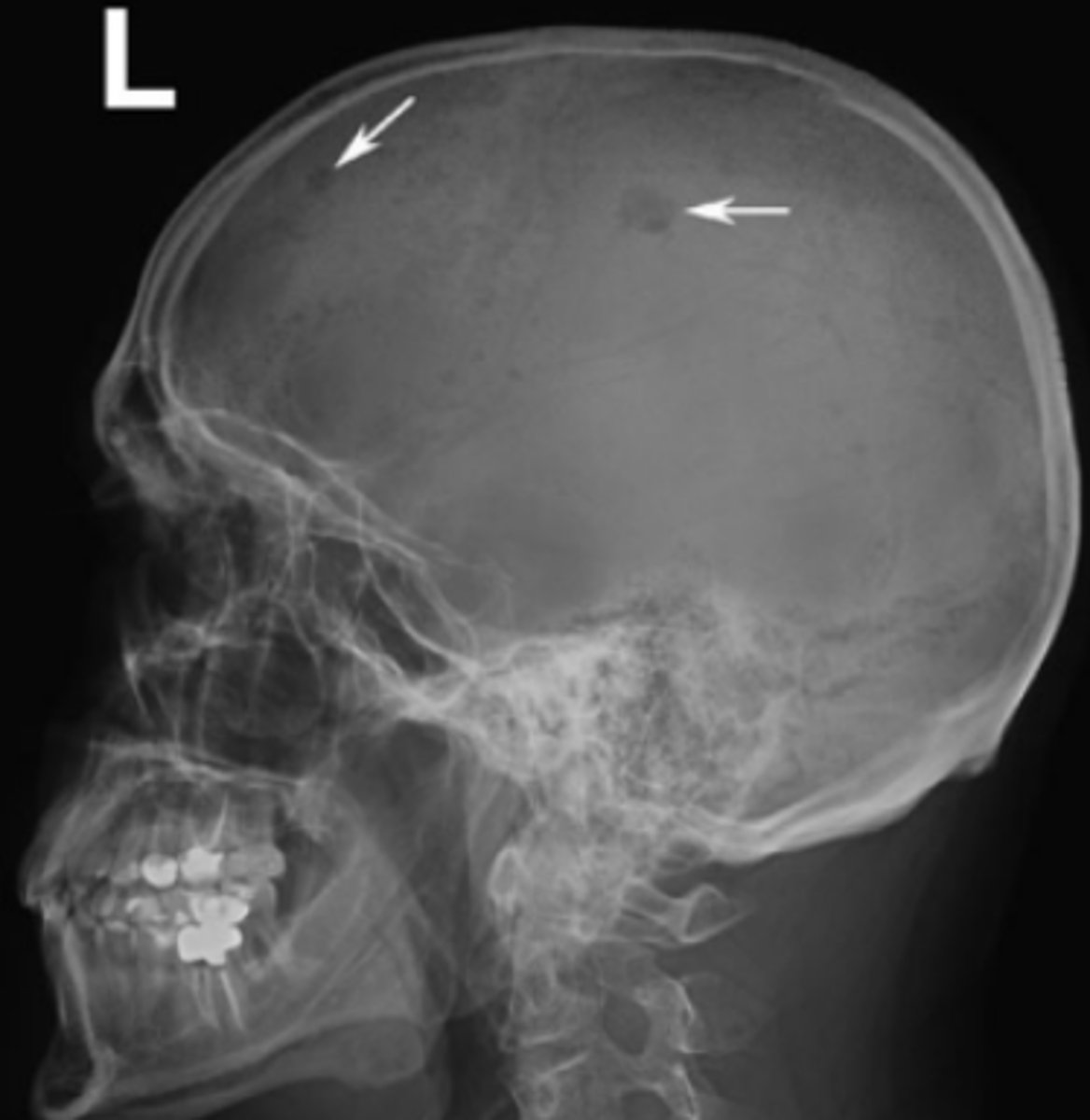
Patient presents to the ER following a fall complaining of pain in the "snuff box". Do you see any thing?
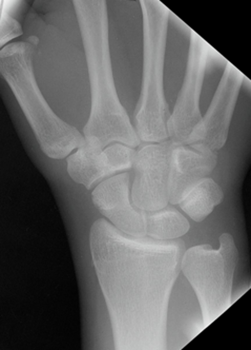
Answer
Possible AVN. Its sclerotic looking
The films were normal now what do you want to do? What other imaging could you also consider?
-Follow up in another 7-10 days with x-ray
-Could also use MRI
Here it is 3 months later
Proximal mid and distal poles are sclerotic
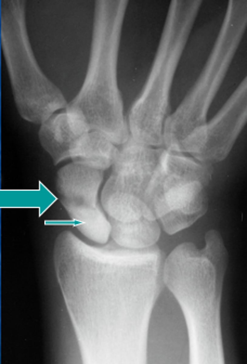
Whats happening in this one?
Answer
-This is a colles fracture.
-The comminuted fx is dorsally and radially displaced
-With volar angulation
-Extension into the joint (intra articular)
Dorsal vs volar
-Dorsal: non palm side
-Volar: palm side
Criteria for a fracture to be comminuted
More than one fracture line
What type of fracture is this?
Answer
5th metacarpal fracture. Boxers fracture
What type of fracture is this?
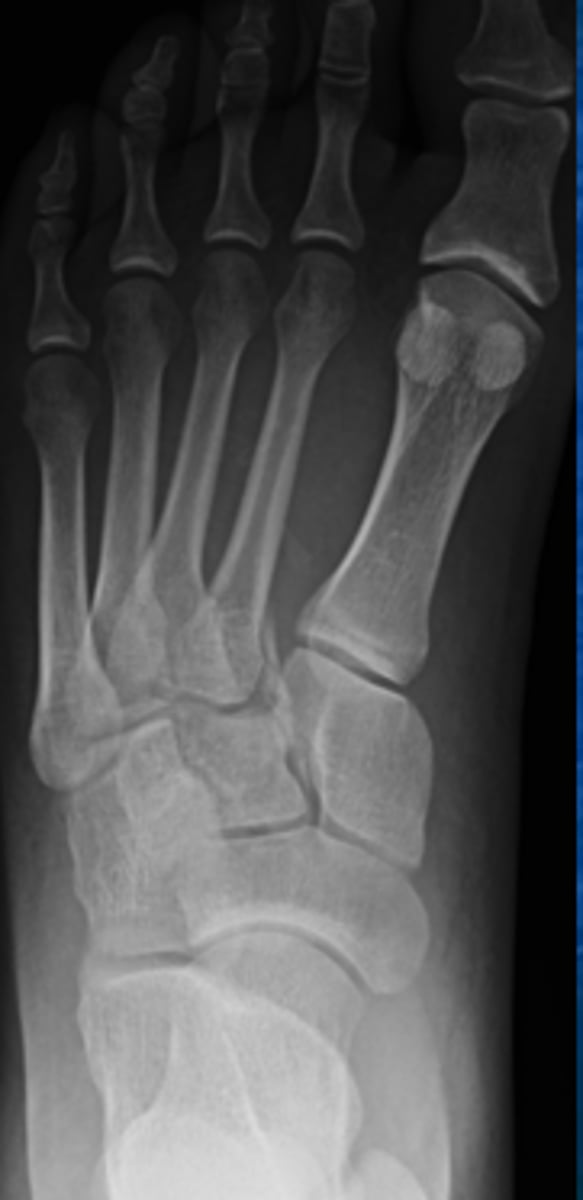
Answer
Lisfranc fracture
A fracture that doesnt heal well of the wrist and foot
Wrist=scaphoid
Foot=lisfranc
What type of fracture is this?
answer
Supracondylar Fracture