Lab 4 - Walking a myelin Dr. Gibson's Shoes
1/43
There's no tags or description
Looks like no tags are added yet.
Name | Mastery | Learn | Test | Matching | Spaced | Call with Kai |
|---|
No analytics yet
Send a link to your students to track their progress
44 Terms
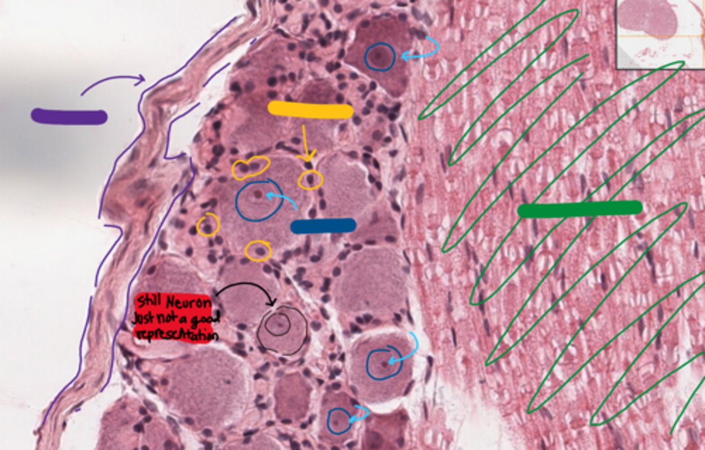
neurons
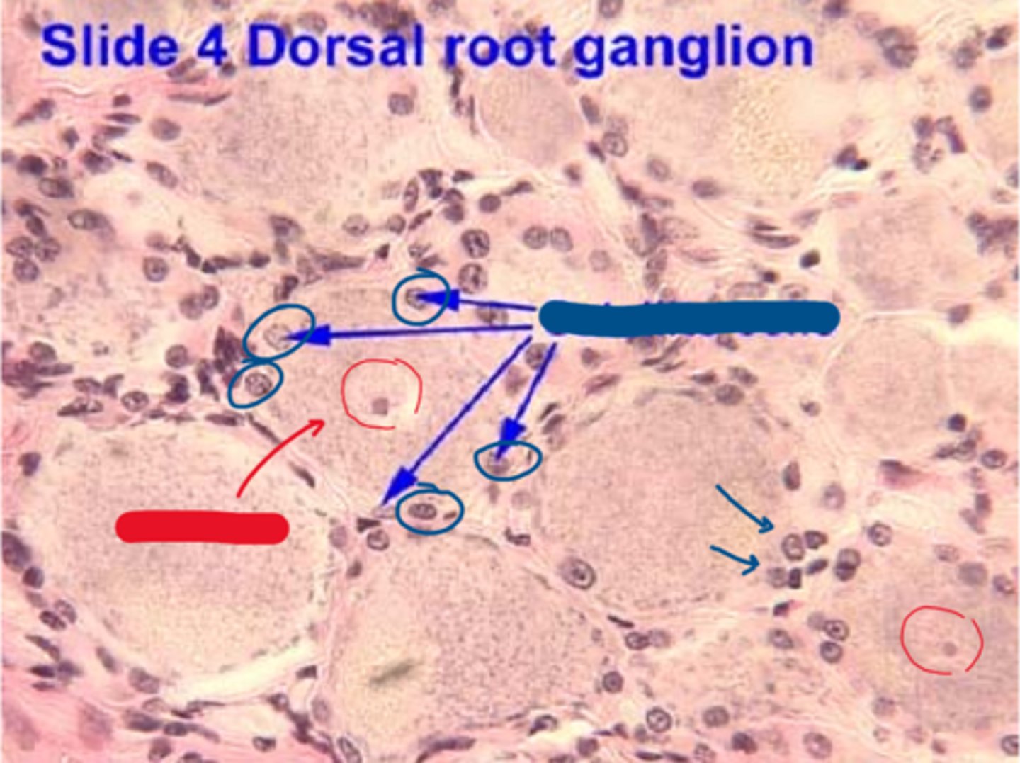
This is blurry but its a dorsal root ganglion I swear.
What tends to be peripherally located in the red areas?
axons
This is blurry but its a dorsal root ganglion I swear.
What tends to be centrally located in the green areas?

cell bodies
red
satellite cells
blue

epineurium
purple
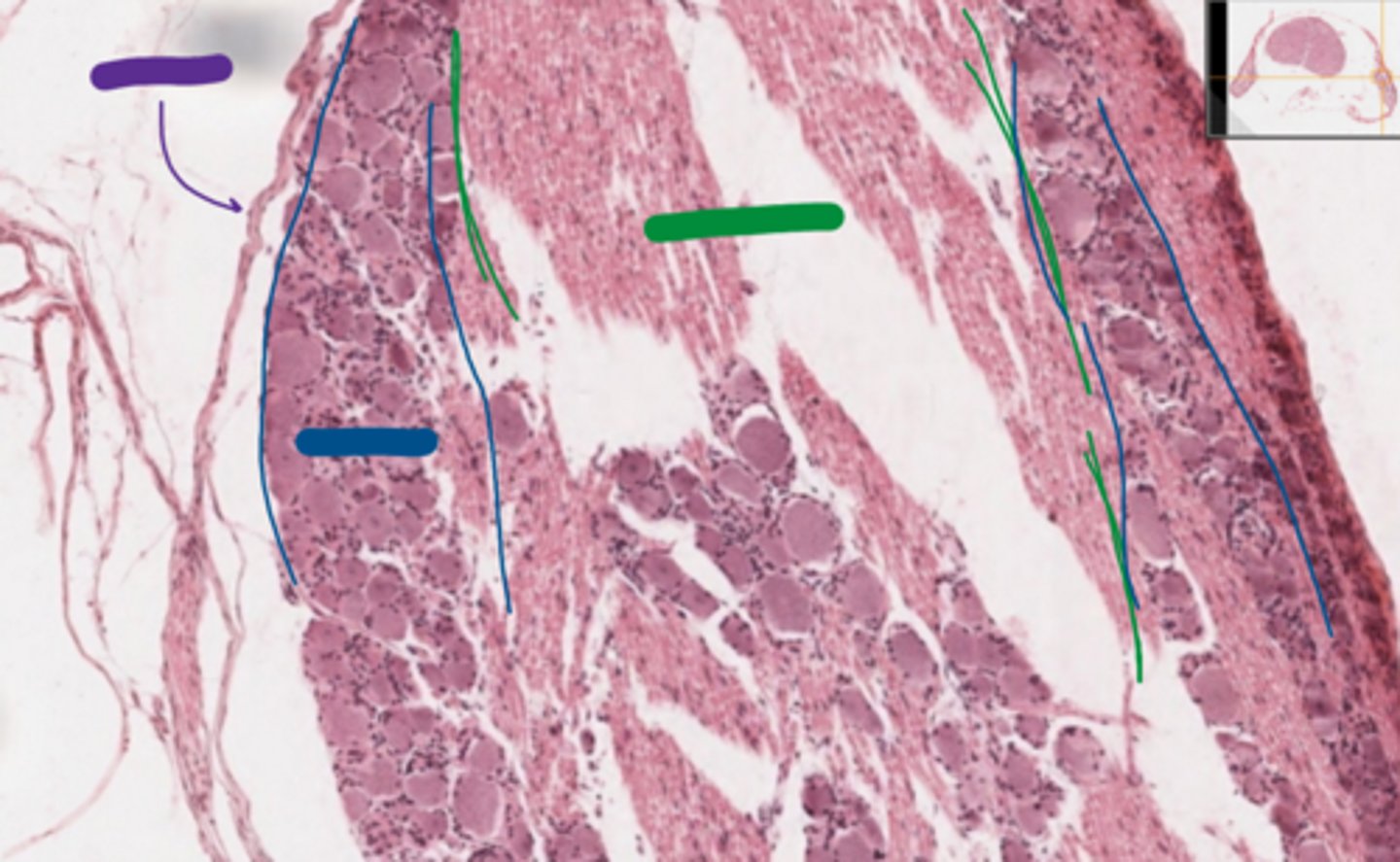
neurons
blue

axons
green

epineurium/perineurium
purple

satellite cells
yellow
axon region
green

neurons
blue

nucleus is NOT centrally located
the neuron indicated in red.
Why is it not a good representation of a dorsal root ganglion neuron?

RER
what organelle is the nissl substance in these neurons made of?

axons
black

myelin sheath
red

LIPID myelin sheath
what was the white space filled with in vivo?
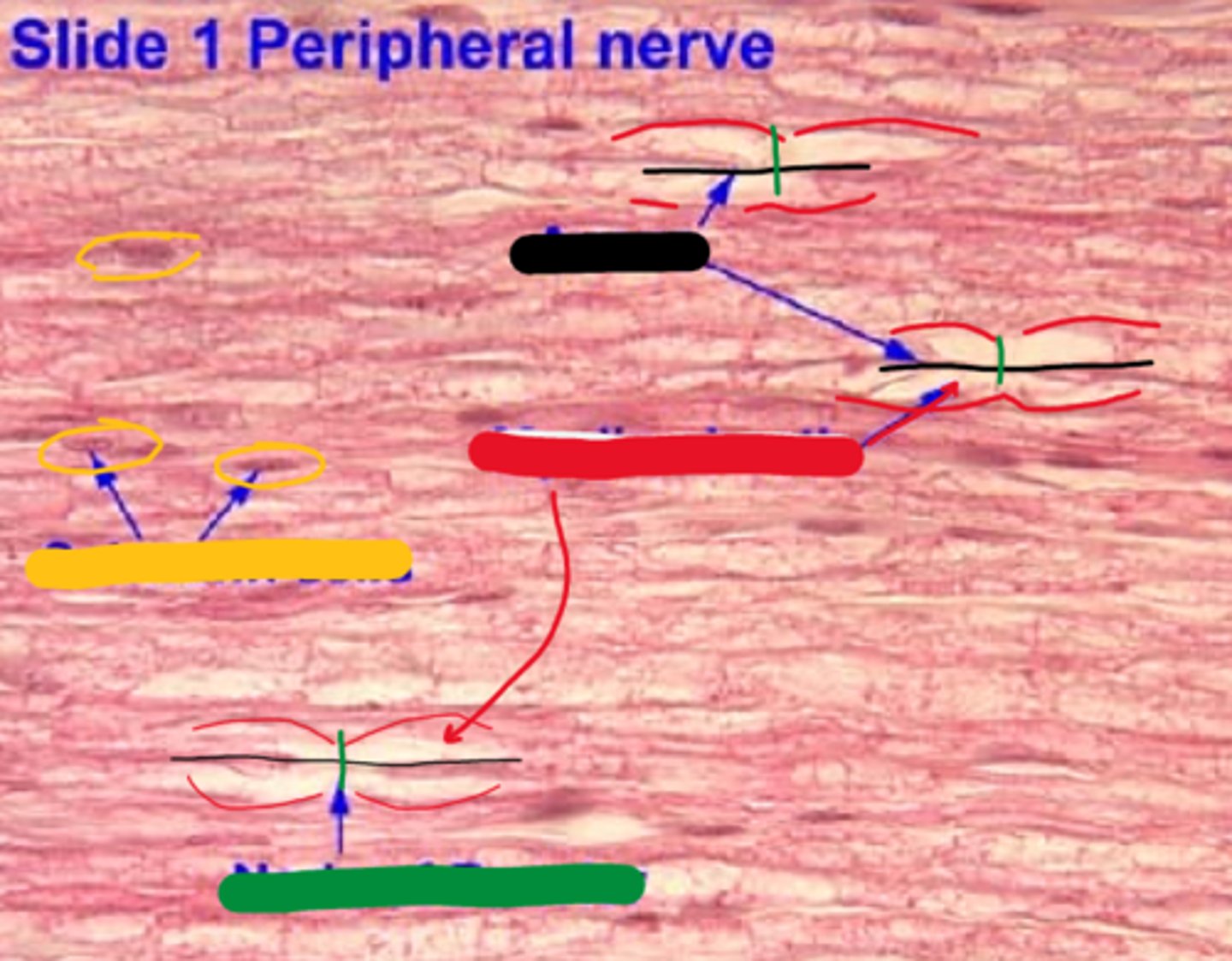
schwan cells
yellow

nodes of ranvier
green

axon
black
myelin sheath
red

node of ranvier
green

schwan cell
yellow

axon
green

myelin sheath
red

node of ranvier
black
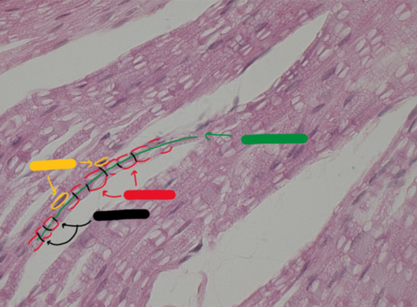
schwann cells
yellow

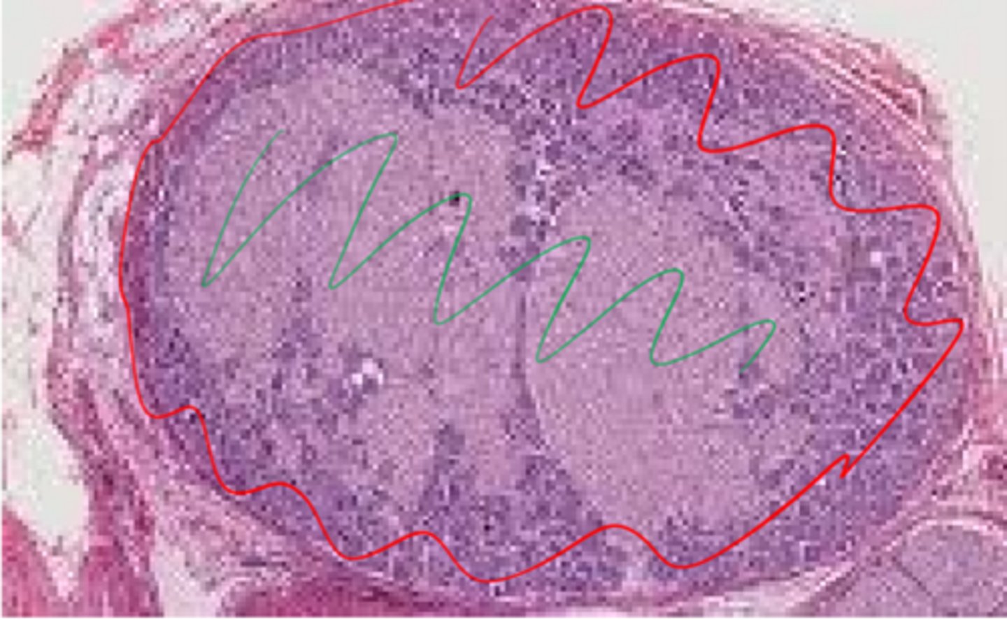
epineurium
red
(nerve in cross section)
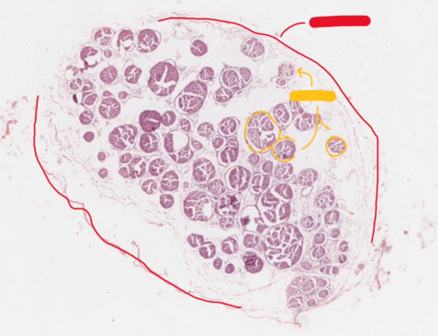
perineurium
yellow
(nerve in cross section)

epineurium
red
(nerve in cross section)
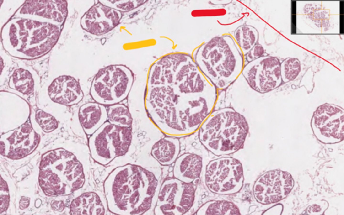
perineurium
yellow
(nerve in cross section)

perineurium
yellow
(nerve in cross section)

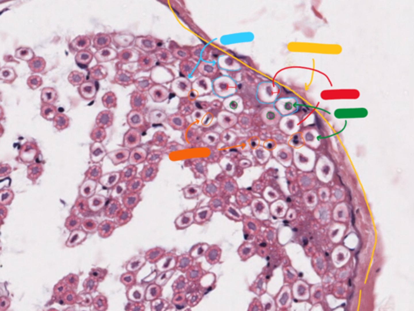
endoneurium
blue - membrane
(nerve in cross section)

myelin sheath
red
(nerve in cross section)

axon
green
(nerve in cross section)

schwan cell
orange - NOTE: ONLY WHEN CELL IS TOUCHING MYELIN
(nerve in cross section)
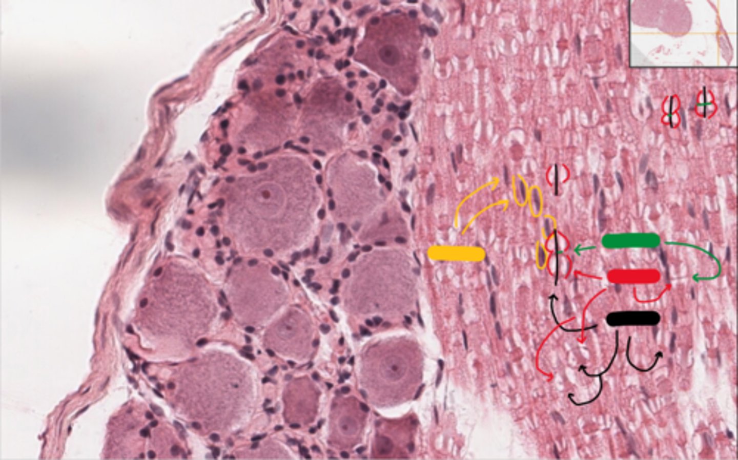
neurons
dark blue

satellite cells
orange

lipofuscin
red

axon region
green
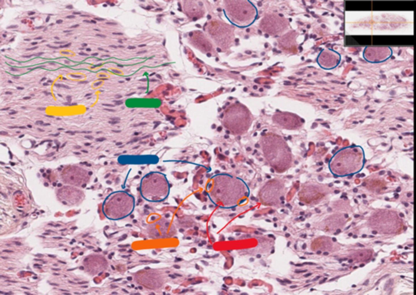
schwan cells
yellow

Myenteric plexus
what is this neuronal structure?
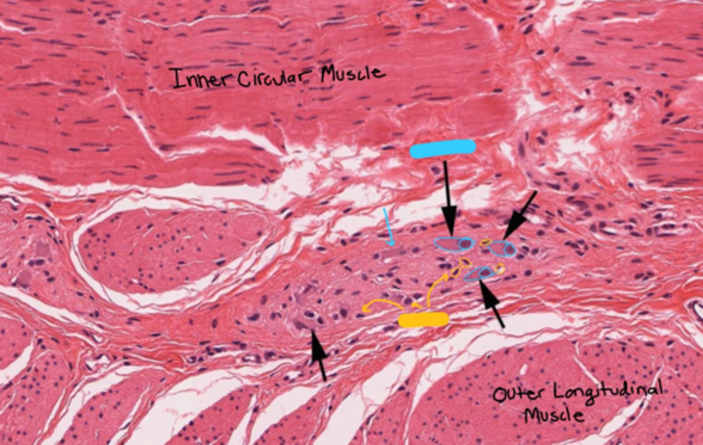
neurons
blue

satellite cells
yellow

noted... maybe
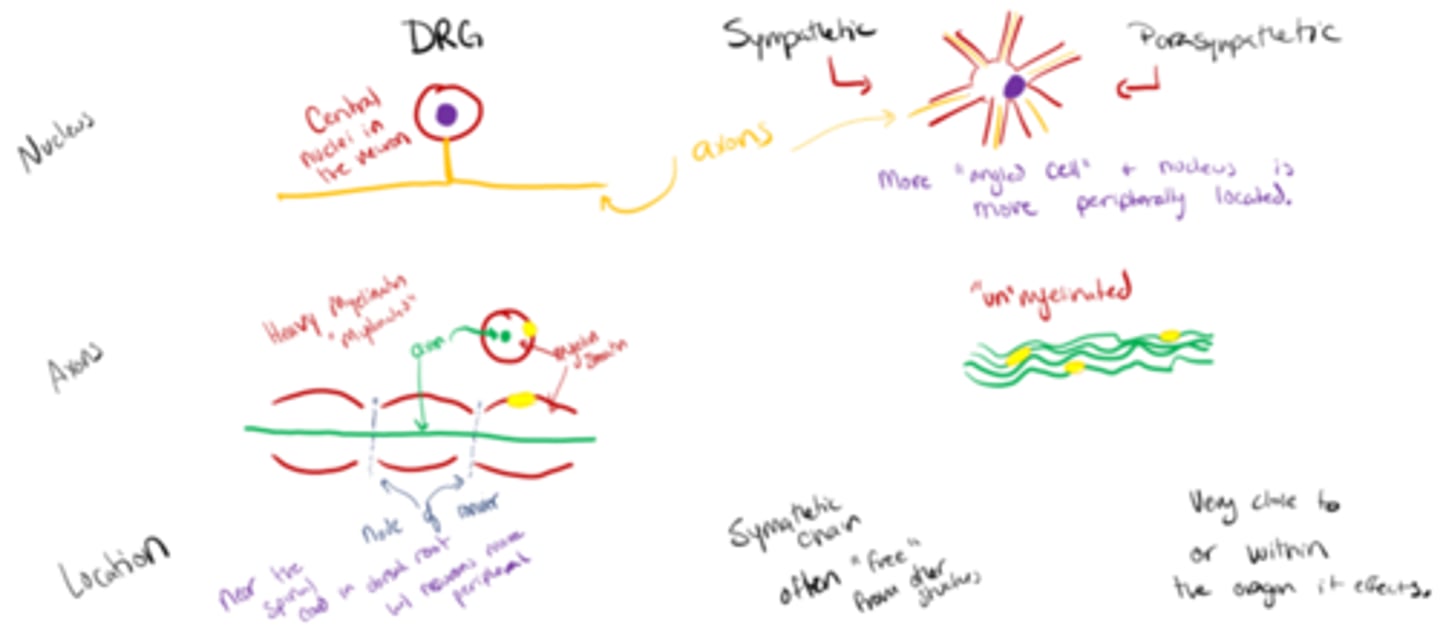
note this slide about the differences in DRG vs sympathetic/parasympathetic neurons...
...like, if you want to