302 Term Test 2
1/247
There's no tags or description
Looks like no tags are added yet.
Name | Mastery | Learn | Test | Matching | Spaced | Call with Kai |
|---|
No analytics yet
Send a link to your students to track their progress
248 Terms
What are the inter-relationships and functions of the circulatory system?
O2 and nutrients in the blood distributed by the heart
De-O2 blood to the lungs
Waste products
Transport system
What lines blood vessels and lymphatics?
An endothelial cells = endothelium (epithelium that lines vessels)
What does simple squamous epithelia do?
Facilitates diffusion and transport
Usually 1 surface is facing a cavity or lumen of an internal duct → Lungs, capillaries
What makes up the endothelium?
Simple squamous epithelia
What are the main components of the circulatory system?
Heart
Pump
Vascular system
Circuit of vessels
Arterial system
Blood away from heart
Venous system
Blood towards heart
Lymphatic system
Drains fluids back to venous system
What is the lymphatic system?
Collects fluids from connective tissues
Vessels consisting of endothelium only
Flattened
No RBCs → Pink
Have no shape
Where do the parts of the heart go?
Pulmonary artery → to lung
Left atrium → oxygenated blood in (from lung)
Right atrium → deoxygenated blood in (from body)
What are the 3 layers of the heart tissue?
Endocardium → Myocardium → Epicardium
What makes up the tissues of the heart?
Endo and epicardium → Simple squamous epithelium
Myocardium → Muscle
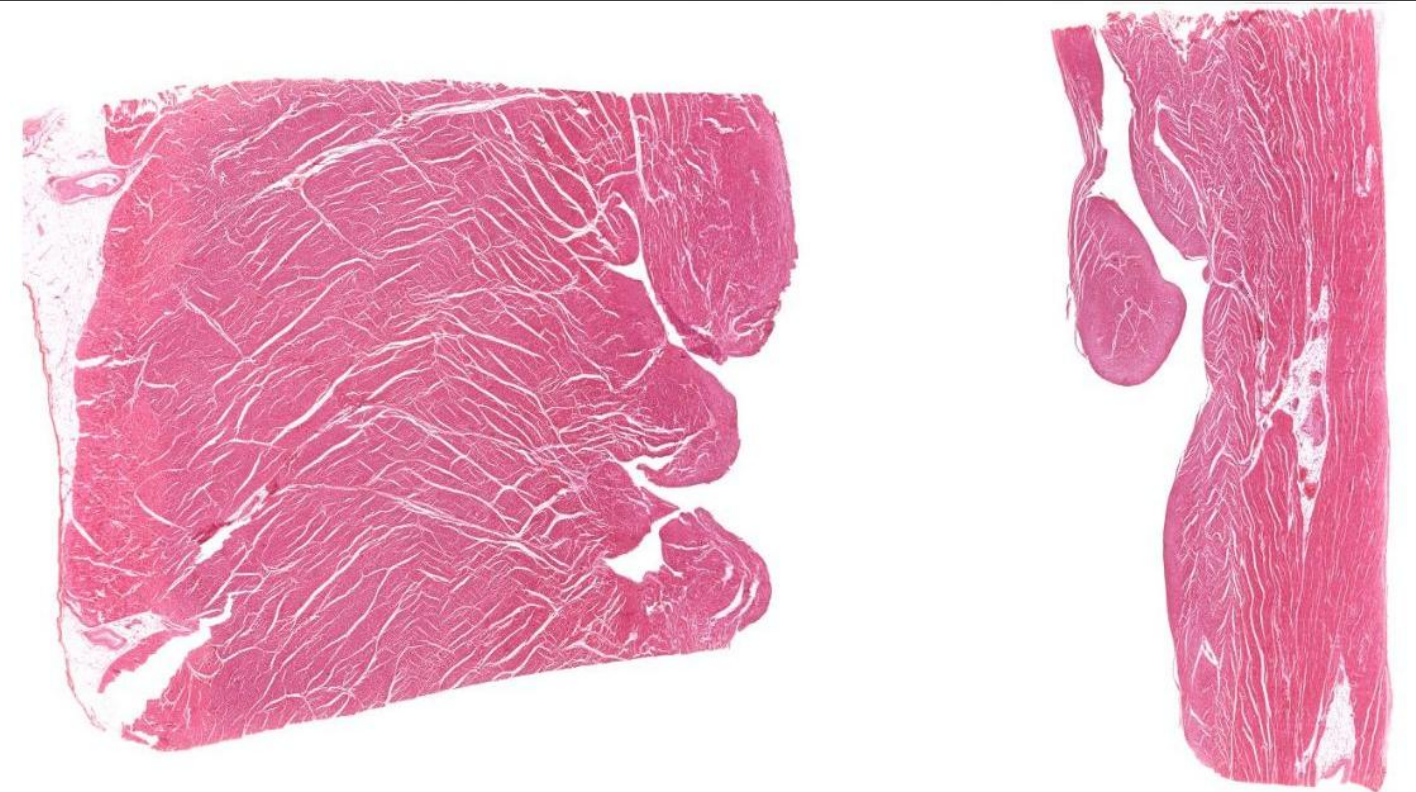
Identify the structure?
Ventricles
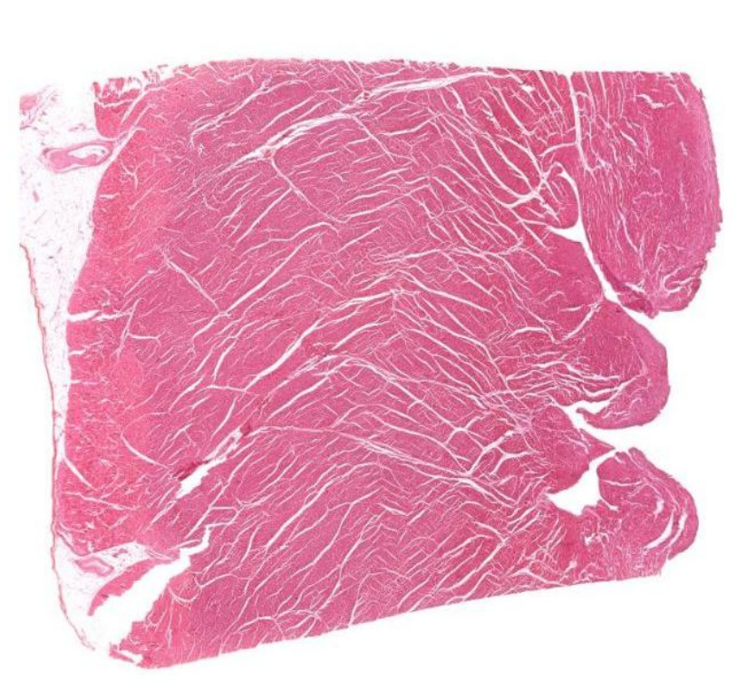
Identify this?
Left ventricle → much more density and more muscular
Identify this?
Right ventricle → much less density
What does the left ventricle do?
Receives oxygenated blood from lungs
Pumps into systemic circulation
BV closer to heart, have characteristics to match that force
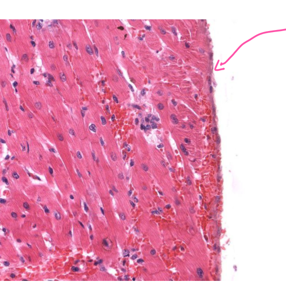
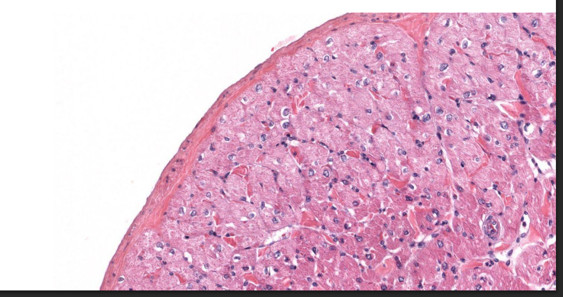
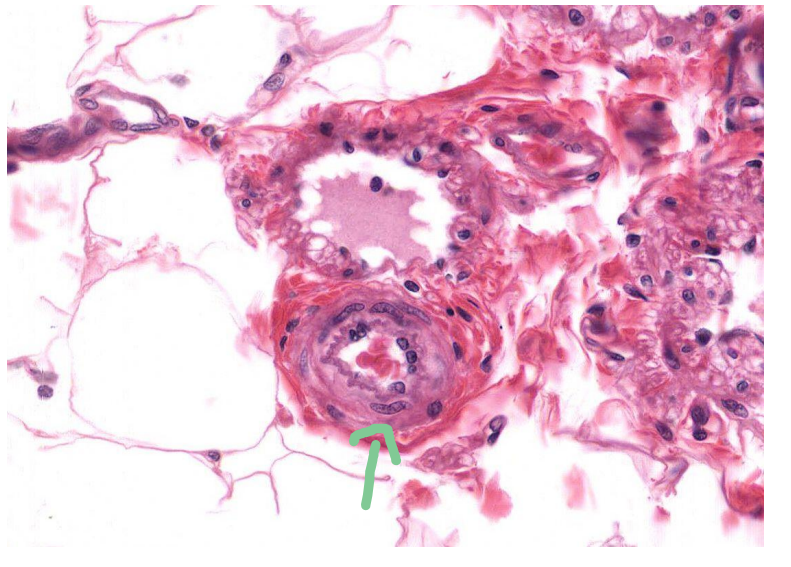
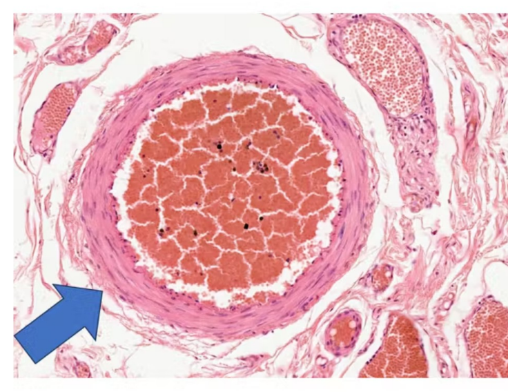
What tissue is indicated by the arrow?
Simple squamous epithelium of endocardium of the ventricle
Supported by a few fibres of connective tissues
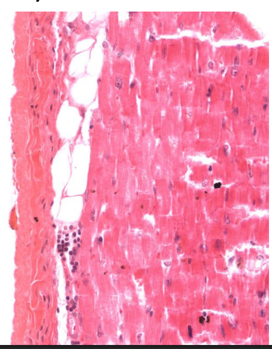
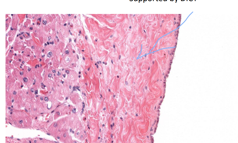
What tissue is this?
Endocardium of the Atrium
Inner surface
Simple squamous epithelium
Supported by thin layer of DICT
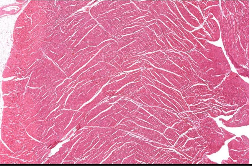
What structure is this?
Myocardium of the left ventricle
What is special about the myocardium?
Cardiac muscle
Thickest layer
Ventricle folds = trabecular carnae

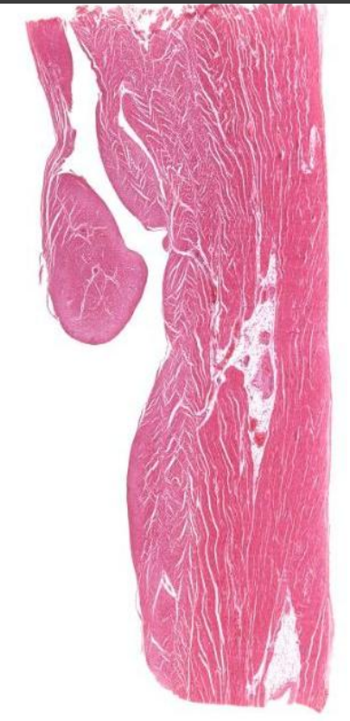
Identify this structure and main component
Atrium and main density is myocardium
Identify this structure
Epicardium of the ventricle
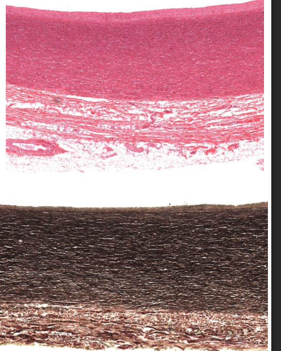
What makes up the epicardium?
Outer mesothelial cells (often missing)
Supported by DICT
Loose CT = unilocular adipose
Identify the structure and the tissue indicated by the arrow
Epicardium of the atrium
What are the layers of the Blood vessels?
Tunica Intima (Coat, inside, endothelium)
Tunica Media (Middle / smooth muscle)
Tunica Adventitia (Externa → CT, Dense)
How do the walls of CV system and the GI tract differ?
CV have 1 layer of muscle while GI tract have 2 or 3.
What are 2 ways to identify veins vs arteries?
Tunica media of arteries are thicker
Arteries are more circular and shaped.
Do veins and arteries occur together?
Yes, commonly with a nerve → neurovascular bundle
What makes up a large (elastic) Artery (Aorta)?
Close to heart
Elastic nature acts as a buffer
T. intima
Endothelium + Elastin + Collagen
T. Media (Thick)
Mostly elastic fiber
Collagen
Smooth muscle
Adventitia (thin)
vaso vasorum → own blood supply
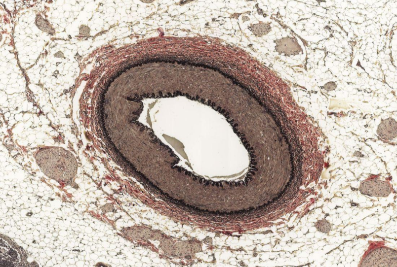
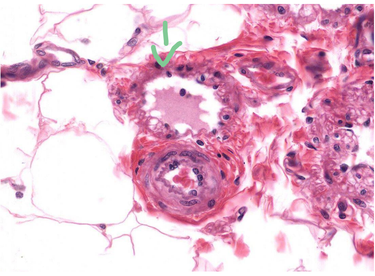
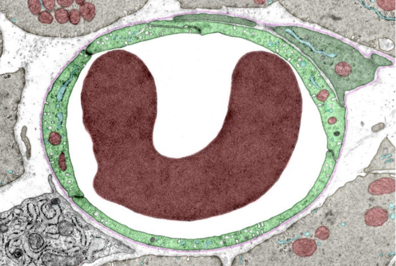
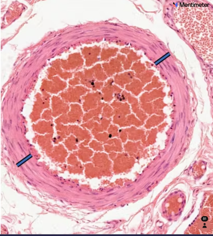
Identify these two vessels?
Both are Elastic / Large Arteries
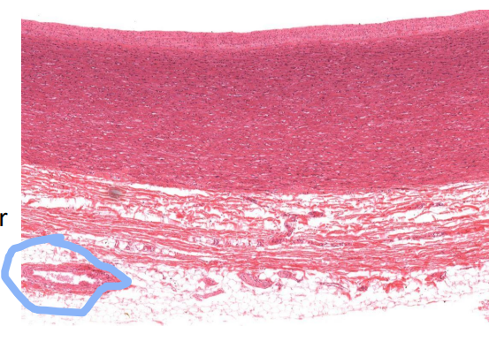
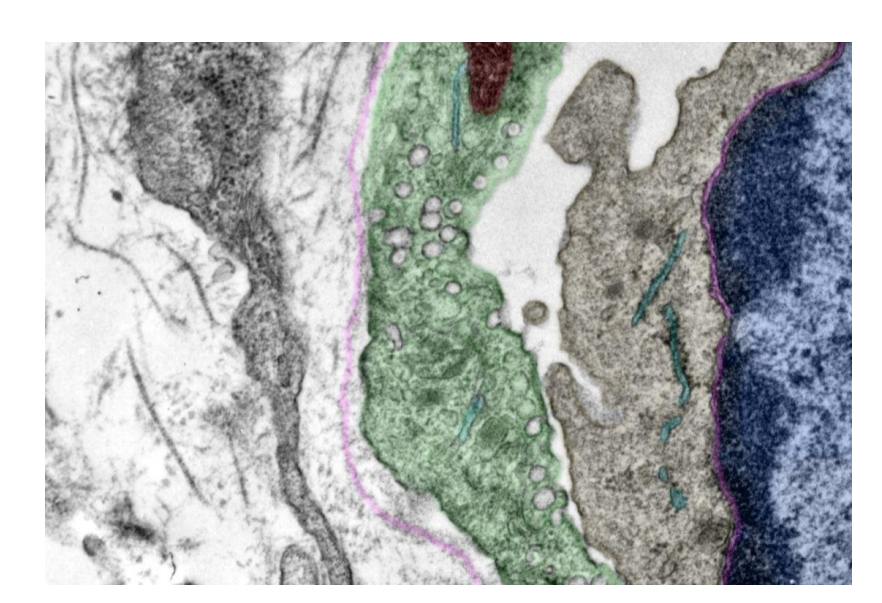
Identify structure circled
Vaso vasorum

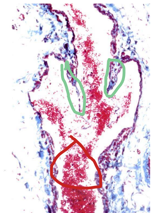
What is indicated by the lines?
Blue: T. Media
Green: T. adventitia
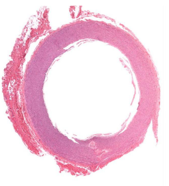
Identify this vessel?
Large Artery
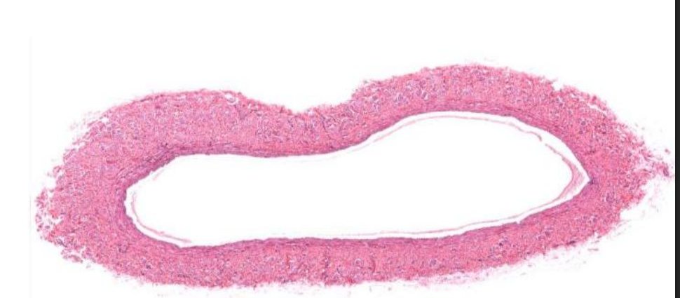
Identify this vessel
Large Vein
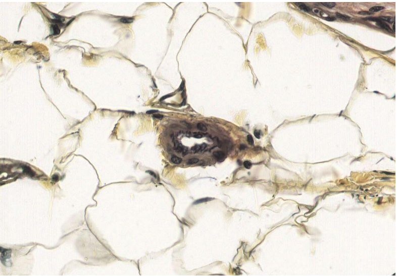
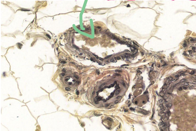
Identify this
Vaso vasorum
Identify the vessel?
Medium Arteries
What is a distinctive feature of medium arteries?
Very muscular but also has an IEL and EEL on either side of TM.

Identify this vessel
Medium vein
What is a distinctive feature of Medium veins?
Has an IEL but no EEL has (acts like an IEL but isn’t)
Identify this vessel
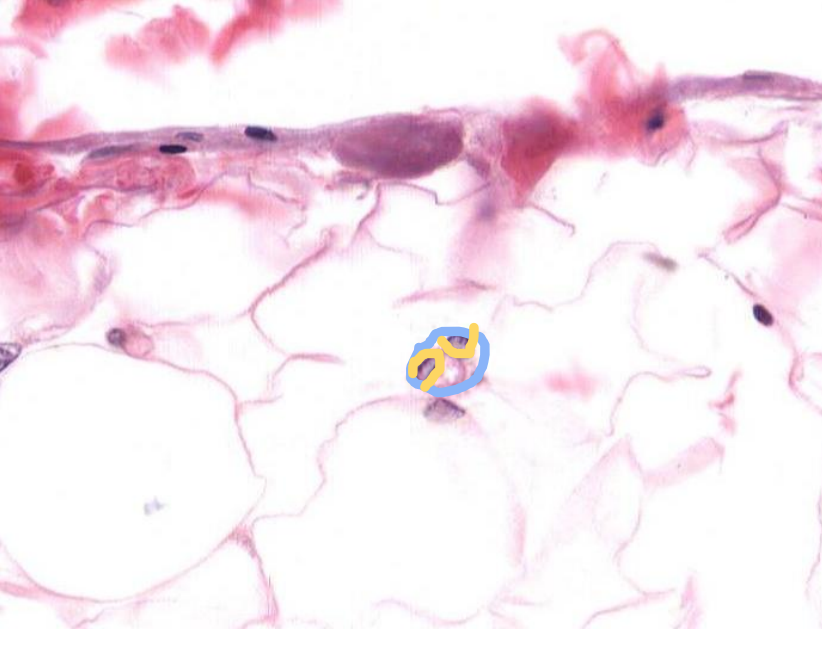
Arteriole
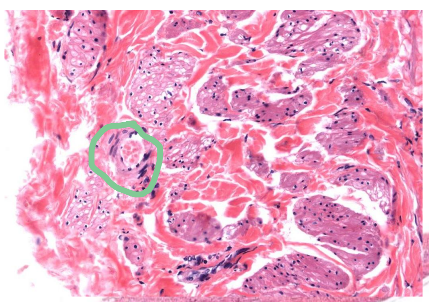
Identify the vessel indicated by the arrow
Arteriole
Identify the vessel indicated by the arrow
Venule
Identify the vessel indicated by the arrow?
Venule
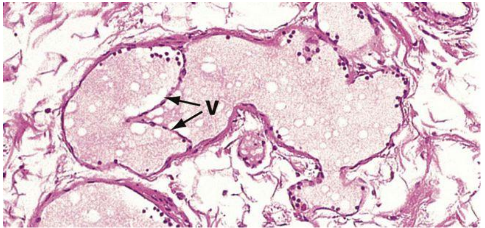
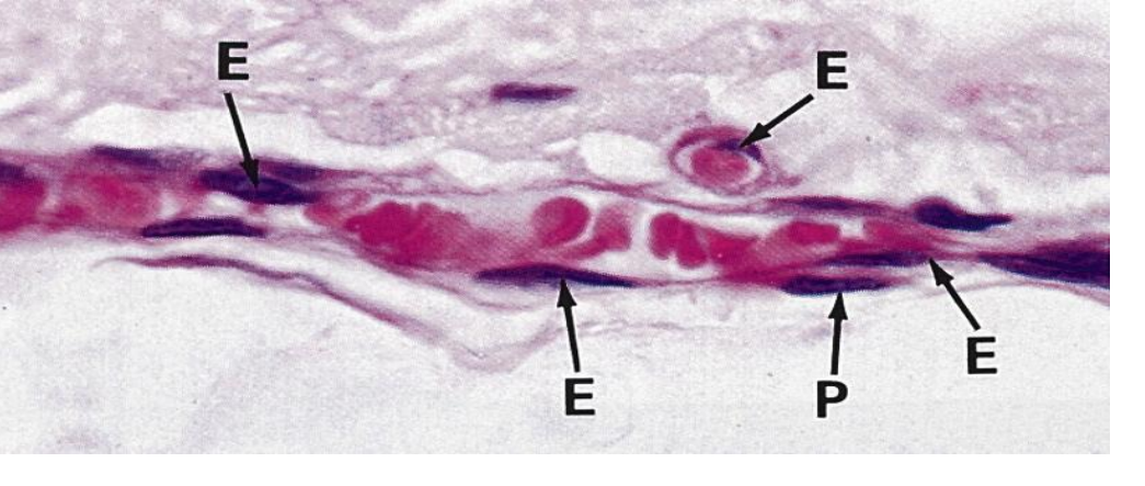
Identify the parts of this structure
A photo of a vein
Green: Valves
Red: RBC
What are valves?
Only seen in veins or lymphatic vessels
Extension of TI.
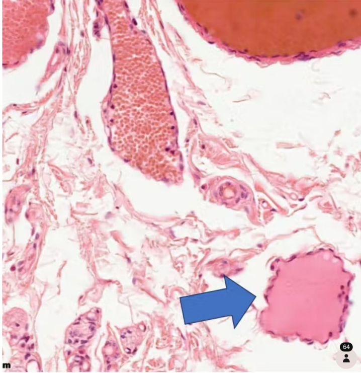
Identify the structure
Lymphatic vessel
What are the types of capillaries?
Continuous, Fenestrated, Discontinuous → name refers to epithelium
What is continuous capillaries?
All cells are connected and equal thickness
What is Fenestrated capillaries?
Windowed → small gaps → better diffusion
In Kidney
What is Discontinuous capillaries?
Sinusoidal → big gaps → best diffusion
In Liver
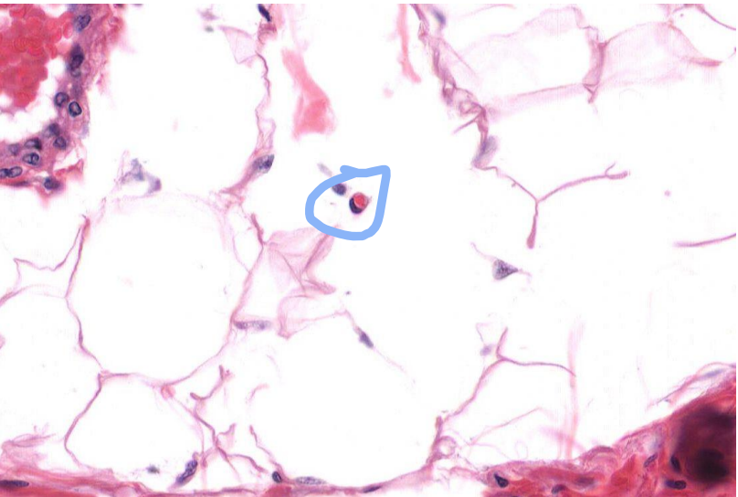
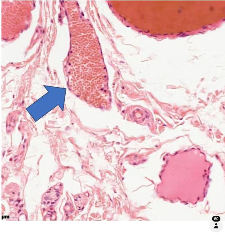
Identify this vessel
Capillary → roughly same size as an RBC (Middle)
Identify the white circles
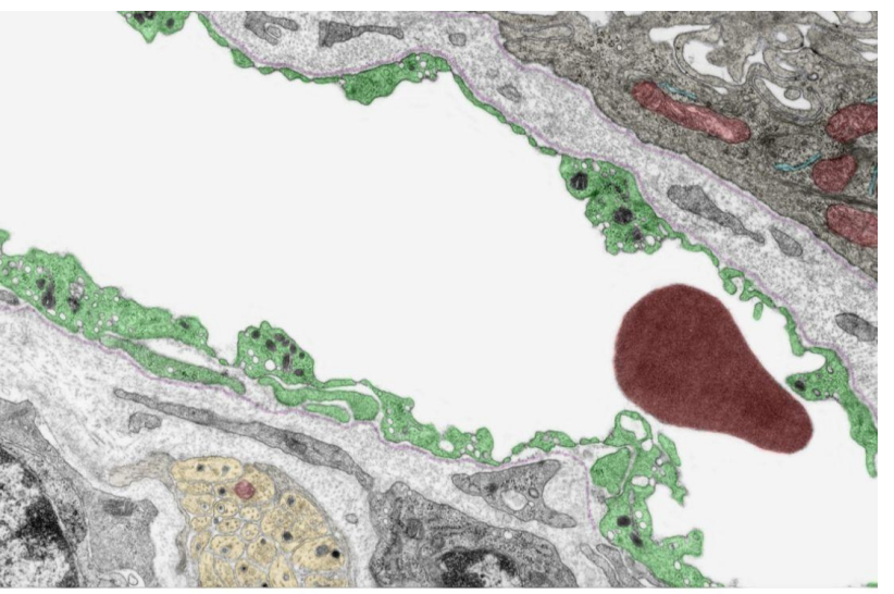
Pinocytic vesicles → allows for molecular transport from lumen
Identify this vessel
Capillary → one RBC thick
Identify the structure indicated by the circle
Capillary
identify the vessel
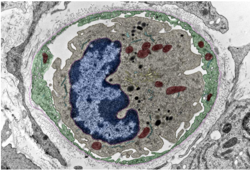
Capillary (blue) with Pericyte (Yellow)
Identify the vessel
Fenestrated capillary
Identify the vessel
Fenestrated capillary
Identify the vessel
Medium Artery
Identify the layer indicated by the line
Tunica media
Identify the vessel
Lymphatic vessel
Identify the vessel
Venule
What is the integument system?
Skin and its appendages
What are the appendages of the integument system?
Adnexa
Nails
Hair follicles
Glands
Sebaceous
eccrine
apocrine
What is the function of the integument system?
Protection
Temperature regulation
Moisture control
Sensory
How does the integument system temperature regulate?
Vasoconstrict blood vessels → keeps heat in
Vasodilation blood vessels → allows heat out
Thermogenesis → sweats to evaporate water and cools us
How does the integument system protect?
Melanin absorb UV
Metabolic: Vitamin D
Nails provide physical support → tools, prevents injury
Prevents water loss
Provides barrier against “wetting”
Glabrous skin (hairless and thick)
Nerve dependent → not osmosis (Skin wrinkles based on nerves to offer more grip)
How does the integument system offer sensory function?
Largest sensory organ in the body → 16% of body weight
Has many many nerves
What does the appearance of skin tell you?
It is an indicator for health
What are the main components of the integument system?
Epidermis
Papillary dermis
Reticular dermis
Hypodermis
Eccrine sweat glands
Adipose cells
Hair follicle
Sebaceous gland
Apocrine sweat gland
Pacinian corpuscle
Erector muscle
Blood vessels
Sensory nerve ending
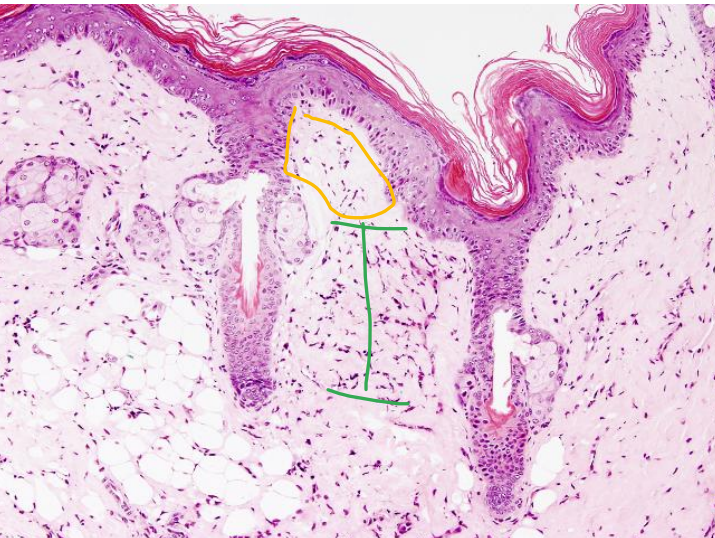
Dermal Papillae
Rete Ridges
What are the different accessory cells in the integument system?
Melanocyte
Langerhans cell
Merkel cell
What does a melanocyte do?
Pigment cell
In the stratum basale
What does a Langerhans cell do?
Macrophage derivative
In the Stratum spinosum
What does a merkel cell do?
Attached to free (non-myelinated) nerve ending
Sensitive to touch (mechanoreceptors)
Touch
in the stratum basale
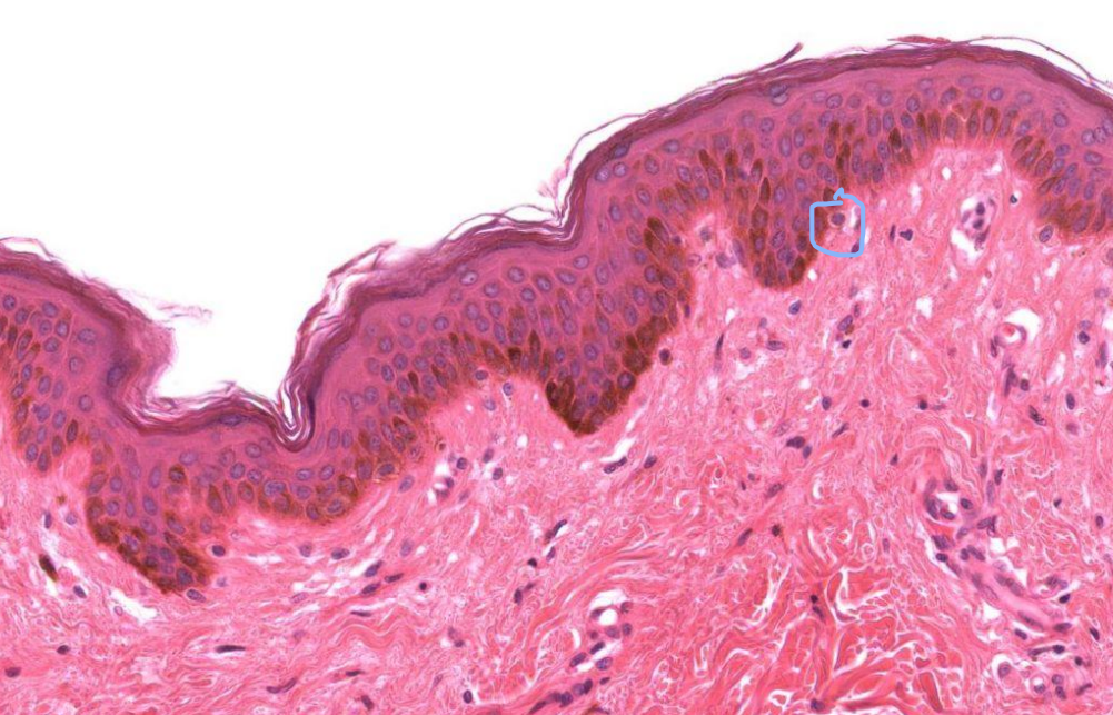
What are the layers of the skin?
Epidermis
Epithelium
Dermis
Connective tissue
Hypodermis
Adipose tissue
Connective tissue
What is the epidermis?
Has keratin
Offers protection
Has folds
Rete ridges (dips down)
Dermal Papillae (dips up)
What are the layers of the Epithelium - Strata in order from top to bottom?
Stratum corneum
Stratum granulosum
Stratum spinosum
Stratum basale
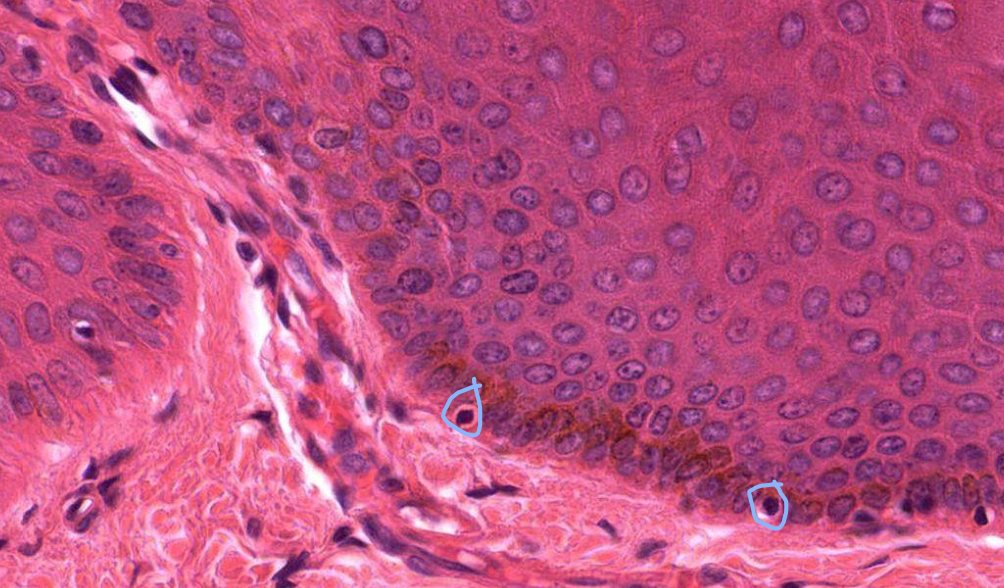
What are the different parts of the Stratum basale?
Single cell layer
Mitotic cells → regenerates, dividing layer (reason why it looks multilayered)
Basal lamina → bellow and connects to CT
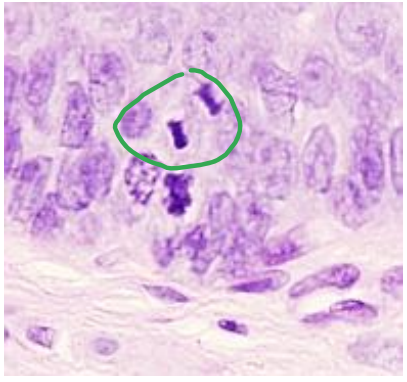
What is happening and where?
Cells are dividing in the stratum basale
What is happening and where?
Cells are becoming keratinocytes
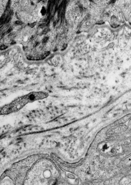
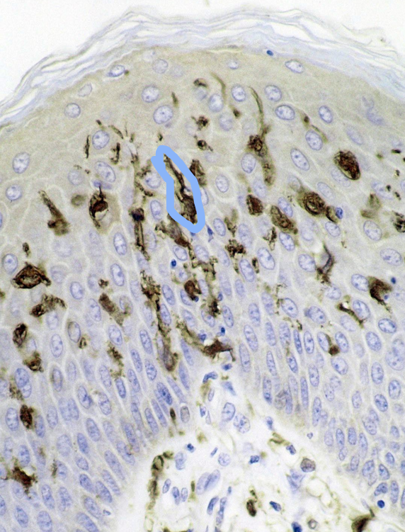
Identify
an EM of the Dermo-epidermal junction
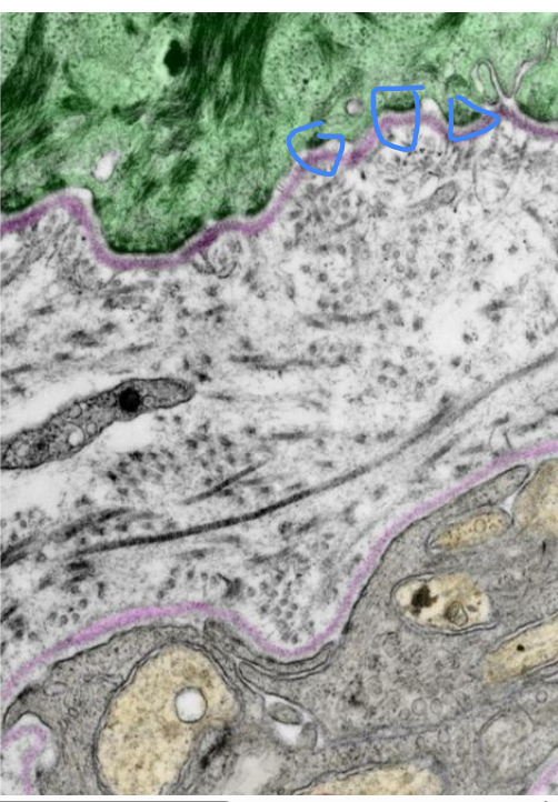
Identify structure that is circled?
Hemidesmosomes that are connecting to the basement membrane (At the dermo-epidermal junction)

Identify the layers indicated by the blue line
Stratum Spinosum
What are key features of the Stratum spinosum?
Keratinocytes
2-10 cells thick
Connected through desmosomes (connects cells to cells)
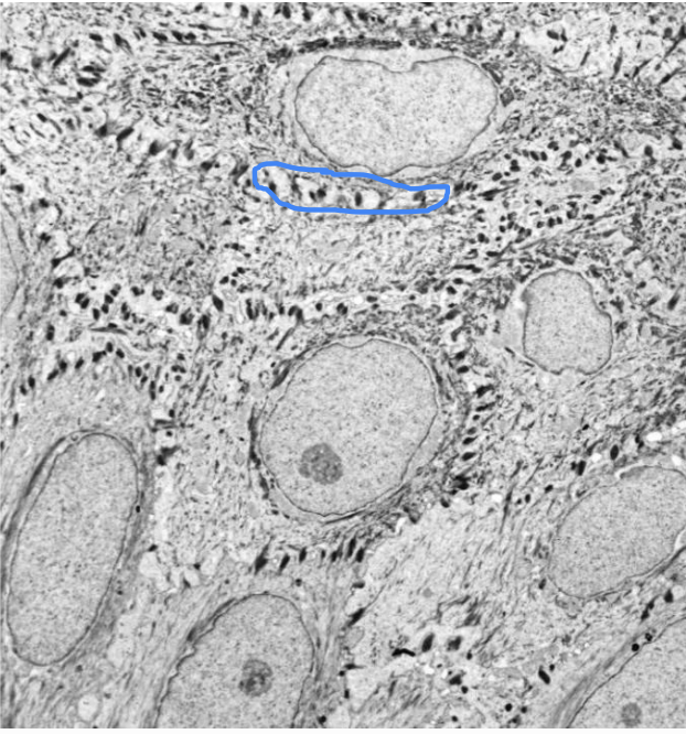
Identify the blue circles
Desmosomes
Where is this, and what is the arrow pointed at?
Stratum Spinosum
Langerhans cells
What are melanocytes?
Transient cell → clear
Dies in stratum spinosum
Makes pigment (melanin)
UV protection nucleus
Expose UV light
Increase melanin production
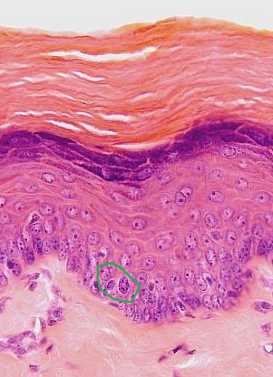
Identify the circled cell
Melanocyte (no distinct colour)
What is the circled cell?
Melanocyte
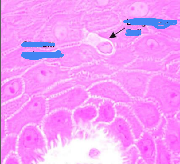
What are Langerhans cells?
Irregularly shaped dendritic cells
Antigen presenting
Allergic reaction

What is circled?
Langerhans cells
Identify the cell
Langerhans cell
What are key features of the Granulosum?
3-5 layers
Losing Nuclei and cytoplasmic organelles
Keratinized squames
Granules = secretion for water sealant
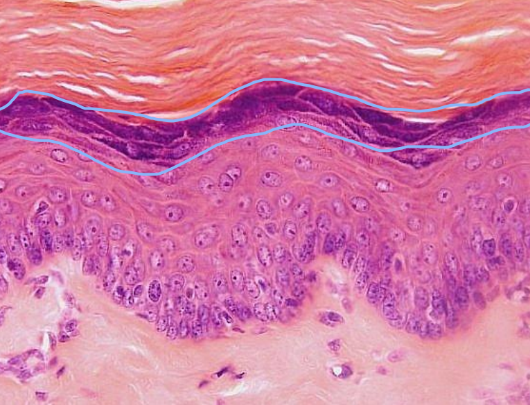
What layer is circled here?
Stratum Granulosum
What is the Stratum Corneum?
Dead cells
Flattened scales
Densely packed keratin
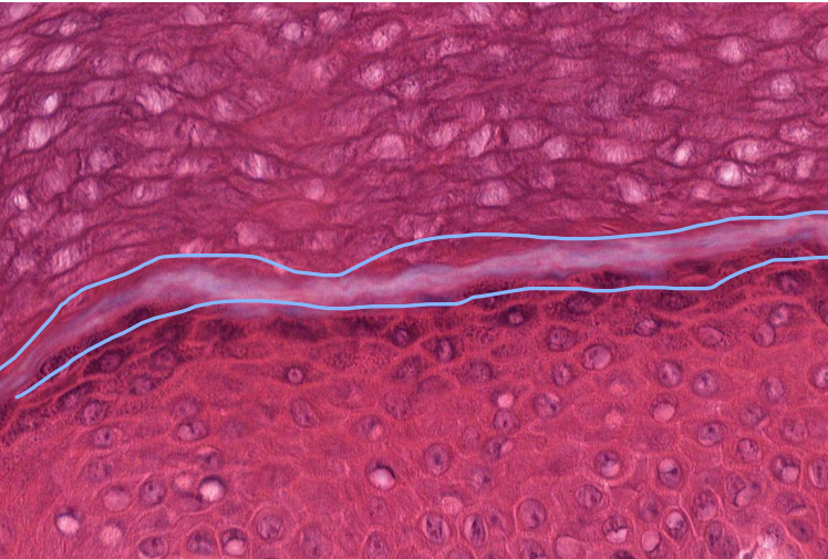
What is the Stratum Lucidum?
A layer in thick skin
Soles of feet and palms of hands
Glabrous skin → devoid of hair

What is circled here?
Sweat glands
What is layer is circled here?
Stratum Lucidum
What does Stratum Lucidum do?
Act as a water barrier
Made up of anucleated keratinocytes
Name the layers top to bottom?
Dermis, epidermis, Stratum Lucidum, Corneum
What is the dermis?
Loose and dense irregular connective tissue
Structural support
protection
Blood vessels (stop in dermis)
Nerves
Sensory receptors
Epidermal specializations
Hair
Sweat glands
Sebaceous glands
What makes up the connective tissue of the dermis?
Papillary dermis → Bumps into epidermis
Reticular dermis → rest on dermis
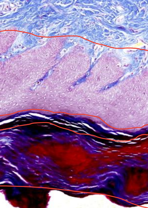
Identify the two layers?
Yellow: Papillary dermis
Green: Reticular dermis
What is the papillary dermis?
Loose connective tissue
Highly vascular
Dermal Papillae
More cells