iRAs (copy)
0.0(0)
Card Sorting
1/167
Earn XP
Description and Tags
Last updated 10:32 PM on 5/5/23
Name | Mastery | Learn | Test | Matching | Spaced | Call with Kai |
|---|
No analytics yet
Send a link to your students to track their progress
168 Terms
1
New cards
D
Which of the following structures is derived from the embryonic prosencephalon (forebrain)?
A. midbrain
B. cerebellum
C. pons
D. cerebral cortex
E. medulla oblongata
A. midbrain
B. cerebellum
C. pons
D. cerebral cortex
E. medulla oblongata
2
New cards
C
Which of the following structures is derived from the embryonic rhombencephalon (hindbrain)?
3
New cards
D
In conventional human radiological imaging (e.g., MRI, PET, CT) of the head, the axial plane is synonymous with the horizontal plane. Which of the following statements concerning the axial or horizontal plane is MOST ACCURATE?
A. The axial or horizontal plane is parallel to the coronal plane.
B. The axial or horizontal plane is in the plane of the face.
C. The axial or horizontal plane is parallel to the sagittal plane.
D. The axial or horizontal plane is more-or-less orthogonal (not quite a right angle, but close-- within about 30 degrees) to the longitudinal axis of the spinal cord.
E. The axial or horizontal plane is more-or-less orthogonal to the horizon when standing.
4
New cards
C
Which of the following pairs of directional terms is synonymous when applied to the identified region of the central nervous system?
A. in the forebrain, rostral & posterior
B. in the forebrain, ventral & anterior
C. in the forebrain, dorsal & superior
D. in the brainstem, ventral & posterior
E. in the spinal cord, caudal & posterior
A. in the forebrain, rostral & posterior
B. in the forebrain, ventral & anterior
C. in the forebrain, dorsal & superior
D. in the brainstem, ventral & posterior
E. in the spinal cord, caudal & posterior
5
New cards
E
Which of the following external spaces provides a major landmark dividing one cerebral lobe from another within a single hemisphere?
A. superior frontal sulcus
B. cingulate sulcus
C. calcarine sulcus
D. intraparietal sulcus
E. parieto-occipital sulcus
A. superior frontal sulcus
B. cingulate sulcus
C. calcarine sulcus
D. intraparietal sulcus
E. parieto-occipital sulcus
6
New cards
D
The insula is hidden from view when the brain is seen from its surface. The insula is covered by an operculum (operculum means covering) that folds into an external space, which in turn becomes an important landmark in the brain. The insula is found at the depth of which important space in the brain?
A. parieto-occipital sulcus
B. central sulcus
C. longitudinal fissure
D. lateral (Sylvian) fissure
E. interpeduncular fossa
A. parieto-occipital sulcus
B. central sulcus
C. longitudinal fissure
D. lateral (Sylvian) fissure
E. interpeduncular fossa
7
New cards
E
Which of the following statements concerning the central sulcus is MOST ACCURATE?
A. The central sulcus terminates laterally in or very near the longitudinal fissure.
B. The central sulcus is the principal landmark that divides the two cerebral hemispheres from one another.
C. The central sulcus terminates medially in or very near the lateral (Sylvian) fissure.
D. The central sulcus is formed by gyral formations that harbor the primary visual cortex in the human brain.
E. The central sulcus is formed by gyral formations that harbor the somatic sensory and motor divisions of the cerebral cortex in the human brain.
8
New cards
A
Which two lobes of the cerebral hemisphere feature three parallel, longitudinal gyri on their lateral aspect?
A. frontal and temporal lobes
B. parietal and occipital lobes
C. frontal and parietal lobes
D. temporal and occipital lobes
E. temporal and parietal lobes
A. frontal and temporal lobes
B. parietal and occipital lobes
C. frontal and parietal lobes
D. temporal and occipital lobes
E. temporal and parietal lobes
9
New cards
A
Which two lobes of the cerebral hemisphere feature three parallel, longitudinal gyri on their lateral aspect?
A. frontal and temporal lobes
B. parietal and occipital lobes
C. frontal and parietal lobes
D. temporal and occipital lobes
E. temporal and parietal lobes
A. frontal and temporal lobes
B. parietal and occipital lobes
C. frontal and parietal lobes
D. temporal and occipital lobes
E. temporal and parietal lobes
10
New cards
E
Which of the following spaces are roughly orthogonal in the human brain?
A. lateral (Sylvian) fissure & superior temporal sulcus
B. inferior frontal sulcus & inferior temporal sulcus
C. central sulcus & the parieto-occipital sulcus
D. precentral sulcus & postcentral sulcus
E. parieto-occipital sulcus & calcarine sulcus
A. lateral (Sylvian) fissure & superior temporal sulcus
B. inferior frontal sulcus & inferior temporal sulcus
C. central sulcus & the parieto-occipital sulcus
D. precentral sulcus & postcentral sulcus
E. parieto-occipital sulcus & calcarine sulcus
11
New cards
E
Which of the following structures is hidden from view when the brain is seen from its ventral surface?
A. midbrain
B. hypothalamus
C. parahippocampus gyrus
D. olfactory bulbs
E. thalamus
A. midbrain
B. hypothalamus
C. parahippocampus gyrus
D. olfactory bulbs
E. thalamus
12
New cards
B
] In the cerebral cortex, which layer of the cortex receives the primary input from specific thalamic relay nuclei?
A. layer 1
B. layer 4
C. layer 6
D. layer 2/3
E. layer 5
A. layer 1
B. layer 4
C. layer 6
D. layer 2/3
E. layer 5
13
New cards
C
What are the important functions of the canonical columnar microcircuit in the cerebral cortex?
A. amplify direct (monosynaptic) input from primary sensory neurons, compute new physiological properties, communicate with other neural circuits
B. amplify direct input from the thalamus, faithfully relay the same physiological properties that are computed in the thalamus, communicate with other neural circuits
C. amplify direct input from the thalamus, compute new physiological properties, communicate with other neural circuits
D. amplify direct input from primary sensory neurons, compute new physiological properties, communicate directly (through monosynaptic connections) with muscle fibers in skeletal muscle
E. amplify direct input from the thalamus, compute new physiological properties, communicate directly with visceral organs
14
New cards
D
Two important functional regions of the cerebral cortex that we will study in more detail later in the course are critical for language function. Based on the overview of these regions in "Lateral Surface of the Human Brain", and your general background in neuroscience, which of the following statements concerning these regions is MOST ACCURATE?
A. For most people, language lateralization is complete and the regions in the non-dominant cerebral hemisphere have no functional role in language processing.
B. For most people, Broca’s and Wernicke’s areas are localized to the right hemisphere.
C. A cortical stroke that afflicted a person with weakness in the right foot and ankle is also likely to produce an obvious impairment of language function.
D. A cortical stroke that afflicted a person with weakness in the right arm and right lower face is also likely to produce an obvious impairment of language function.
E. A cortical stroke that afflicted a person with blindness for the left half of the visual world is also likely to produce an obvious impairment of language function.
15
New cards
D
Which of the following arteries typically contributes to the anterior circulation of the brain?
A. basilar artery
B. superior sagittal sinus
C. anterior inferior cerebellar artery
D. middle cerebral artery
E. posterior inferior cerebellar artery
A. basilar artery
B. superior sagittal sinus
C. anterior inferior cerebellar artery
D. middle cerebral artery
E. posterior inferior cerebellar artery
16
New cards
A
The posterior circulation of the brain is typically derived from which of the following arteries?
A. vertebral arteries
B. internal carotid arteries
C. superior sagittal sinus
D. posterior communicating arteries
E. anterior communicating artery
A. vertebral arteries
B. internal carotid arteries
C. superior sagittal sinus
D. posterior communicating arteries
E. anterior communicating artery
17
New cards
B
At the base of the brain, the anterior and posterior circulations meet at an anastomotic ring of arterial blood supply called the “circle of Willis” (in honor of the great British physician-scientist of the 17th century, Thomas Willis). Which of the following vessels is critical for allowing for the passage of blood across the sagittal midline of this anastomotic ring?
A. anterior choroidal artery
B. anterior communicating artery
C. posterior communicating artery
D. anterior cerebral artery
E. posterior cerebral artery
A. anterior choroidal artery
B. anterior communicating artery
C. posterior communicating artery
D. anterior cerebral artery
E. posterior cerebral artery
18
New cards
C
Which of the following functional regions of the cerebral cortex is supplied by the anterior cerebral artery?
A. representation of the contralateral face in somatic sensory cortex
B. representation of the contralateral hand in somatic sensory cortex
C. representation of the contralateral foot in motor cortex
D. representation of the contralateral visual hemifield in the visual cortex
E. Broca's area
A. representation of the contralateral face in somatic sensory cortex
B. representation of the contralateral hand in somatic sensory cortex
C. representation of the contralateral foot in motor cortex
D. representation of the contralateral visual hemifield in the visual cortex
E. Broca's area
19
New cards
E
Which of the following gyral structures is supplied by the posterior cerebral artery?
A. superior frontal gyrus
B. medial orbital gyri
C. anterior cingulate gyrus
D. paracentral lobule
E. occipito-temporal gyrus (also known as the fusiform gyrus)
A. superior frontal gyrus
B. medial orbital gyri
C. anterior cingulate gyrus
D. paracentral lobule
E. occipito-temporal gyrus (also known as the fusiform gyrus)
20
New cards
A
A person presents with weakness and numbness in her right arm and the lower right side of her face, with all other functions being "within normal limits". As a clinician (or at least a budding functional neuroanatomist), you would suspect a possible stroke involving a branch of which artery?
A. left middle cerebral artery
B. right vertebral artery
C. left anterior choroidal artery
D. left posterior cerebral artery
E. left anterior cerebral artery
A. left middle cerebral artery
B. right vertebral artery
C. left anterior choroidal artery
D. left posterior cerebral artery
E. left anterior cerebral artery
21
New cards
D
Which of the following gyral structures is supplied by the middle cerebral artery?
A. cuneus gyrus
B. occipito-temporal gyrus (also known as the fusiform gyrus)
C. inferior temporal gyrus
D. superior temporal gyrus
E. cingulate gyrus
A. cuneus gyrus
B. occipito-temporal gyrus (also known as the fusiform gyrus)
C. inferior temporal gyrus
D. superior temporal gyrus
E. cingulate gyrus
22
New cards
C
An older person with a history of hypertension and cigarette smoking has recently been injured in a car crash on 9th Street while attempting to pull out of a parallel parking space. [This task requires looking back to the left.] The patient says that he just “never saw the car coming” in the lane of traffic. Although the patient is being evaluated for treatment of his orthopedic injuries, and no somatic sensory or motor impairments were discovered, the attending clinician should also wonder if there may have been a prior stroke that contributed to this patient’s accident. Which cerebral artery should the clinician consider most seriously when exploring whether a prior stroke contributed to this accident?
A. left middle cerebral artery
B. right anterior cerebral artery
C. right posterior cerebral artery
D. left vertebral artery
E. right posterior inferior cerebellar artery
A. left middle cerebral artery
B. right anterior cerebral artery
C. right posterior cerebral artery
D. left vertebral artery
E. right posterior inferior cerebellar artery
23
New cards
A
A person suddenly develops signs and symptoms that resemble “Parkinsonism” (difficulty expressing voluntary movement), but only on one side of the body. Knowing that such “hypokinetic” movement disorders have something to do with the basal ganglia and that you suspect the patient had a stroke, which cerebral artery should you consider as possibly being involved in this stroke?
A. a lenticulostriate branch of the middle cerebral artery
B. a long circumferential branch of a vertebral artery
C. an amygdalo-hippocampal branch of the anterior choroidal artery
D. a paramedian perforating branch of the proximal vertebral artery
E. a long circumferential branch of the basilar artery
24
New cards
B
In which structure does cerebrospinal fluid first mix with venous blood?
A. cavernous sinus
B. superior sagittal sinus
C. superior petrosal sinus
D. the great vein of Galen
E. inferior sagittal sinus
A. cavernous sinus
B. superior sagittal sinus
C. superior petrosal sinus
D. the great vein of Galen
E. inferior sagittal sinus
25
New cards
A
Which of the following sequences (indicated by >) correctly captures the drainage of venous blood from the dorsal aspects of the cerebral hemispheres?
A. superior sagittal sinus > transverse sinus > sigmoid sinus > internal jugular vein
B. cavernous sinus > transverse sinus > sigmoid sinus > internal jugular vein
C. superior sagittal sinus > sigmoid sinus > transverse sinus > internal jugular vein
D. sigmoid sinus > transverse sinus > inferior sagittal sinus > internal jugular vein
E. great vein of Galen > straight sinus > superior sagittal sinus > internal jugular vein
26
New cards
E
How is blood supplied to the spinal cord?
A. via a pair of anterior spinal arteries and a single posterior spinal artery
B. via major descending branches of the posterior cerebral artery
C. via major descending branches of the anterior inferior cerebellar arteries
D. via extravascular blood that circulates freely in the subarachnoid space
E. via a pair of posterior spinal arteries and a single anterior spinal artery
27
New cards
A
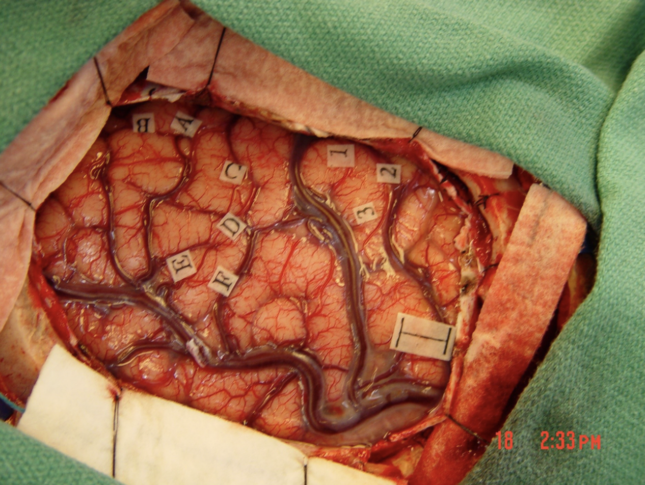
The photograph below is from a neurosurgical case in Duke University Hospital. The neurosurgeon has just completed intra-operative mapping of key functional sites related to somatic sensation, motor control, and language processing (the small pieces of papers with letters and numbers indicate those sites and the scale bar = 1 cm). The patient remained awake and conversant throughout this phase of the operation. In order to perform this functional analysis intraoperatively, which layer(s) of the meninges must remain intact (after the other layer(s) have been dissected away)?
A. pia mater only need remain intact
B. dura mater, arachnoid mater, pia mater all must remain intact
C. arachnoid mater and pia mater both must remain intact
D. arachnoid mater only need remain intact
E. dura mater only need remain intact
A. pia mater only need remain intact
B. dura mater, arachnoid mater, pia mater all must remain intact
C. arachnoid mater and pia mater both must remain intact
D. arachnoid mater only need remain intact
E. dura mater only need remain intact
28
New cards
E
Two dorm-mates are “horsing around” one evening—and while one has the other in a “head-lock” (putting pressure with biceps and forearm on the lower head and neck)—the “head-locked” student suddenly experiences strange sensations and alterations of consciousness. Seeing this happen, you quickly intervene because you are concerned for the possible occlusion (by head-lock) of one or both of the common carotid arteries and the perfusion of which of the following cerebral vessels typically derived from the common carotid arteries?
A. anterior cerebral artery, posterior cerebral artery
B. middle cerebral artery, posterior cerebral artery
C. middle cerebral artery, basilar artery
D. vertebral artery, basilar artery
E. anterior cerebral artery, middle cerebral artery
A. anterior cerebral artery, posterior cerebral artery
B. middle cerebral artery, posterior cerebral artery
C. middle cerebral artery, basilar artery
D. vertebral artery, basilar artery
E. anterior cerebral artery, middle cerebral artery
29
New cards
B
A middle-aged university professor was exercising by riding his bicycle along back roads in Orange County, when he rather suddenly felt some odd sensations in his right arm and face (tingling, “pins-and-needles”, feelings of swelling or tightness). After a few minutes of experiencing these sensations, he became faint and realized that he needed to rest by the side of the road. He got off his bike and sat down on the grassy shoulder when he noticed that the odd feelings were subsiding, but he felt unusually weak in his right arm and there was now numbness over the affected body parts. This event was very short-lived and he continued his ride after resting there for about 20 minutes. Although he did not know it at the time, this individual had a “transient ischemic attack” (TIA).
Thinking back on what you have learned elsewhere in the neuroscience curriculum, what would be the impact of a TIA on the resting membrane potential of neurons in the affected region?
A. hyperpolarization precisely to the Nernst equilibrium potential for potassium
B. depolarization just beyond the threshold potential for firing action potentials
C. hyperpolarization, but not precisely to the Nernst equilibrium potential for potassium
D. depolarization precisely to the Nernst equilibrium potential for sodium
E. depolarization precisely to zero millivolts
30
New cards
A
Which of the following structures that may be seen on sections through the forebrain is telencephalic (i.e., derived from the embryological telencephalon)?
A. hippocampus
B. fourth ventricle
C. third ventricle
D. hypothalamus
E. thalamus
A. hippocampus
B. fourth ventricle
C. third ventricle
D. hypothalamus
E. thalamus
31
New cards
A
Which of the following structures that may be seen on sections through the forebrain is diencephalic (i.e., derived from the embryological diencephalon)?
A. hypothalamus
B. amygdala
C. corpus callosum
D. basal ganglia
E. cerebral cortex
A. hypothalamus
B. amygdala
C. corpus callosum
D. basal ganglia
E. cerebral cortex
32
New cards
C
Which of the following deep gray matter structures is a component of the basal ganglia?
A. hippocampus
B. basal forebrain nuclei
C. caudate nucleus
D. hypothalamus
E. thalamus
A. hippocampus
B. basal forebrain nuclei
C. caudate nucleus
D. hypothalamus
E. thalamus
33
New cards
D
Which bundle of white matter separates the thalamus from the putamen (and globus pallidus)?
A. cerebral peduncle
B. fornix (also known as fimbria/fornix)
C. anterior commissure
D. internal capsule
E. external capsule
A. cerebral peduncle
B. fornix (also known as fimbria/fornix)
C. anterior commissure
D. internal capsule
E. external capsule
34
New cards
D
In the lab today, you will have the opportunity to observe a brain cutting. First, you will hemisect a brain (if that has not been already done) in the midsagittal plane. Then, you will take one hemisphere and make slabs from it in the coronal (or axial) plane. Consider now the hemisphere that you will section in the coronal plane and focus in your mind’s eye on the coronal slab containing a mammillary body. Got the slab visualized?
Now the question … which of the following structures would you expect to see in just one location in that slab from just one hemisphere?
[Hint: there are several structures that will appear in at least two locations in that slab.]
A. lateral ventricle
B. choroid plexus
C. fornix (and its root off the dorsal-lateral margin of the hippocampus known as the fimbria)
D. amygdala
E. caudate nucleus
35
New cards
A
Cerebrospinal fluid (CSF) is produced in each of the ventricles. How does the CSF get from the system of ventricles in the brain to the subarachnoid space?
A. CSF flows from the fourth ventricle to the subarachnoid space via the median and lateral apertures in the fourth ventricle (also known as the foramina of Magendie and Luschka, respectively).
B. CSF flows from the lateral ventricles into the subarachnoid space via the foramina of Monro (also known as the interventricular foramina).
C. CSF flows from the third ventricle to the subarachnoid space via the great cerebral vein of Galen.
D. CSF flows from the third ventricle to the subarachnoid space via the aqueduct of Sylvius (also known as the cerebral aqueduct).
E. CSF flows from the lateral ventricles to the subarachnoid space via the arachnoid granulations (also known as arachnoid villi).
36
New cards
E
If you read and wrestled with the Lab Challenge—internal capsule and deep gray matter, Blumenfeld Chapter 16, and/or the tutorial notes associated with the videos, then you should be well prepared for our virtual lab this week, and well prepared to address the next few questions.
Which pair of structures is located on the medial side of the internal capsule?
A. putamen & insula
B. putamen & globus pallidus
C. amygdala & hippocampus
D. nucleus accumbens & putamen
E. caudate nucleus & thalamus
Which pair of structures is located on the medial side of the internal capsule?
A. putamen & insula
B. putamen & globus pallidus
C. amygdala & hippocampus
D. nucleus accumbens & putamen
E. caudate nucleus & thalamus
37
New cards
D
Which pair of structures is located on the lateral side of the internal capsule?
A. globus pallidus & caudate nucleus
B. pineal gland & mammillary body
C. fornix & anterior commissure
D. globus pallidus & putamen
E. caudate nucleus & thalamus
A. globus pallidus & caudate nucleus
B. pineal gland & mammillary body
C. fornix & anterior commissure
D. globus pallidus & putamen
E. caudate nucleus & thalamus
38
New cards
A
Which of the following structures is located between the internal capsule and the putamen?
A. globus pallidus
B. nucleus accumbens
C. caudate nucleus
D. hypothalamus
E. thalamus
A. globus pallidus
B. nucleus accumbens
C. caudate nucleus
D. hypothalamus
E. thalamus
39
New cards
A
Which structure is located mostly or entirely anterior to the other?
A. with the exception of a small region of overlap, the head of the caudate nucleus is anterior to the globus pallidus
B. the thalamus is anterior to the nucleus accumbens
C. the anterior commissure is anterior to the nucleus accumbens
D. the temporal horn of the lateral ventricle is anterior to the frontal horn of the lateral ventricle
E. with the exception of small region of overlap, the hippocampus is anterior to the amygdala
40
New cards
A
Suppose you are holding a hemisphere in your hand. You can see into the lateral ventricle in this hemisphere -- because the septum pellucidum has been removed -- and notice something worth touching. So you insert your pinky finger (your fifth digit) into the frontal horn of the lateral ventricle and gently palpate the bulging structure you discover just lateral to the ventricle. (This is something you might try to do in the lab today.) If you were to do this, what structure would you be palpating?
A. caudate nucleus
B. nucleus accumbens
C. globus pallidus
D. putamen
E. thalamus
A. caudate nucleus
B. nucleus accumbens
C. globus pallidus
D. putamen
E. thalamus
41
New cards
B
Does CSF flow directly from one lateral ventricle to the other?
A. yes; the interventricular foramen of Monro allows passage of CSF from one lateral ventricle to the other
B. no, in a typical brain, the septum pellucidum forms a diffusion barrier between the two lateral ventricles
C. no; the falx cerebri forms a diffusion barrier between the two lateral ventricles
D. yes; the massa intermedia (interthalamic adhesion) allows passage of CSF from one lateral ventricle to the other
E. yes; in a typical brain, CSF diffuses freely from one lateral ventricle to the other
42
New cards
D
Identify the ACCURATE layering of meninges from outer to (>) inner:
A. pia mater > dura mater > arachnoid mater
B. dura mater > pia mater > arachnoid mater
C. pia mater > arachnoid mater > dura mater
D. dura mater > arachnoid mater > pia mater
E. arachnoid mater > pia mater > dura mater
A. pia mater > dura mater > arachnoid mater
B. dura mater > pia mater > arachnoid mater
C. pia mater > arachnoid mater > dura mater
D. dura mater > arachnoid mater > pia mater
E. arachnoid mater > pia mater > dura mater
43
New cards
B
Which structure produces cerebrospinal fluid?
A. arachnoid granulations
B. choroid plexus
C. pineal gland
D. cisterna magna
E. pituitary gland
A. arachnoid granulations
B. choroid plexus
C. pineal gland
D. cisterna magna
E. pituitary gland
44
New cards
D
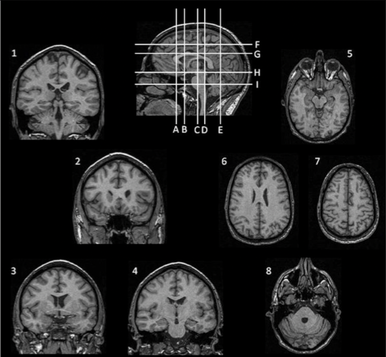
For the next two questions, match the images numbered 1-8 with the lines labeled A-I, which indicate possible locations of the sections through the head.
Identify the INCORRECT matching of image to section level.
A. image 6 = level G
B. image 3 = level B
C. image 1 = level D
D. image 8 = level E
E. image 5 = level H
45
New cards
E
Match the images numbered 1-8 with the lines labeled A-I, which indicate possible locations of the sections through the head.
Identify the CORRECT matching of image to section level.
[Note: the previous question asked for the incorrect match; this question is asking for the CORRECT match.]
A. image 6 = level F
B. image 4 = level I
C. image 2 = level H
D. image 7 = level G
E. image 3 = level B
Identify the CORRECT matching of image to section level.
[Note: the previous question asked for the incorrect match; this question is asking for the CORRECT match.]
A. image 6 = level F
B. image 4 = level I
C. image 2 = level H
D. image 7 = level G
E. image 3 = level B
![Match the images numbered 1-8 with the lines labeled A-I, which indicate possible locations of the sections through the head.
Identify the CORRECT matching of image to section level.
[Note: the previous question asked for the incorrect match; this question is asking for the CORRECT match.]
A. image 6 = level F
B. image 4 = level I
C. image 2 = level H
D. image 7 = level G
E. image 3 = level B](https://knowt-user-attachments.s3.amazonaws.com/a4ec7c5a218c4714ba448bc9acb68293.jpeg)
46
New cards
D
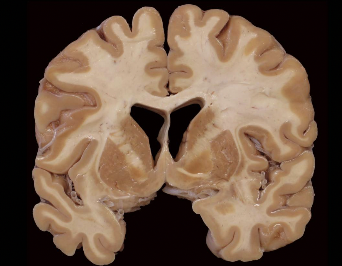
Inspect this photograph below and take a moment to orient yourself to the plane of section and the approximate position of this slab in the brain. Got it?
Given where you think this slab came from in the brain, which of the following gyri or lobules are visible in this slab?
A. precuneus gyrus
B. inferior parietal lobule
C. superior parietal lobule
D. superior frontal gyrus
E. paracentral lobule
Given where you think this slab came from in the brain, which of the following gyri or lobules are visible in this slab?
A. precuneus gyrus
B. inferior parietal lobule
C. superior parietal lobule
D. superior frontal gyrus
E. paracentral lobule

47
New cards
C
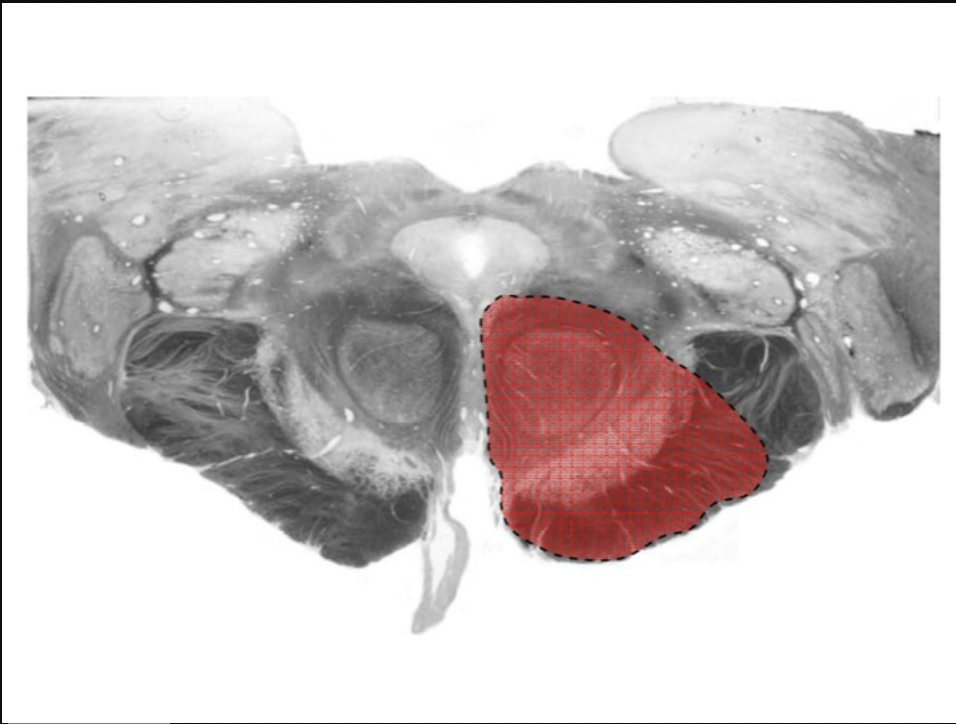
Consider the distinctive dark brown streaks that are obvious just dorsal to the cerebral peduncles in the photograph below. (We will look for similar dark brown streaks in the brains that we will examine in the lab today.) What are these streaks?
A. locus coeruleus
B. posterior cerebral arteries
C. substantia nigra
D. choroid plexus
E. basal forebrain nuclei
A. locus coeruleus
B. posterior cerebral arteries
C. substantia nigra
D. choroid plexus
E. basal forebrain nuclei
48
New cards
B
You know a friend with what is sometimes called a “lazy eye”. More properly termed, this condition is known as “strabismus” (from the Greek, meaning “to squint”). This condition involves a misalignment of the two eyes, and sometimes a tendency to squint. This is a serious neurological problem as the brain may actively suppress – make blind – the information derived from the “lazy” eye. In the case of your friend, their LEFT eye is deviated outward, which is a condition called “exotropia”. Unilateral damage to which cranial nerve (CN) in the cranium or in the orbit could account for this impairment?
A. left CN II
B. left CN III
C. left CN VI
D. right CN VI
E. right CN III
A. left CN II
B. left CN III
C. left CN VI
D. right CN VI
E. right CN III
49
New cards
B
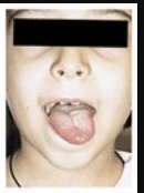
The patient pictured below was asked to “stick your tongue straight out”, but the tongue deviated to the left. Unilateral damage to which cranial nerve could account for this movement impairment?
A. right trigeminal nerve
B. left hypoglossal nerve
C. left glossopharyngeal nerve
D. left abducens nerve
E. right facial nerve
A. right trigeminal nerve
B. left hypoglossal nerve
C. left glossopharyngeal nerve
D. left abducens nerve
E. right facial nerve
50
New cards
D
Where would you find the olive (inferior olive)?
A. on the dorsal aspect of the pons forming an outward bulge in the floor of the fourth ventricle
B. on the ventral-lateral surface of the mesencephalon (midbrain), just lateral to CN III
C. on the dorsal-lateral surface of the caudal medulla oblongata, just medial to CN XI
D. on the ventral-lateral surface of the rostral medulla oblongata, just lateral to CN XII
E. on the dorsal aspect of the cervical enlargement of the spinal cord, in register with the underlying cuneate tract
51
New cards
D
Which peduncles (“stalks” of white matter) originate in the pons?
A. the cerebral peduncles
B. the medullary peduncles
C. the superior cerebellar peduncles
D. the inferior cerebellar peduncles
E. the middle cerebellar peduncles
A. the cerebral peduncles
B. the medullary peduncles
C. the superior cerebellar peduncles
D. the inferior cerebellar peduncles
E. the middle cerebellar peduncles
52
New cards
C
Where would you find the inferior colliculus?
A. on the dorsal aspect of the pons forming an outward bulge in the floor of the fourth ventricle
B. on the dorsal aspect of the mesencephalon (midbrain), just inferior to CN IV
C. on the dorsal aspect of the mesencephalon (midbrain), just inferior to the superior colliculus
D. on the dorsal aspect of the caudal medulla oblongata, just inferior to the fourth ventricle
E. on the dorsal aspect of the pons, in register with the roots of CN V
53
New cards
E
Which of the following ACCURATELY accounts for the number of pairs of spinal nerves connected to the human spinal cord in a given longitudinal region of the cord?
A. There are 5 cervical nerves.
B. There are 8 thoracic nerves.
C. There are 12 lumbar nerves.
D. The cauda equina comprises 8 sacral and 1 coccygeal nerves.
E. There are 8 cervical nerves.
A. There are 5 cervical nerves.
B. There are 8 thoracic nerves.
C. There are 12 lumbar nerves.
D. The cauda equina comprises 8 sacral and 1 coccygeal nerves.
E. There are 8 cervical nerves.
54
New cards
E
What would you expect to find in the dorsal roots of the cervical enlargement of the spinal cord?
A. axons conveying efferent somatic motor commands to the upper extremities
B. axons conveying efferent preganglionic parasympathetic signals to the pelvic viscera
C. axons conveying efferent preganglionic sympathetic signals to ganglia in the paravertebral sympathetic chain
D. axons conveying efferent somatic motor commands to muscles of the neck
E. axons conveying afferent somatic sensory signals from the upper extremities
55
New cards
C
What would you expect to find in the ventral roots of the lumbosacral enlargement of the spinal cord?
A. axons conveying afferent visceral sensory signals from the pelvic viscera
B. axons conveying afferent somatic sensory signals from the lower extremities
C. axons conveying efferent somatic motor commands to the lower extremities
D. axons conveying efferent preganglionic sympathetic signals to ganglia in the paravertebral sympathetic chain
E. axons conveying efferent somatic motor commands to contralateral muscles (on the side of the body that is opposite the ventral root in question)
56
New cards
D
Which of the following structures of the brainstem are largely present in more than one embryological subdivision (i.e., midbrain and pons, pons and medulla, or midbrain, pons and medulla)?
A. red nucleus
B. oculomotor nucleus
C. middle cerebellar peduncle
D. spinal trigeminal nucleus
E. inferior olivary nucleus
A. red nucleus
B. oculomotor nucleus
C. middle cerebellar peduncle
D. spinal trigeminal nucleus
E. inferior olivary nucleus
57
New cards
A
Identify the most accurate statement regarding the longitudinal organization of the spinal cord.
A. The lateral horns are a characteristic feature of the thoracic spinal cord.
B. There is more neural circuitry in the gray matter of the thoracic segments than in the cervical enlargement of the spinal cord.
C. The spinal cord achieves its greatest cross-sectional area in the lower lumbar segments (L3-5) of the spinal cord.
D. There is progressively more white matter in the spinal cord from one segment to the next in a caudal progression.
E. In the lumbar enlargement, the dorsal column comprises two subdivisions on each side of the midline.
58
New cards
B
Where in the brainstem are the posterior (dorsal) column nuclei?
[Hint: “tegmentum” refers to the core of the brainstem, between the ventricular system dorsally and the basal structures ventrally.]
A. dorsal tegmentum of the rostral (superior half) pons
B. dorsal tegmentum of the caudal (inferior half) medulla
C. dorsal tegmentum of the ponto-medullary junction
D. dorsal tegmentum of the midbrain
E. tectum of the midbrain
59
New cards
C
Where in the brainstem are the vestibular nuclei?
A. dorsal-lateral tegmentum of the midbrain
B. dorsal-medial tegmentum of the rostral pons
C. dorsal-lateral tegmentum of the ponto-medullary junction
D. dorsal-medial tegmentum of the caudal medulla
E. tectum of the midbrain
A. dorsal-lateral tegmentum of the midbrain
B. dorsal-medial tegmentum of the rostral pons
C. dorsal-lateral tegmentum of the ponto-medullary junction
D. dorsal-medial tegmentum of the caudal medulla
E. tectum of the midbrain
60
New cards
E
Where in the brainstem is the pontine gray matter, known as the “pontine nuclei”?
A. dorsal-medial tegmentum of the rostral pons
B. dorsal-lateral tegmentum of the rostral pons
C. dorsal-medial tegmentum of the ponto-medullary junction
D. gray matter encircling the walls of the fourth ventricle
E. basilar or ventral region (“basis pontis”) of the pons
A. dorsal-medial tegmentum of the rostral pons
B. dorsal-lateral tegmentum of the rostral pons
C. dorsal-medial tegmentum of the ponto-medullary junction
D. gray matter encircling the walls of the fourth ventricle
E. basilar or ventral region (“basis pontis”) of the pons
61
New cards
D
What is the brainstem nucleus that accounts for the outward bulge just lateral to the nerve roots of CN XII?
A. gracile nucleus
B. cuneate nucleus
C. spinal accessory nucleus
D. inferior olivary nucleus
E. spinal trigeminal nucleus
A. gracile nucleus
B. cuneate nucleus
C. spinal accessory nucleus
D. inferior olivary nucleus
E. spinal trigeminal nucleus
62
New cards
A
Which of the following cranial nerve nuclei is found in the medulla?
A. dorsal motor nucleus of vagus
B. abducens nucleus
C. facial motor nucleus
D. trigeminal motor nucleus
E. chief (main, principal) sensory nucleus of the trigeminal complex
A. dorsal motor nucleus of vagus
B. abducens nucleus
C. facial motor nucleus
D. trigeminal motor nucleus
E. chief (main, principal) sensory nucleus of the trigeminal complex
63
New cards
C
Which of the following cranial nerve nuclei is found in the pons?
A. spinal accessory nucleus
B. oculomotor nucleus
C. superior vestibular nucleus
D. nucleus ambiguus
E. Edinger-Westphal nucleus
A. spinal accessory nucleus
B. oculomotor nucleus
C. superior vestibular nucleus
D. nucleus ambiguus
E. Edinger-Westphal nucleus
64
New cards
A
Which of the following cranial nerve nuclei is found in the midbrain?
A. Edinger-Westphal nucleus
B. trigeminal motor nucleus
C. chief sensory (principal) nucleus of the trigeminal complex
D. abducens nucleus
E. nucleus ambiguus
A. Edinger-Westphal nucleus
B. trigeminal motor nucleus
C. chief sensory (principal) nucleus of the trigeminal complex
D. abducens nucleus
E. nucleus ambiguus
65
New cards
B
Identify the MOST ACCURATE pairing of cranial nerve to origin (i.e., location of the neuronal cell bodies that grew the axons in the cranial nerve; please don’t miss that clue about what we mean when we speak of “origin”).
A. CN III / origin = retina
B. CN VII / origin = facial motor nucleus
C. CN V / origin = chief sensory nucleus of the trigeminal complex
D. CN VI / origin = trochlear nucleus
E. CN XII / origin = extrinsic muscles of the tongue
A. CN III / origin = retina
B. CN VII / origin = facial motor nucleus
C. CN V / origin = chief sensory nucleus of the trigeminal complex
D. CN VI / origin = trochlear nucleus
E. CN XII / origin = extrinsic muscles of the tongue
66
New cards
A
Which of the following cranial nerve nuclei is a somatic motor nucleus and is, therefore, found along the dorsal midline of the brainstem tegmentum?
A. abducens nucleus
B. Edinger-Westphal nucleus
C. facial motor nucleus
D. trigeminal motor nucleus
E. nucleus ambiguus
A. abducens nucleus
B. Edinger-Westphal nucleus
C. facial motor nucleus
D. trigeminal motor nucleus
E. nucleus ambiguus
67
New cards
E
Which of the following cranial nerve nuclei is a branchial motor nucleus and is, therefore, found in an intermediate position in the lateral brainstem tegmentum?
A. Edinger-Westphal nucleus
B. dorsal motor nucleus of vagus
C. hypoglossal nucleus
D. abducens nucleus
E. facial motor nucleus
A. Edinger-Westphal nucleus
B. dorsal motor nucleus of vagus
C. hypoglossal nucleus
D. abducens nucleus
E. facial motor nucleus
68
New cards
A
The lesion outlined in the dashed line (enclosing the red area) over the brainstem section involves which of the following structures?
A. substantia nigra
B. trigeminal motor nucleus
C. roots of the abducens nerve
D. medullary pyramid
E. pontine nuclei
A. substantia nigra
B. trigeminal motor nucleus
C. roots of the abducens nerve
D. medullary pyramid
E. pontine nuclei
69
New cards
E
The lesion outlined in the dashed line (enclosing the red area) over the brainstem section involves which of the following structures?
A. medial lemniscus
B. medullary pyramid
C. hypoglossal nucleus
D. inferior olivary nucleus
E. spinal trigeminal nucleus
A. medial lemniscus
B. medullary pyramid
C. hypoglossal nucleus
D. inferior olivary nucleus
E. spinal trigeminal nucleus
70
New cards
B
As Blumenfeld summarizes the functions of neurotransmitters in your reading for today, he highlights the important function of neuromodulation that compliments the more obvious fast, excitatory and inhibitory neurotransmission. Which of the following pairings of neuromodulator and anatomical nucleus ACCURATELY accounts for the source (the location of the relevant cell bodies) of the chemical substance in question?
A. acetylcholine; source = locus coeruleus of the dorsal pontine tegmentum
B. dopamine; source = ventral tegmental area of the midbrain
C. norepinephrine; source = raphe nuclei of the midline brainstem
D. serotonin; source = substantia nigra pars compacta of the ventral midbrain tegmentum
E. histamine; source = basal forebrain nuclei (e.g., nucleus of the diagonal band of Broca; nucleus basalis of Meynert)
71
New cards
D
As painful as this may be, imagine walking barefoot on the beach and stepping on a broken seashell. You immediately have a sharp, jolting pain that emanates from the sole of your foot. What pathway allows you to experience this immediate, sharp, painful sensation (sometimes called “first pain”) with somatotopic precision?
A. posterior (dorsal) column-medial lemniscal pathway
B. spinomesencephalic tract
C. spinoreticular tract
D. spinothalamic tract
E. dorsal spinocerebellar tract
A. posterior (dorsal) column-medial lemniscal pathway
B. spinomesencephalic tract
C. spinoreticular tract
D. spinothalamic tract
E. dorsal spinocerebellar tract
72
New cards
A
Just after stepping on that seashell, you sit down in the sand and you immediately grab and squeeze the bottom of your foot. That sharp "first" pain seems to be diminished by this action within a second or two of applying that compressive squeeze. You have just “closed the gate” to the passage of pain signals (or so the classical gate control theory would say). What was the neural mechanism that closed the “gate”?
A. You activated large diameter primary afferents that excited inhibitory interneurons, which in turn reduced the excitability of projection neurons in the dorsal horn of the spinal cord.
B. You activated large diameter secondary afferents in the medial lemniscus that excited inhibitory interneurons (in the thalamic reticular nucleus), which in turn reduce the excitability of projection neurons in the ventral posterior lateral nucleus of the thalamus.
C. You activated large diameter tertiary afferents in the internal capsule that excited inhibitory interneurons in cortical layer IV, which in turn reduced the excitability of pyramidal neurons throughout the relevant columns of the primary somatic sensory cortex.
D. You activated neural networks in the insula that suppressed the feelings of distress associated with the unexpected and somewhat traumatic event of stepping on the seashell.
E. You activated neural circuits in the brainstem reticular formation that fed back into the dorsal horn of the spinal cord and produce a persistent state of analgesia.
73
New cards
B
Despite squeezing the injured foot, a more dull, aching pain that is less well localized sets in a few minutes after the initial wave of ("first") pain has passed. What is the principal site that integrates and distributes signals that manifest as this dull, aching pain (sometime called “second pain”) that often leads to feeling badly and maybe somewhat emotionally upset because of the pain?
A. ventral horn of the spinal cord
B. reticular formation of the brainstem
C. postcentral gyrus
D. ventral posterior lateral nucleus of the thalamus
E. insula
A. ventral horn of the spinal cord
B. reticular formation of the brainstem
C. postcentral gyrus
D. ventral posterior lateral nucleus of the thalamus
E. insula
74
New cards
C
Maybe it is the company you're with there on the beach or maybe it's the beautiful weather. Whatever the case, you are determined to resume your walk. As you do so, you are more focused on your company, the beauty of the coastline, the diving birds, the sunshine, etc., and as you press on with more positive thoughts in mind, the pain seems to subside. This analgesic effect is the product of central integration (and feedback modulation) of ascending nociceptive information with “top-down” contextual signals. What brainstem structure is especially important in the central integration of these signals and the source of descending input to other brainstem sites, which in turn, project to the dorsal horn of the spinal cord?
A. spinal trigeminal nucleus
B. locus coeruleus
C. periaqueductal gray
D. mesencephalic trigeminal nucleus
E. cuneate nucleus
A. spinal trigeminal nucleus
B. locus coeruleus
C. periaqueductal gray
D. mesencephalic trigeminal nucleus
E. cuneate nucleus
75
New cards
E
Where is the decussation (midline crossing) of the posterior (dorsal) column medial lemniscal system?
A. in the posterior commissure of the midbrain
B. in the corpus callosum
C. in the ventral (anterior) white commissure of the spinal cord
D. in the pyramidal decussation of the caudal medulla
E. in the internal arcuate fibers of the caudal medulla
A. in the posterior commissure of the midbrain
B. in the corpus callosum
C. in the ventral (anterior) white commissure of the spinal cord
D. in the pyramidal decussation of the caudal medulla
E. in the internal arcuate fibers of the caudal medulla
76
New cards
D
Which thalamic nucleus is the target of the medial lemniscus?
A. lateral geniculate nucleus (LGN)
B. ventral posterior medial nucleus (VPM)
C. medial geniculate nucleus (MGN)
D. ventral posterior lateral nucleus (VPL)
E. ventral lateral nucleus (VL)
A. lateral geniculate nucleus (LGN)
B. ventral posterior medial nucleus (VPM)
C. medial geniculate nucleus (MGN)
D. ventral posterior lateral nucleus (VPL)
E. ventral lateral nucleus (VL)
77
New cards
A
Which thalamic nucleus is the target of the trigeminal lemniscus?
A. ventral posterior medial nucleus (VPM)
B. lateral geniculate nucleus (LGN)
C. medial geniculate nucleus (MGN)
D. ventral posterior lateral nucleus (VPL)
E. ventral lateral nucleus (VL)
A. ventral posterior medial nucleus (VPM)
B. lateral geniculate nucleus (LGN)
C. medial geniculate nucleus (MGN)
D. ventral posterior lateral nucleus (VPL)
E. ventral lateral nucleus (VL)
78
New cards
C
Now that you know quite a lot about what somatic sensory tracts are present in various white matter structures in the spinal cord and brain, consider the following question. Which of the following structures when lesioned is most likely to produce a pure motor deficit without an accompanying somatic sensory deficit?
Hint: as you will study in more depth in two weeks (and as you should recall from prior lessons), the corticospinal tract conveys motor signals from the motor cortex to the spinal cord.
A. posterior limb of internal capsule
B. corona radiata
C. medullary pyramid
D. dorsal column of spinal cord
E. lateral column of spinal cord
79
New cards
A
Which of the following structures when lesioned is most likely to produce a pure somatic sensory deficit without an accompanying motor deficit?
A. dorsal column of spinal cord
B. posterior limb of internal capsule
C. corona radiata
D. medullary pyramid
E. lateral column of spinal cord
A. dorsal column of spinal cord
B. posterior limb of internal capsule
C. corona radiata
D. medullary pyramid
E. lateral column of spinal cord
80
New cards
B
Perforating branches of which major artery supplies blood directly to the thalamus?
A. anterior inferior cerebellar artery
B. posterior cerebral artery
C. vertebral artery
D. basilar artery
E. posterior inferior cerebellar artery
A. anterior inferior cerebellar artery
B. posterior cerebral artery
C. vertebral artery
D. basilar artery
E. posterior inferior cerebellar artery
81
New cards
A
Perforating branches of which major artery supplies blood directly to the medulla?
A. vertebral artery
B. anterior inferior cerebellar artery
C. posterior cerebral artery
D. basilar artery
E. superior cerebellar artery
A. vertebral artery
B. anterior inferior cerebellar artery
C. posterior cerebral artery
D. basilar artery
E. superior cerebellar artery
82
New cards
E
Consider the experience of holding a piece of ice in your fingertips. To help your imagination with this question, feel free to help yourself to ice that maybe near (there’s an ice maker in the kitchen area near our classroom). Now, let it drip and melt and get just a bit messy in your fingers. Appreciate the fact that this single stimulus is uncomfortably cold, wet, smooth, slippery, and it has a particular shape. Very likely, many of the major (and some that are not so major) somatic sensory receptors in your fingers and hand were activated by this stimulus.
Now, here’s the question …
What do you think this experience tells you about the organization and/or function of the somatic sensory system?
A. although there are many different kinds of somatic sensory receptors, their central axons must converge on the same projection neurons in the dorsal horn of the spinal cord
B. although there are many different kinds of somatic sensory tracts in the spinal cord, they converge on the same nuclei of the caudal brainstem
C. although there are many different kinds of somatic sensory tracts in the brainstem, they converge on the same neurons in the ventral-posterior complex of the thalamus
D. because of the many submodalities of somatic sensation—each with its dedicated receptors, afferent axons, and central tracts—unified percepts are emergent phenomena that cannot be explained by spatial and/or temporal association
E. there must be some spatial and/or temporal means—most likely in higher-order somatic sensory cortex—for associating disparate signals and generating a unified perception of such stimuli
83
New cards
A
Reflect on what you now know about the organization of somatic sensory pathways in the brainstem and consider the following question: is it possible for a focal, unilateral injury to the brainstem to produce disocciated sensory loss?
Note:
dissociated sensory loss involves loss of pain on one side of the body and loss of mechanosensation in the mirror-symmetrical body parts on the opposite side of the body
focal implies that the injury is small -- typically, much less than half the medial-lateral dimension of the CNS at any given brainstem level
A. No; because pathways representing mirror-symmetric body parts are spatially segregated in the brainstem and not subject to focal, unilateral injury.
B. No; because dissociated sensory loss is associated with cortical injury.
C. No; because dissociated sensory loss is associated with thalamic injury.
D. Yes; because dissociated sensory loss is associated with injury to either the brainstem or the spinal cord.
E. Yes; because dissociated sensory loss may result from any focal unilateral injury anywhere that interrupts two or more somatic sensory pathways.
84
New cards
B
Which neuronal cell bodies grew the axons in the optic nerves?
A. projection neurons in the lateral geniculate nucleus
B. retinal ganglion cells
C. interneurons in the suprachiasmatic nucleus of the hypothalamus
D. retinal photoreceptors
E. pyramidal neurons in the striate cortex
A. projection neurons in the lateral geniculate nucleus
B. retinal ganglion cells
C. interneurons in the suprachiasmatic nucleus of the hypothalamus
D. retinal photoreceptors
E. pyramidal neurons in the striate cortex
85
New cards
D
What type of cell makes myelin in the optic nerve?
A. Schwann cells
B. astrocytes
C. fibroblasts
D. oligodendrocytes
E. microglia
A. Schwann cells
B. astrocytes
C. fibroblasts
D. oligodendrocytes
E. microglia
86
New cards
E
Retinal ganglion cells project to a variety of structures in the brain. (Recall, after all, that the retina is derived embryologically from the forebrain.) Can you name four targets of retinal ganglion cell axons? Four of these targets are in the list below. Your challenge in this question is to identify which structure on this list is the “odd-ball” that DOES NOT receive direct input from retinal ganglion cells.
A. suprachiasmatic nuclei of the hypothalamus
B. pretectum
C. superior colliculus
D. lateral geniculate nucleus
E. oculomotor nucleus
A. suprachiasmatic nuclei of the hypothalamus
B. pretectum
C. superior colliculus
D. lateral geniculate nucleus
E. oculomotor nucleus
87
New cards
A
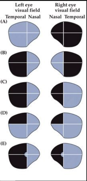
Which of these diagrams of the visual fields would be characteristic of a patient with right optic neuritis (inflammation of the optic nerve is commonly seen in patients with multiple sclerosis), which would severely impair action potential propagation along the affected optic nerve?
Note: the blackened areas represent regions of the visual field NOT seen by the subject.
A. (A)
B. (B)
C. (C)
D. (D)
E. (E)
Note: the blackened areas represent regions of the visual field NOT seen by the subject.
A. (A)
B. (B)
C. (C)
D. (D)
E. (E)

88
New cards
E
Which of these diagrams of the visual fields would be characteristic of a patient with a vascular lesion of the RIGHT anterior two-thirds of the occipital cortex?
A. (A)
B. (B)
C. (C)
D. (D)
E. (E)
A. (A)
B. (B)
C. (C)
D. (D)
E. (E)

89
New cards
B
Which of these diagrams of the visual fields would be characteristic of a patient with a traumatic head injury during a motor vehicle accident that caused sheering forces along the midline of the brain? This patient experienced sufficient sheering forces to sever white matter structures that cross the midline.
A. (A)
B. (B)
C. (C)
D. (D)
E. (E)
A. (A)
B. (B)
C. (C)
D. (D)
E. (E)

90
New cards
A
Which of the illustrated visual field deficits would be characteristic of a lesion anterior to the optic chiasm?
A. (A)
B. (B)
C. (C)
D. (D)
E. (E)
A. (A)
B. (B)
C. (C)
D. (D)
E. (E)

91
New cards
C
Which of the illustrated visual field deficits would be described as a complete LEFT homonymous hemianopia (without macular sparing)?
A. (A)
B. (B)
C. (C)
D. (D)
E. (E)
A. (A)
B. (B)
C. (C)
D. (D)
E. (E)

92
New cards
C
Which of the illustrated visual field deficits would be most likely to result from a lesion of the entire RIGHT optic tract?
A. (A)
B. (B)
C. (C)
D. (D)
E. (E)
A. (A)
B. (B)
C. (C)
D. (D)
E. (E)

93
New cards
D
Which of these diagrams of the visual fields would be characteristic of a person with a demyelinating lesion in the RIGHT Meyer's loop?
A. (A)
B. (B)
C. (C)
D. (D)
E. (E)
A. (A)
B. (B)
C. (C)
D. (D)
E. (E)
94
New cards
A
There is a person (patient “L.M.”) who happens to enjoy a cup of tea most days; however, since recovering from a serious illness, she tends to spill her tea. The reason she spills her tea is that she does not appreciate the movement of tea filling her tea cup as she pours it out. She also has great difficulty judging the movement of traffic when she crosses a street at a crosswalk. Which of the following best explains her visual impairment?
A. She has bilateral lesions in her lateral temporo-parieto-occipital associational cortex (involving the region of her angular gyri).
B. The parvocellular layers of her lateral geniculate nuclei (bilaterally) have degenerated.
C. She is blind in her dominant eye.
D. She has torn the center of her optic chiasm.
E. She has a large lesion in her inferior occipitotemporal association cortex (involving the fusiform gyrus) in her right hemisphere.
95
New cards
B
My colleague passes through airport security and recognizes his friend who is waiting to meet him. To my colleague’s surprise, he walks up to meet his friend, but his friend does not seem to recognize him until he says “Hey … it’s me!”. Being a neuroscientist himself, my colleague now suspects that his friend has suffered a brain injury that has produced prosopagnosia, which is failure to recognize the identity of a familiar face (a.k.a. “face-blindness”). Which of the following best explains this person’s prosopagnosia?
A. He has bilateral lesions in his lateral temporo-parieto-occipital associational cortex (involving the region of the angular gyri).
B. He has a large lesion in his inferior occipitotemporal association cortex (involving the fusiform gyrus) in his right hemisphere.
C. The magnocellular layers of his lateral geniculate nuclei (bilaterally) have degenerated.
D. He is blind in his non-dominant eye.
E. He has torn the center of his optic chiasm.
96
New cards
C
You examine a patient that you suspect has a neurological impairment. One simple test that is a component of the neurological exam is the test of the pupillary light reflex. When you test this patient, you discover that shining light in the right eye produces constriction of the right pupil, but not the left. Similarly, shining light in the left eye produces constriction of the right eye, but not the left. These findings would be consistent with which neurological lesion?
A. lesion of the left optic nerve
B. lesion of the right oculomotor nerve
C. lesion of the left oculomotor nerve
D. lesion of the right optic nerve
E. lesion of the left lateral tegmentum of the medulla
A. lesion of the left optic nerve
B. lesion of the right oculomotor nerve
C. lesion of the left oculomotor nerve
D. lesion of the right optic nerve
E. lesion of the left lateral tegmentum of the medulla
97
New cards
E
You are serving an EMS shift and are called to the scene of a motor vehicle accident. Unfortunately, the driver involved appears to be severely injured. As you evaluate this unconscious individual, you perform the pupillary light reflex and discover that the pupils are “fixed and dilated”, meaning that they are unresponsive to light shone in either eye. What is the BEST interpretation of this clinical sign?
A. the retinas in both eyes became completely detached during the accident
B. the center of the optic chiasm was torn during the accident
C. the corpus callosum was severed during the accident
D. the visual cortex was likely damaged in both hemispheres during the accident
E. the medial tegmentum of the pretectum and/or adjacent midbrain was likely damaged during the accident
98
New cards
B
Which of the following statements BEST expresses the concept of lower motor neurons?
Remember, we are always trying to identify the very best response among the options presented below. In this case, identify the statement that most fully, specifically, and unequivocally best expresses the concept of a lower motor neuron.
A. Lower motor neurons grow axons into mixed spinal nerves.
B. Lower motor neurons innervate the motor endplates of striated muscle fibers.
C. Lower motor neurons are the output neurons in layer 5 of the primary motor cortex.
D. Lower motor neurons are autonomic preganglionic neurons.
E. Lower motor neurons receive monosynaptic inputs from upper motor neurons.
99
New cards
D
Which of the following statements BEST expresses the concept of upper motor neurons?
A. Upper motor neurons are supraspinal neurons (localized superior to the spinal cord) that participate in motor control.
B. Upper motor neurons are alpha and gamma motor neurons that participate in motor control.
C. Upper motor neurons are the output neurons in layer 5 of the primary motor cortex that make monosynaptic connections with lower motor neurons.
D. Upper motor neurons are brainstem and cortical neurons that directly and indirectly (through local circuit neurons) govern the output of lower motor neurons.
E. Upper motor neurons are frontal lobe neurons that plan the kinematic strategies for achieving movement goals.
100
New cards
E
Which of the following pathways in the human central nervous system is MOST IMPORTANT for governing the skilled movements of the hands?
A. rubrospinal tract
B. anterior (ventral) corticospinal tract
C. vestibulospinal tract
D. reticulospinal tract
E. lateral corticospinal tract
A. rubrospinal tract
B. anterior (ventral) corticospinal tract
C. vestibulospinal tract
D. reticulospinal tract
E. lateral corticospinal tract